251
Radiol Bras. 2012 Set/Out;45(5):251–258
Endovascular treatment of thoracic aortic diseases: a single
center result analysis
*
Tratamento endovascular das doenças da aorta torácica: análise dos resultados de um centro
Eduardo Rafael Novero1, Patrick Bastos Metzger2, Javier Obregon1, Vanessa Luciene Abreu de Marco3, Fabio Henrique Rossi4, Samuel Martins Moreira5, Nilo Mitsuru Izukawa6, Antonio Massamitsu Kambara7
Objective: To analyze treatment outcomes in a consecutive series of patients submitted to endovascular treatment of thoracic aortic disease. Technical success, therapeutic success, morbimortality, rate of perioperative complications and reinterventions were taken into consideration. Materials and Methods: The present retrospective study was developed in a reference center in the period from January 2010 to July 2011, involving patients submitted to endovascular treatment of thoracic aortic disease. The study population was divided into two groups: group 1 (G1) – true thoracic aortic aneurysms, aortic ulcer and pseudoaneurysm; group 2 (G2) – chronic type B aortic dissection. Results: Out of a total of 55 patients, 29 belonged to the G1 and 26 to the G2 group. Mean ages were 66.8 ± 10 and 56.4 ± 7 years, respectively. The technical and therapeutic success reached respectively 86.3% and 68.6% in G1 and 100% and 74% in G2. The perioperative mortality rate was 10.3% in G1 and 19.3% in G2. The reintervention rate was 10.3% in G1 and 15.3% in G2. Conclusion: In the present study, the endovascular treatment of thoracic aortic disease proved to be a feasible method associated with acceptable rate of complications.
Keywords: Aneurysm; Vascular graft; Atherosclerosis.
Objetivo: Analisar os resultados do tratamento de uma série consecutiva de pacientes submetidos a tratamento en-dovascular de doenças da aorta torácica. Foram observados o sucesso técnico, o sucesso terapêutico, a morbimorta-lidade e a taxa de complicações perioperatórias e de reintervenções. Materiais e Métodos: Estudo retrospectivo, realizado em um centro de referência, no período de janeiro de 2010 a julho de 2011, em que foram analisados pacientes submetidos a correção endovascular de doenças da aorta torácica. A população foi dividida em dois grupos: grupo 1 (G1) – aneurismas de aorta torácica verdadeiros, úlcera aórtica e pseudoaneurisma; grupo 2 (G2) – dissecção aórtica tipo B crônica. Resultados: Em um total de 55 pacientes tratados, 29 pertenciam ao G1 e 26, ao G2. As idades médias foram 66,8 ± 10 e 56,4 ± 7 anos, respectivamente. Os sucessos técnico e terapêutico foram, respectiva-mente, 86,3% e 68,6% no G1 e 100% e 74% no G2. A mortalidade perioperatória foi 10,3% no G1 e 7,6% no G2, com taxa de mortalidade anual de 10,3% no G1 e de 19,3% no G2. As taxas de reintervenções foram 10,3% e 15,3%, respectivamente. Conclusão: Em nosso estudo, o tratamento endovascular das doenças da aorta torácica demons-trou ser um método viável e associado a aceitáveis taxas de complicações.
Unitermos: Aneurisma; Prótese vascular; Aterosclerose. Abstract
Resumo
* Study developed at Instituto Dante Pazzanese de Cardiolo-gia, São Paulo, SP, Brazil.
1. Interventional Cardiologists, MDs, Foreign Trainees at Centro de Intervenções Endovasculares (CIEV) – Instituto Dante Pazza-nese de Cardiologia, São Paulo, SP, Brazil.
2. Vascular Surgeon, MD, Trainee at Centro de Intervenções Endovasculares (CIEV) – Instituto Dante Pazzanese de Cardiolo-gia, São Paulo, SP, Brazil.
3. Cardiologist, MD, Resident of Vascular Echography, Hospi-tal do Servidor Público Estadual “Francisco Morato de Oliveira” (HSPE), São Paulo, SP, Brazil.
4. PhD, Physician Assistant, Section of Vascular Surgery, Member of Centro de Intervenções Endovasculares (CIEV) – Ins-tituto Dante Pazzanese de Cardiologia, São Paulo, SP, Brazil.
5. Vascular Surgeon, Physician Assistant, Section of Radiology, Member of Centro de Intervenções Endovasculares (CIEV) – Ins-tituto Dante Pazzanese de Cardiologia, São Paulo, SP, Brazil.
6. PhD, Head of Sector of Vascular Surgery and Centro de Intervenções Endovasculares (CIEV) – Instituto Dante Pazzanese de Cardiologia, São Paulo, SP, Brazil.
Novero ER, Metzger PB, Obregon J, Marco VLA, Rossi FH, Moreira SM, Izukawa NM, Kambara AM. Endovascular treatment of thoracic aortic diseases: a single center result analysis. Radiol Bras. 2012 Set/Out;45(5):251–258.
diameter present 80% of risk for rupture and death within one year of follow-up. The aneurysm diameter is the most relevant factor in the determination of such risk(1,2).
Aortic dissection is the most feared con-dition of the aorta and is twice as frequent as aortic rupture. The mortality rate asso-ciated with dissection ranges between 1% and 2% per hour, during the first 48 hours. Risk factors frequently associated with such condition include hypertension, car-diovascular and respiratory diseases. Dis-section is three times more frequent in men than in women. The most common entry
7. PhD, Head of Section of Radiology and Centro de Interven-ções Endovasculares (CIEV) – Instituto Dante Pazzanese de Cardiologia, São Paulo, SP, Brazil.
Mailing Address: Dr. Patrick Bastos Metzger. Instituto Dante Pazzanese de Cardiologia. Rua Doutor Dante Pazzanese, 500, Vila Mariana. São Paulo, SP, Brazil, 04012-180. E-mail: [email protected]
Received April 6, 2012. Accepted after revision August 9, 2012.
INTRODUCTION
tear sites are the ascending aorta, some centimeters distal from the aortic valve, and the descending aorta, below the origin of the subclavian artery. Acute dissection dif-ferentiates from chronic dissection in cases where the diagnosis is established within 14 days from the symptoms onset. In cases where a type B dissection progresses with ischemic complications, surgical or endo-vascular intervention is mandatory. Ap-proximately 30% of patients with type B dissection develop complications. The mortality associated with surgical correc-tion of type B acute disseccorrec-tion ranges from 30% to 80% in cases of renal or mesenteric ischemia. Endovascular therapy can offer techniques of reperfusion of ischemic vas-cular beds in complicated dissections, with lower risks than those in open surgery(3).
Aortic dissection and TAAs represent conditions with sometimes catastrophic consequences. Despite the developments in diagnostic methods, operative techniques and intra- and postoperative management, such conditions still constitute major chal-lenges for endovascular surgeons(4).
The concept that aortic diseases could be corrected by endoluminal prostheses po-sitioned within the aorta by means of cath-eters inserted through the femoral artery has emerged over the past few years. As the high morbimortality rate related to surgical interventions in cases of thoracic aneurysm dissection is considered, the endovascular technique becomes a very attractive option. Endovascular devices allow for a mini-mally invasive approach, with the flow being maintained without clamping(5).
Other advantages include lower blood transfusion rate, shorter hospital stay, shorter intensive care unit (ICU) stay and lower costs. The main disadvantage is that such a technique predisposes the patient to a higher number of reinterventions in the medium and long terms(4).
The endovascular intervention involv-ing the thoracic aorta has been poorly com-pared with open surgery in controlled and randomized clinical studies, but medium and long term post-implantation results have been encouraging, with lower rates of early morbimortality as compared with open surgery(6).
According to some authors, the endo-vascular treatment of true TAAs poses a
greater challenge than that performed in patients with chronic type B dissection, with lower rates of therapeutic success and higher mortality(6,7).
The present study is aimed at evaluat-ing the clinical results obtained with a con-secutive series of patients submitted to endovascular correction of TAAs and chronic type B aortic dissection, analyzing the technical and therapeutic success, morbimortality, complications and rein-tervention rate.
MATERIALS AND METHODS
The present retrospective, longitudinal and observational study was developed in a reference center for cardiovascular disor-ders, in the period between January, 2010 and July, 2011, evaluating 55 patients sub-mitted endovascular repair of thoracic aorta. Such a population was divided into two groups, as follows: group 1 (G1) – patients with true TAA, penetrating ulcer of thoracic aorta and pseudoaneurysm; group 2 (G2) – patients with type B aortic dissection and intramural hematoma. Both groups included patients whose indication for endovascular procedure was treatment of leakage of previous endoprosthesis, in-dependently of the underlying disease.
The present study included both male and female patients, with or without tho-racic symptoms, with indication of aortic correction for: a) TAA with > 55 mm in di-ameter, correction of type I or type III
leak-age; b) chronic type B dissection with an-eurysmal dilation > 55 mm or correction of type I or III leakage; c) thoracic aorta pseudoaneurysm; d) penetrating ulcer of thoracic aorta with > 2 cm in diameter and 1 cm in depth.
Exclusion criteria were the following: patients with proximal aortic neck with < 15 mm in length; distal aortic neck with < 15 mm in length from the celiac trunk; presence of thrombus or calcifications with > 50% of the neck diameter; external iliac arteries with < 7 mm in diameter; serum creatinine levels > 2.0 mg/dl or creatinine clearance < 30 ml/min.
Those patients who remained in the present study population according to the inclusion and exclusion criteria, were con-sidered as having a favorable anatomy and were referred to endovascular treatment. The evaluation of cardiac and/or anesthetic risks assessments was not taken into con-sideration for inclusion/exclusion of pa-tients.
In all of the cases, the diagnosis and the therapeutic schedule were based on com-puted tomography angiography, and the role of preoperative arteriography remained as an optional diagnostic method.
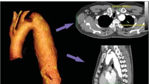
Computed tomography images recon-struction was performed with the aid of the Osirix® software with 3D viewer and
multiplanar reconstruction (MPR) modes in order to obtain diameter, angle and length of the aneurysms proximal and dis-tal aortic necks (Figure 1).
4. Complications – a) intraoperative complications: those occurred in the hemo-dynamics room during the intervention; b) intra-hospital complications: those oc-curred during hospital stay, out of the he-modynamics room and within 30 days af-ter the inaf-tervention.
The following events were considered as being complications: local bleeding; presence of inguinal or retroperitoneal he-matoma; inadvertent occlusion of the sub-clavian artery during the device deploy-ment; presence of peripheral embolization of lower limbs as a cause of acute arterial occlusion; occurrence of paraplegia or paraparesis; infections, either in the surgi-cal site, lower respiratory tract or in the utilized endoprosthesis; development of acute renal failure, defined as an increase in creatinine levels greater than twice the value observed previously to the procedure; and death.
5. Reintervention rate – interventions performed for maintenance of appropriate functioning of the endoprosthesis or for resolution of complications associated with the intervention.
Secondary outcomes
1. Hospitalization time – time span be-tween admission and hospital discharge.
2. Procedural time– elapsed time be-tween the start of anesthetic induction and the closure of the operative site.
Leakages
1. Initial or primary leakages – those originated during the initial procedure or those diagnosed within the first 30 postop-erative days.
2. Secondary leakages – those diag-nosed after 30 days from the initial proce-dure.
Definitions
Carotid-subclavian and/or carotid-ca-rotid shunt to extra-anatomic bypass graft performed for anchoring the endopros-thesis in zones 1 and 2, in cases of compro-mise of the left carotid or left subclavian arteries.
RESULTS
The present series included 55 patients submitted to correction of thoracic aortic diseases.
Surgical technique
All procedures were performed in the hemodynamics laboratory of the Centro de Intervenções Endovasculares (CIEV) of Instituto Dante Pazzanese de Cardiologia, in São Paulo, SP, Brazil.
The patients were treated under general anesthesia by inhalation. The antimicrobial prophylaxis was performed with 1.5 g of cefuroxime at the moment of anesthesia induction. The preferred approach was through the common femoral artery with uni- or bilateral open surgical dissection, according to the selected type of interven-tion or endoprosthesis. In case of unfeasibility of such an approach, the ex-ternal iliac artery by retroperitoneal access was utilized as an alternative.
The radiographic control was per-formed with a Siemens Artis Flat Panel apparatus. The following devices were uti-lized: Valiant® (Medtronic; Minneapolis,
MN, USA), Zenith® TX2® (Cook Medical;
Bloomington, IN, USA), TAG® (Gore
Medical; Flagstaff, AZ, USA), Hercules™ (MicroPort; Shangai, China), Relay®
(Bolton Medical; Sunrise, FL, USA), and E®-vita (Jotec GmbH; Hechingen,
Ger-many). Intraoperative arteriography was performed in all the patients. The immedi-ate postoperative follow-up was under-taken in the ICU in all the cases (Figure 2). All the individuals were postoperatively followed-up on an outpatient basis 15, 30, 180 and 360 days after the procedure. Af-ter the first year, the follow-up was per-formed once a year. Computed tomography angiography was performed at the 30th and 360th days of follow-up.
The analyzed outcomes were defined as per the following description:
Primary outcomes
1. Technical success – when the objec-tive of deploying the endoprosthesis in the affected area was achieved, either with or without the presence of leakage or other complications that might negatively influ-ence the progression of the aortic disease.
2. Therapeutic success – when the de-ployment of the endoprosthesis occurred without leakage or other complications that might affect the favorable course of the aortic disease.
3. Perioperative mortality – number of deaths recorded within 30 days after the procedure.
Figure 2. Intraprocedural aortography by left brachial approach (A) and final outcome of endovascular repair of thoracic aorta aneurysm (B). Absence of intraoperative leakage and preservation of left subcla-vian artery.
Group 1 comprised 29 patients. The pri-mary diseases in this population were true TAAs in 26 cases (89.6%), penetrating ul-cer in 2 cases (6.8%), and pseudoaneurysm in 1 case (3.4%). The mean age was 66.8% ± 10 years, and 18 patients were men (69%). The patients were asymptomatic in 20 cases (68.9%) and were incidentally diagnosed with basis on findings at routine examinations. The predominant risk factor was hypertension in 28 patients (96.8%), 19 patients were smokers (65.5%) and 12 (41.3%) presented dyslipidemia. Diabetes was present in 4 cases (13.7%). Obesity, with body mass index > 40, was present in 2 patients (6.89%) of the population (Table 1). Among the present comorbidities, the authors have found a high incidence of is-chemic cardiopathy (58.6%), acute renal failure (17.2%) and, in lesser degree, di-lated cardiopathy in one case (3.4%). Also, a significant percentage (20.6%) of patients previously submitted to endovascular aor-tic aneurysm correction was observed.
Chronic obstructive pulmonary disease was present in two cases (6.9%) (Table 2).
The indications for endovascular treat-ment in this population were distributed as follows: true TAAs in 23 cases (79.3%), leakage in previously implanted endopros-thesis in 3 cases (10.3%), penetrating ulcer in 2 cases (6.8%), and pseudoaneurysm in 1 case (3.4%).
Among the treated aneurysms, 17 were thoracic (10 saccular, and 7 fusiform), and 9 were thoracoabdominal (3 cases were type I, 3 type II, 2 type III and 1 case was type V, according to the Saffi classifica-tion).
The treatment was elective in all of the cases. Inhalation anesthesia was utilized in all cases, with liquor drainage in those pa-tients with indication for aortic reinter-vention, in those with previous abdominal or thoracic aortic surgery, and in those pa-tients with preoperatively documented vis-ceral or supra-aortic trunk occlusion. Tech-nical success was obtained in 86.2% of the
cases, i.e. in 25 patients it was possible to deploy the endoprosthesis in the desired location. In four cases, the endoprosthesis could not reach the desired location be-cause of technical or anatomical difficul-ties (calcification or tortuosity).
Therapeutic success was achieved in 58.6% of the cases, i.e., in 17 patients the endoprosthesis was deployed without leak-age or other events that might negatively affect the outcome of the intervention. The only cause of therapeutic failure was leak-age or its persistence, according to the in-dication for the intervention.
The complication rate was 48.2%, with the following most frequent intra-operative complication: femoral lesion in 7 cases (24.5%) and peripheral embolism in 1 case (3.4%). Intra-hospital postoperative com-plications included infection in 2 cases (6.8%) – both surgical wound infection –, acute renal failure in 2 patients (6.8%), paraplegia in 1 case (3.4%), and retroperi-toneal hematoma in 1 case (3.4%) (Table 3). Perioperative mortality rate was 10.3%. One death occurred due to aneurysm rup-ture during the deployment of the endo-prosthesis; two other deaths occurred as a consequence of sepsis by respiratory tract infection (Table 4). No death was observed 30 days after surgery. The total primary leakage rate was 27.5%, with type Ia leak-age being observed in 4 cases, and type Ib in 3 cases. Type IV leakage was present in 1 case (3.4%). There was no case of type III leakage and endoprosthesis migration during follow-up of the patients. The rate of reinterventions within one year was 10.3%, due to treatment of type I leakage (Table 4). One-year survival during follow-up was 89.7%. One death occurred due vascular causes.
The mean procedure duration was 78 minutes (ranging from 54 to 115 minutes), and the mean hospitalization time was 4.2 days with a variation of 7 days.
The most utilized endoprosthesis de-vices were the following: Valiant® in 13
cases (45%), Zenith® TX2® in 3 cases
(10.4%), TAG® in 3 cases (10.4%),
Her-cules™ in 3 cases (10.4%) and E®-vita in
3 cases (10.4%).
The rate of carotid-carotid shunt and/or carotid-subclavian shunt was 6.8%, i.e., 2 cases required such shunting.
Table 1 Clinical data (n = 55).
Characteristics of the population
Mean age (years)
Male patients
Symptomatic disease
True aneurysm
Pseudoaneurysms
Aortic ulcer
Aortic dissection with TAA
Severe obesity (BMI > 40)
Smoking (active or non-smoker for at least one year
Diabetes mellitus
Hypertension
Dyslipidemia
Value
Group 1 (n = 29)
66.8 ± 10
18
9
26 (89.6%)
1 (3.4%)
2 (6.8%)
0
2 (6.8%)
19 (65.5%)
4 (13.7%)
28 (96.8%)
12 (41.3%)
Group 2 (n = 26)
56.4 ± 7
16
19
0
0
0
26 (100%)
2 (7.2%)
12 (46.1%)
6 (23%)
26 (100%)
9 (34.6%)
TAA, thoracic aortic aneurysm; BMI, body mass index.
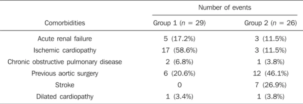
Table 2 Comorbidity data (n = 55).
Comorbidities
Acute renal failure
Ischemic cardiopathy
Chronic obstructive pulmonary disease
Previous aortic surgery
Stroke
Dilated cardiopathy
Number of events
Group 1 (n = 29)
5 (17.2%)
17 (58.6%)
2 (6.8%)
6 (20.6%)
0
1 (3.4%)
Group 2 (n = 26)
3 (11.5%)
3 (11.5%)
1 (3.8%)
12 (46.1%)
7 (26.9%)
The endoprosthesis anchoring zones were distributed as follows: 25 cases (86.2%) in zone 3, two cases in zone 4 (6.9%), one in zone 2 and one in zone 1 (Figure 3).
The indications for endovascular treat-ment in this population were distributed as follows: type B dissection in 15 cases (57.6%) and leakage in previously im-planted endoprosthesis in 11 cases (42.3%). In this group, all the patients were submitted to elective treatment.
General inhalation anesthesia was uti-lized in all cases, with liquor drainage in those patients with indication for aortic reintervention, in those with previous his-tory of thoracic or abdominal surgery, and in the patients who presented preopera-tively documented visceral or supra-aortic trunk obstruction. The technical success reached 100%. The endoprostheses were deployed in the desired site in all cases.
Therapeutic success was achieved in 74% of cases, i.e., in 20 patients the endoprosthesis was deployed without leak-age or other events that might negatively affect the outcome of the intervention. The sole cause of therapeutic failure was leak-age or its persistence, according to the in-dication for the intervention.
The rate of complications was 30.7%, and the most frequent intra-operative com-plications were femoral lesion in 2 cases (7.6%) and involuntary occlusion of the subclavian artery in 1 case (3.8%). Intra-hospital postoperative complications were the following: infections in 2 cases (7.6%), retroperitoneal hematoma in 1 case (3.8%), acute renal failure in 1 patient (3.8%), and paraplegia in 1 case (3.8%) (Table 3).
The perioperative mortality rate was 7.6%. Two deaths occurred: one because of sepsis caused by to lower respiratory tract infection, and another caused by cardiop-ulmonary arrest secondary to Chagas-re-lated diChagas-re-lated cardiopathy (Table 3). Three deaths occurred after 30 days from surgery, two because of recurrent dissection, and one because of acute pulmonary edema. The total rate of primary leakage was 23%, with type Ia leakage being observed in all such cases. Types II and III leakages as well as endoprosthesis migration were not ob-served during follow-up of such patients. The rate of reintervention within one year was 15.3%, as a result of type I leakage treatment (Table 4). The one-year survival rate during follow-up was 80.7%.
The mean duration of procedures was 67 minutes (ranging between 49 and 104
Table 3 Intra and perioperative complications (n = 55).
Outcome
Intraoperative complications
Peripheral embolization
Femoral artery injury
Subclavian occlusion
Intra-hospital complications
Paraplegia
Infection
Retroperitoneal hematoma
Acute renal failure
Death
Number of events
Group 1 (n = 29)
1 (3.4%)
7 (24.5%)
0
1 (3.4%)
2 (6.8%)
1 (3.4%)
2 (6.8%)
3 (10.3%)
Group 2 (n = 26)
1 (3.8%)
2 (7.6%)
1 (3.8%)
1 (3.8%)
2 (7.6%)
1 (3.8%)
1 (3.8%)
2 (7.6%)
Table 4 Primary leakage data (n = 55).
Types of endoleaks
Total
Type Ia
Type Ib
Type II
Type III
Type IV
Reintervention rate
Number of events
Group 1 (n = 29)
8 (27.5%)
4 (13.7%)
3 (10.3%)
0
0
1 (3.4%)
3 (10.3%)
Group 2 (n = 26)
6 (23%)
0
0
0
0
0
4 (15.3%)
Figure 3. Endoprosthesis anchoring zones in tho-racic aorta aneurysms.
Group 2 included 26 patients treated for chronic type B aortic dissection.
minutes), and the mean hospitalization time was 9.9 days with a variation of 8 days.
The most utilized endoprosthesis de-vices were the following: Valiant® in 10
cases (38.5%), Zenith® TX2® in 7 cases
(27%), TAG® in 6 cases (23%), Relay® in
2 cases (7.7%), and Hercules™ in 1 case (3.8%).
The rate of carotid-subclavian and/or carotid-carotid shunting was 23%, i.e., in 6 cases such shunt was necessary.
The endoprosthesis anchoring zones were distributed as follows: 6 cases in zone 2 (23%), 14 cases (53.8%) in zone III, and 6 cases in zone 4 (23%) (Figure3).
DISCUSSION
Despite the recognition of its indication, the endovascular treatment of chronic aor-tic disease presents results that are variable and dependent upon the analyzed popula-tion(7). Physiopathological and anatomic
differences between aneurysms and aortic dissections determine operative techniques translating into different results. Because of the clinical instability and weakness of the aortic wall in thoracic aortic dissection, the utilization of proximal compliant bal-loon after endoprosthesis implant is not recommended. On the other hand, in the case of aneurysms, the anatomic distortion translated into greater tortuosity of the aor-tic arch and higher incidence of stenosis by peripheral atherosclerotic plaques fre-quently limit the smooth advancement of the deployment system in this population. Brazilian authors have demonstrated a rate of technical success of 97% in 92 pa-tients treated for true TAAs, and 98% in 130 patients treated for type B aortic dis-section(7). The EUROSTAR study has
dem-onstrated a rate of primary technical suc-cess of 89% in patients with aortic dissec-tion(8).
In the present study, the rate of techni-cal success reached 86.2% for G1 and 100% for G2. In 4 cases, the devices could not be deployed in the descending thoracic aorta of patients with true aneurysms be-cause of limitations of the aortic and iliac-femoral axis anatomy.
The rate of therapeutic success was 58.6% for G1 and 73% for G2. Brazilian
authors have presented a rate of clinical success of 71% in cases of aneurysm and 84% in cases of dissection(7).
Type I leakage was the most frequent event associated with endovascular correc-tion of aortic aneurysm and disseccorrec-tion and may eventually lead to clinical failure. The rates reported in the literature range from 0% to 44%(9,10). In the present study
popu-lation, leakage was the only cause for thera-peutic failure. The rate of primary leakage in G1 was 27.5%, while in G2, it was 23%. It is known that a proximal neck smaller than 2 cm in the left subclavian artery and the presence of an entry tear located on the small curvature of the aortic arch constitute predisposing factors of leakage and endoprosthesis kinking observed in the angiographic follow-up.
The evolution of type I leakage is con-troversial. Some authors believe that there is necessity of deployment of an additional endoprosthesis, eventual embolization or surgical conversion. However, there are reports on spontaneous thrombosis without any other type of treatment, despite the ini-tial presence leakage during the procedure. The period of spontaneous resolution ranges from one week to eight months. In case of persistence of such leakage, correc-tive therapeutic interventions must be per-formed(11).
In the population with type I leakage,
the authors found five patients in G1 and two in G2 with low-volume leakages at ar-teriography, which spontaneously resolved between the third and sixth months of fol-low-up. Two G1 patients presented neck length < 2 cm. Such data translates into better rates of therapeutic success without precipitated interventions, and also into smaller future reinterventions.
No secondary leakage was observed in both groups. Considering the cases of spon-taneous resolution, the annual rates of reinterventions were 10.3% for G1 and 15.3% for G2. The VALOR study has pre-sented annual leakage rate of 17%, with type I leakage occurring in 6.3% of the cases, type II in 9.5%, and type III in 1.9%(12).
In the Group1, three patients (10.3%) presented leakage as indication for endo-vascular procedure. Among them, one pa-tient had a previous history of aortic
sur-gery, and two had previously undergone endovascular treatment. Upon the analysis of the type of treated leakage, the author observed that two patients had type Ia leak-age persisting after intervention, while one patient had type Ib leakage with therapeu-tic success after intervention. In the Group 2, 11 patients (42.3) had leakage as indica-tion for the endovascular treatment; four of them with previous aortic surgical treat-ment and seven with previous endovascular treatment. As regards leakage types, seven cases were of type Ia, with therapeutic suc-cess in three of them and persistence of leakage in four patients; and four cases presented type Ib leakage, all of them be-ing successfully corrected.
The authors have observed that the rate of therapeutic success was lower in cases of type Ia leakage than in cases of type Ib leakage, a phenomenon that was more no-ticeable in G1. Thus, in G1, type Ia leak-ages could not be corrected, while in G2 only 43% of the leakages were corrected. In order to explain such data, the authors observed that, in G1, the percentage of endoprosthesis anchoring in zones 1 and 2 was 6.8%. In 6.8% of the patients carotid-subclavian and/or carotid-carotid shunt was performed, which allowed for endo-prosthesis anchoring in zone 1 or 2 with greater safety, thus occluding the subcla-vian and/or the left common carotid arter-ies. In G2, 23% of the endoprostheses were anchored in zone 2. All such patients were submitted to carotid-subclavian shunting. Probably, the anchoring zone 2 was underutilized for correcting type Ia leak-ages in G1. Coverage of the subclavian artery is generally well tolerated and rarely the patients develop dizziness or left upper limb claudication. The VALOR study has demonstrated carotid-subclavian shunt in 5% of cases. Many groups are electively and belatedly submitted to such a shunting, in case symptoms manifest(12).
Avoiding aortic clamping with the endovascular technique reduces the com-plications by ischemia of the target organ, as well as cardiac (1% versus 10%), respi-ratory (8% versus28%) and renal compli-cations, as compared with open surgery(13).
French) and iliac-femoral atherosclerosis is very frequently observed in procedures with the endovascular technique.
In the VALOR study, the patients with true TAAs presented complications in 41% of cases(12). In the series published by the
Arizona Heart Institute, mild complication occurred in only 38% of patients with dis-section(14).
The present study has found a compli-cation rate of 48.2% in G1 and 30.7% in G2. The population treated for true TAAs shows femoral injury in 24.1% of cases, a rate considerably higher than in G2, with 7.6%. Peripheral atherosclerosis in this population plays a role not only in this type of injury, but also in the incidence of pe-ripheral embolization caused by the pro-longed manipulation of guidewires or of the device itself within the aortic arch. The endovascular correction of TAAs is asso-ciated with risk for medullary ischemia and permanent paraplegia. The main predictive factor is the extent of the aortic segment covered by the endoprosthesis(15).
Accord-ing to the Gore TAG protocol, which has compared open surgery versus endovas-cular treatment in TAAs, the incidence of paraplegia is lower in endovascular treat-ment (14% versus 3%)(16). Surgically
treated dissections have a paraplegia inci-dence of 19%, but with the endovascular technique the incidence is around 0–10% (mean = 1.8%)(17–19). The EUROSTAR
reg-istry presents paraplegia incidence of 4% in patients treated for aneurysms, and 0.8% for aortic dissections treated with endo-vascular approach(8).
In the analyzed population, two cases of permanent paraplegia were observed, as follows: one in G1 (3.4%) and another in G2 (3.8%). No case of stroke was observed in either group. Previous or simultaneous surgery, coverage of extensive aortic seg-ment and perioperative hypotension repre-sent risk factors for paraplegia after TAA correction. Prophylactic cerebrospinal fluid drainage may be useful in high-risk pa-tients, i.e., those patients presenting asso-ciated TAA and abdominal aortic aneu-rysm, previous history of either open or endovascular abdominal aneurysm correc-tion, need for iliac arterial conduit for endoprosthesis deployment in patients with a history of transient lower limb paralysis
and in the impossibility of cerebrospinal fluid drainage in less than one hour after endoprosthesis deployment in cases where paraparesis or paraplegia are expected. The main disadvantage of the preoperative drainage is the potential development of spinal hematoma in cases where heparin is administered during the intervention. In such an event, the cerebrospinal fluid drainage must be suspended(20,21). In the
present study, 17% of the G1 population received continuous spinal anesthesia cath-eter, while in G2, only 10% of the patients received it.
Outcomes after endoprosthesis implant in thoracic aortic diseases are favorable both at short and medium terms, with sig-nificantly lower rates of early mortality as compared with open surgery.
The VALOR(12) trial presents
intraop-erative mortality rate of 2.1%, while Stone et al. have presented a rate of 10.4% in patients treated for TAA(22). The
EURO-STAR registry presents intra-hospital mor-tality rate of 10%, increasing to 28% in patients submitted to emergency aneurysm repair(8). A protocol for Talent thoracic stent
graft for aneurysm repair presented intra-hospital mortality in 55 of cases(23).
Ac-cording to Stone et al., the intraoperative mortality reaches 15.1% in this popula-tion(22). In the present study, the G1
popu-lation presented one death attributed to endovascular intervention and two deaths caused by sepsis resulting from postopera-tive lower respiratory tract infection. The perioperative mortality rate in this popula-tion reaches 10.3%, falling within the variation of values reported in the litera-ture. The Talent thoracic stent graft system clinical study has demonstrated one-year survival rate of 91%. In the present study, G1 presented a survival rate of 89.6%, as no death was recorded within the period of 30 days to one year of follow-up.
Differently, the EUROSTAR registry re-ports a rate of intra-hospital mortality of 8.4% in patients treated for dissection(8).
The overall mortality rate of surgical repair for type B dissection is around 30%, reach-ing 50% in cases where such patients un-dergo surgery in emergency situations(3).
The EUROSTAR registry presented one-year survival rate of 90% in patients with dissection, while in the series from the
Arizona Heart Institute the one-year sur-vival rate was 85%(8,14). The G2 population
presented a perioperative mortality rate of 7.6%, with one-year survival of 80.7%. Three deaths occurred after 30 days from intervention: two cases for redissection and one for acute pulmonary edema. The present study demonstrated that the sur-vival rate in G2 is similar to that in other studies, and lower than in G1.
It is not clear if endovascular therapy af-fects the survival of patients who evolve to chronic Stanford type B dissection as com-pared with clinical treatment. The IN-STEAD trial has compared the medical treatment versus endograft prosthesis in 140 patients with dissection, and no differ-ence was observed in rates of all-cause mortality. The survival rate was 95.6% in the medical treatment group versus 88.9% in the endovascular treatment group, over a two-year follow-up. The rates of progres-sion and death were equal for both groups, while aortic remodeling reached 91.3% in the patients treated by endovascular ap-proach versus 19.4% in the medical treat-ment group, a fact suggestive of the neces-sity of endovascular treatment for those patients presenting emerging aortic compli-cations. However, at clinical follow-up, there were a reasonable number of patients who had to migrate to endovascular treat-ment(24).
CONCLUSIONS
REFERENCES
1. Bickerstaff LK, Pairolero PC, Hollier LH, et al. Thoracic aortic aneurysms: a population-based study. Surgery. 1982;92:1103–8.
2. Dapunt OE, Galla JD, Sadeghi AM, et al. The natural history of thoracic aortic aneurysms. J Thorac Cadiovasc Surg. 1994;107:1323–33. 3. Trimarchi S, Nienaber CA, Rampoldi V, et al.
Role and results of surgery in acute type B aortic dissection: insights from the International Reg-istry of Acute Aortic Dissection (IRAD). Circu-lation. 2006;114(1 Suppl):I357–64.
4. Palma JH, Buffolo E, Gaia D. Tratamento endo-vascular das doenças da aorta: visão geral. Rev Bras Cir Cardiovasc. 2009;24(2 Supl 1): 40s–44s. 5. Dake MD, Kato N, Mitchell RS, et al. Endovas-cular stent-graft placement for the treatment of acute aortic dissection. N Engl J Med. 1999;340: 1546–52.
6. Matalanis G, Durairaj M, Brooks M. A hybrid technique of aortic arch transposition and ante-grade stent graft deployment for complete arch repair without cardiopulmonary bypass. Eur J Cardiothorac Surg. 2006;29:611–2.
7. Alves CMR, Fonseca JHP, Souza JAM, et al. Tra-tamento endovascular nos aneurismas verdadei-ros e na dissecção aórtica do tipo B: fase intra-hospitalar, seguimento de médio prazo e uma reflexão sobre seleção de pacientes. Rev Bras Cardiol Invas. 2009;17:46–51.
8. Leurs LJ, Bell R, Degrieck Y, et al. Endovascular treatment of thoracic aortic diseases: combined experience from the EUROSTAR and United Kingdom Thoracic Endograft registries. J Vasc Surg. 2004;40:670–80.
9. Chagas Neto FA, Barreto ARF, Reis HF, et al. A importância do diagnóstico por imagem na clas-sificação dos endoleaks como complicação do tratamento endovascular de aneurismas aórticos. Radiol Bras. 2010;43:289–94.
10. Criado FJ, Clark NS, Barnatan MF. Stent-graft repair in the aortic arch and descending thoracic aorta: a 4-year experience. J Vasc Surg. 2002;36: 1121–8.
11. Lepore V, Lönn L, Delle M, et al. Endograft therapy for diseases of the descending thoracic aorta: results in 43 high-risk patients. J Endovasc Ther. 2002;9:829–37.
12. Fairman RM, Criado F, Farber M, et al. Pivotal results of the Medtronic Vascular Talent Thoracic Stent Graft System: the VALOR trial. J Vasc Surg. 2008;48:546–54.
13. Svensson LG, Crawford ES, Hess KR, et al. Vari-ables predictive of outcome in 832 patients un-dergoing repairs of the descending thoracic aorta. Chest. 1993;104:1248–53.
14. Nathanson DR, Rodriguez-Lopez JA, Ramaiah VG, et al. Endoluminal stent-graft stabilization for thoracic aortic dissection. J Endovasc Ther. 2005; 12:354–9.
15. Amabile P, Grisoli D, Giorgi R, et al. Incidence and determinants of spinal cord ischaemia in stent-graft repair of the thoracic aorta. Eur J Vasc Endovasc Surg. 2008;35:455–61.
16. Makaroun MS, Dillavou ED, Kee ST, et al. Endovascular treatment of thoracic aortic aneu-rysms: results of the phase II muiticenter trial of the GORE TAG thoracic endoprosthesis. J Vasc Surg. 2005;41:1–9.
17. Coselli JS, Conklin LD, LeMaire SA.
Thoracoab-dominal aortic aneurysm repair: review and up-date of current strategies. Ann Thorac Surg. 2002; 74:S1881–4.
18. Palma JH, Souza JAM, Alves CMR, et al. Self-expandable aortic stent-grafts for treatment of descending aortic dissections. Ann Thorac Surg. 2002;73:1138–42.
19. Elefteriades JA, Hartleroad J, Gusberg RJ, et al. Long-term experience with descending aortic dissection: the complication-specific approach. Ann Thorac Surg. 1992;53:11–21.
20. Cinà CS, Abouzahr L, Arena GO, et al. Cere-brospinal fluid drainage to prevent paraplegia during thoracic and thoracoabominal aortic aneu-rysm surgery: a systemic review and meta-analy-sis. J Vasc Surg. 2004;40:36–44.
21. Novero ER, Metzger PB, Angelieri FMR, et al. Correção endovascular do aneurisma da aorta abdominal: análise dos resultados de único cen-tro. Radiol Bras. 2012;45:1–6.
22. Stone DH, Brewster DC, Kwolek CJ, et al. Stent-graft versus open-surgical repair of the thoracic aorta: mid-term results. J Vasc Surg. 2006;44: 1188–97.
23. Fattori R, Nienaber CA, Rousseau H, et al. Re-sults of endovascular repair of the thoracic aorta with the Talent Thoracic stent graft: the Talent Thoracic Retrospective Registry. J Thorac Car-diovasc Surg. 2006;132:332–9.