Original Article
Artigo Original
Tatiana Rocha Silva1 Luciana Macedo de Resende2 Marco Aurélio Rocha Santos1
Descritores
Nervo vestibular Potencial Evocado Motor Vestíbulo do Labirinto Doenças Vestibulares Sáculo e Utrículo Keywords
Vestibular Nerve Motor Evoked Potentials Vestibule of the Labyrinth Vestibular Diseases Saccule and Utricle
Correspondence address:
Tatiana Rocha Silva
Rua Boninas, 1070, Pompeia, Belo Horizonte (MG), Brazil, CEP: 30280-220. E-mail: [email protected]
Received: 02/21/2015
Accepted: 06/01/2015
Study carried out at the Audiology Clinic at the São Geraldo Annex from the University Hospital at Universidade Federal de Minas Gerais – UFMG – Belo Horizonte (MG), Brasil.
(1) Graduate Program in Speech-Language Pathology and Audiology Sciences, School of Medicine,Universidade Federal de Minas Gerais – UFMG – Belo Horizonte (MG), Brazil.
(2) Speech Language Pathology and Audiology Department, School of Medicine, Universidade Federal de Minas Gerais – UFMG – Belo Horizonte (MG), Brazil.
Conlict of interests: nothing to declare.
Ocular and cervical vestibular evoked myogenic
potential simultaneous in normal individuals
Potencial evocado miogênico vestibular ocular
e cervical simultâneo em indivíduos normais
ABSTRACT
Purpose: To characterize the recording and analyze the results of the combined cervical and ocular vestibular evoked myogenic potential in individuals without hearing and vestibular complaints. Methods: In this study, 30 individuals without hearing complaints and hearing within normal limits were evaluated. Data were collected through the simultaneous recording of cervical and ocular vestibular evoked myogenic potential.
Results: Differences were observed between the right and left ears for the amplitude of waves P13 and N23 of the cervical vestibular evoked myogenic potential and the latency of wave N10 of the ocular vestibular evoked myogenic potential. For female subjects, there was no difference between the right and left ears for the amplitude of waves P13, N23, N10, and P15; interamplitude in cervical vestibular evoked myogenic potential and interamplitude in ocular vestibular evoked myogenic potential; and latency in waves P13, N23, N10, and P15. For male subjects, there was a difference between the right and left ears for the amplitude of wave P13.
Conclusion: The results of the combined cervical and ocular vestibular evoked myogenic potentials were consistent, because the responses generated by the vestibular evoked myogenic potentials presented an adequate morphology, latency, and amplitude, allowing for the evaluation of the ipsilateral descending vestibular pathways and the contralateral ascending vestibular pathways.
RESUMO
Objetivo: Caracterizar o registro e analisar os resultados do potencial evocado miogênico vestibular cervical e ocular combinado em indivíduos sem queixas auditivas e vestibulares. Métodos: Participaram da pesquisa 30 indivíduos sem queixa auditiva e com audição dentro dos padrões de normalidade. A coleta de dados foi realizada por meio do potencial evocado miogênico vestibular cervical e ocular registrados simultaneamente.
INTRODUCTION
The vestibular evoked myogenic potential (VEMP) is a muscular potential that originates in the sensory cells of the macula of saccule. Among the vestibular organs, the saccule is the most sensitive to intense sound stimulation, which can be justiied by the higher anatomical proximity of this organ to the cochlea(1,2).
The VEMP evaluates the neural pathway of the inferior vestibular nerve and reaches the vestibular nuclei. The lateral vestibular nucleus receives stimulus from the stimulation of the ipsilateral pathway, while the information originating from the opposite side (contralateral pathway) reaches the superior and medial vestibular nuclei. The efferent ibers of these nuclei run all along the lateral and medial vestibulospinal tracts, through the medulla, and go on to the cervical motor nuclei with the purpose of reaching the accessory nerve, which is the access point for the sternocleidomastoid muscle. This potential is called cervical VEMP(3).
The VEMP, then, is an evoked myogenic potential of aver-age latency,which evaluates the muscle response resulting from auditory stimulation. This neural response is a relex arc with three neurons that involve the inner ear, brainstem, and the vestibulospinal pathway. The VEMP is formed by the myo-genic responses activated by sound or galvanic stimulation and recorded with surface electromyography in the presence of muscle contraction. The auditory stimulation with sounds that have an elevated intensity is the most often used technique, with the response being captured in the cervical muscles in the form of muscle contraction(2,4-6).
Recent investigations have shown that VEMP can also be generated from extraocular muscles in response to sounds with an elevated intensity. This is called ocular VEMP. Unlike the cervical VEMP that evaluates the descending ipsilateral vestibu-lar pathway, the ocuvestibu-lar VEMP evaluates the superior vestibuvestibu-lar pathway: the ascending contralateral pathway(7-14).
The cervical VEMP is composed of two sets of biphasic waveforms. The irst biphasic potential has a positive peak (P) with an average latency of 13 milliseconds (ms), followed by a negative peak (N) with an average latency of 23 ms, making it known as P13–N23 (Figure 1). The ocular VEMP is composed of two sets of biphasic waveforms. The irst biphasic potential has a negative peak (N) with an average latency of 10 ms, fol-lowed by a positive peak (P) with an average latency of 15 ms, making it known as N10–P15 (Figure 2).
The VEMP can evaluate disorders in any of the structures of the neural pathway. Thus, owing to its high reproducibility and the peculiarity of observing structures that are not analyzed in the traditional vestibular examinations, the responses of the VEMP can be used clinically, with innumerable applications in diagnostics for vestibular disorders(15).
The irst study done with the cervical and ocular VEMPs, tested simultaneously, was on normal individuals and individ-uals with superior canal dehiscence syndrome(16). The authors
utilized the cervical and ocular VEMP techniques simultane-ously on normal individuals and individuals with unilateral vestibular hypofunction(17).
The technique employed for the combined cervical and ocular VEMPs generates information that permits the evalua-tion of the cervical vestibular system. However, if the simulta-neous test can or cannot be substituted by the tests performed separately is not yet a consensus. Therefore, methodological questions should be cleared up, seeing that the standardization of records of these potentials is an essential criterion for the reproducibility and sensitivity of the examination.
This study was justiied by the possibility of determining the applicability of the simultaneous ocular and cervical VEMPs technique, contributing to the standardization of the technique and its utilization in an otoneurological diagnosis.
The objective of the study was to characterize the record-ing and analyze the results of the combined cervical and ocular VEMPs in individuals without hearing and vestibu-lar complaints.
METHODS
The procedures of this study were approved by the Research Ethics Committee from Universidade Federal de Minas Gerais (UFMG), under protocol NºCAAE 32505314.0.0000.5149 (Resolution 466/12 from the Conselho Nacional de Saúde–CONEP).
This was a descriptive study, with qualitative and quan-titative analyses. Thirty individuals were invited to take part in the study. The sample, then, was composed of 15 female and 15 male participants, between the ages of 18 and 53 years.
The participants in this study were selected at the UFMG School of Medicine and at the Audiology Clinic at the São Geraldo Annex from the University Hospital at UFMG through nonprobability, convenience sampling. The participants in
P13 13.60 -16.76 µV
27.38 µV
N23 21.20
Figure 1. Line obtained through the recording of the cervical vestibular evoked myogenic potential
N10 10.60 1.81 µV
0.34 µV
P15 15.30
this study were personally told about the objectives of the study, the absence of health hazards, the guarantee of coni-dentially regarding their identity or any other characteristics, which might make it possible to identify them, and the sched-ule for the study. After all the participants were well informed about the study, they signed the informed consent.
The data were collected at the Audiology Clinic at the São Geraldo Annex from the University Hospital at UFMG. All the participants were subjected to a basic audiological evalu-ation. This evaluation was composed of anamnesis, otoscopy, pure tone audiometry, speech audiometry, tympanometry, and acoustic relex.
In anamnesis, the participants provided information such as personal data, audiology history, and aspects related to overall health. To perform the otoscopy, the Heine®, Mini 2000, otoscope
was used. The pure tone audiometry and the speech audiometry were performed in a sound booth and with a one-channel audi-ometer, model AD 229b, by Interacoustics®, utilizing TDH-39
headphones and a B-71 bone vibrator. The tympanometry and the acoustic relex were performed using a middle ear analyzer, model AZ7, by Interacoustics®.
The inclusion criteria utilized were: participants with-out hearing complaints and/or prior ear diseases and with an audiological evaluation within the normal limits. An audio-logical evaluation within the normal limits was considered to have pure tone air conduction audiometry up to 25 dB at frequencies of 250 to 8,000 Hz and pure tone bone con-duction audiometry up to 15 dB at frequencies of 500 to 4,000 Hz with a difference between the air and bone con-duction lower than or equal to 10dB, Type A tympanogram, and acoustic relexes at frequencies of 500, 1,000, 2,000, and 4,000 Hz. For the pure tone audiometry, the guidelines established by Silman and Silverman(18) were considered,
and for the tympanogram, the guidelines established by Jerger(19)were considered.
The following subjects were excluded from the study: patients with neurological diseases, neoplasms, ear infections, tympanic membrane perforation, a history of craniocerebral trauma, prior otologic surgery, and who were unable to perform cervical rotation movements and ocular movements.
After the basic audiological evaluation, participants were referred to the electrophysiological evaluation through the VEMP.
The VEMP was conducted in a comfortable and quiet setting with equipment from Labat®, utilizing two channels.
The stimulus was presented by way of insert earphones, model ER 3A, with disposable foam ear tips. A hearing stimulus with 120-dB nHL tone bursts was used. In this study, a ilter with a bandpass from 10 to 1,500 Hz was used. To obtain results, 100 stimuli were presented, at a frequency of 500 Hz, at a rate of 4 stimuli per second. The window for analysis was 50 ms. Each individual was submitted to, at least, two stimuli on each side, to verify the replication of the potential. The impedance values were veriied before each recording, having to be less than 5 KΩ.
To perform the VEMP, the participant’s skin was cleaned with pure alcohol followed by an abrasive paste; a small
amount of conductive paste was placed on the surface elec-trodes, and they were set with adhesive tape. To record the ocular VEMP, the active electrode (negative electrode) from channel 1 was placed around 1 cm below the bottom eyelid, and the reference electrode (positive electrode) was placed at approximately 1 cm from the active electrode. To simulta-neously record the cervical VEMP, the active electrode from channel 2 was placed on the side opposite to channel 1, on the front edge of the upper-third of the sternocleidomastoi-deus, and the reference electrode in the furcula. A ground electrode was placed on theforehead (Fpz). This way, the position of the electrodes allowed the recording of the ocu-lar and cervical VEMPsto be simultaneous, channel 1 being utilized to register the ocular VEMP and channel 2 to regis-ter the cervical VEMP.
During the examination, the participants were asked to sit on the chair and keep the rotation of the head toward the side opposite the stimulated ear, provoking a contraction in the sternocleidomastoideus muscle. At the same time, they were instructed to look at a ixed target located on the wall of the testing room and, right after, toward a ixed point located above this target, which formed a vertical viewing angle of approxi-mately 30 degrees above the horizontal plane formed by the participants’ head. Subsequently, the contralateral recording of the cervical and ocular VEMPs was performed with the same technique.
After the data were collected, they were tabulated and sub-mitted for statistical analysis. The statistical analysis was done using the software BioSat, version 5.0. Initially, a descriptive analysis was done, which included measurements for central tendency (average and median), dispersion (standard deviation), and position (maximum and minimum). The normality of the samples was observed through the Kolmogorov–Smirnov and Shapiro–Wilk tests. In addition to the descriptive analysis, an inference statistic was done, through the Student t-test and the Mann–Whitney test to compare the two samples independently. The χ2-test was applied for comparison of frequencies obtained
through the calculation of the asymmetry rate. A signiicance level of 5% (p≤0.05) was adopted.
RESULTS
The average age of the population studied was 31.8 (stan-dard deviation of 8.08) years. For female subjects, the aver-age aver-age was 35.07(standard deviation of 7.8) years, and for malesubjects, the average age was 28.6 (standard deviation of 7.25) years.
shown for the central tendency values with stimulus in the right and left ears.
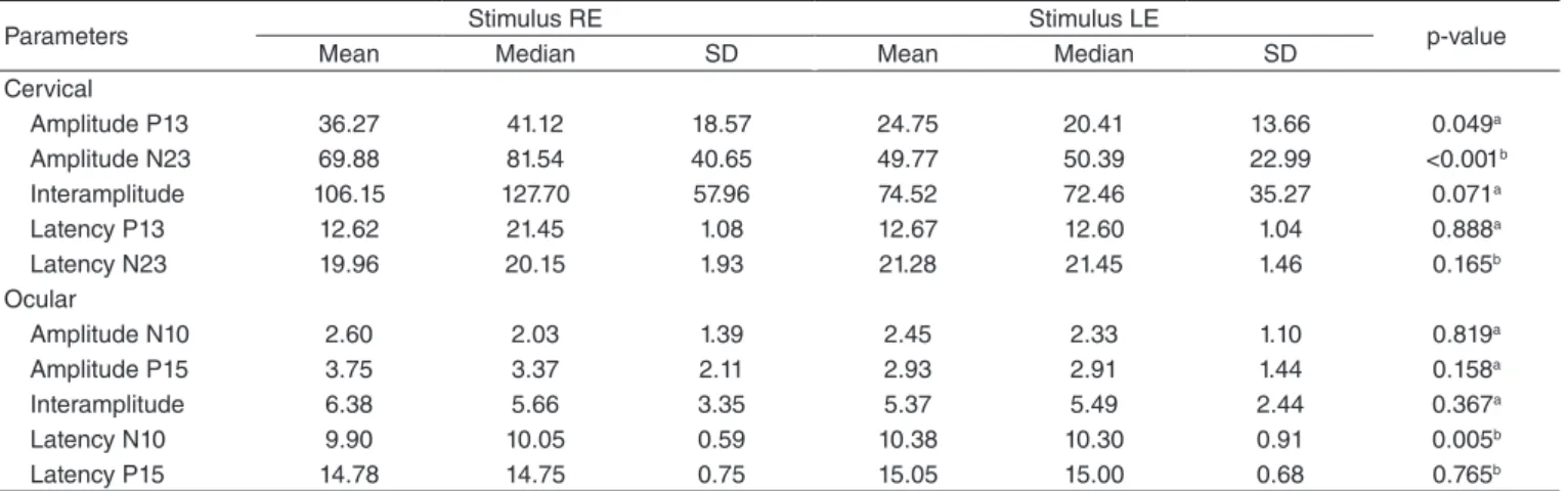
In the inferential statistical analysis, when considering the entire sample, it was found, through Table 3, that there was a difference between the right and left ears, for cervical VEMP, for the amplitude of waves P13 and N23 and, for the ocular VEMP, for the latency of wave N10.
In femalesubjects, for cervical VEMP, it was found that there was no difference between the right and left ears for the amplitude of waves P13 (p=0.967), N23 (p=0.067),interamplitude (p=0.917), and for the latency of waves P13 (p=0.519) and N23 (p=0.124). For the ocular VEMP, there was also no difference between the right and left ears for the amplitude of waves N10 (p=0.787), P15 (p=1.000), interamplitude (p=0.787), and for the latency of waves N10 (p=0.091) and P15 (p=0.259).
Table 4 shows that, in male subjects, there was a difference between the right and left ears, for the cervical VEMP, in the amplitude of wave P13.
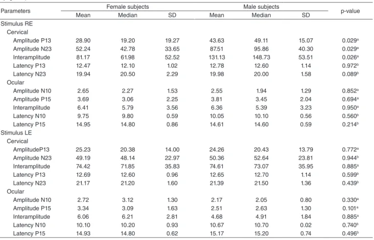
In the comparative analysis, between female subjects and male subjects, it was found in Table 5 that there was a differ-ence between the genders studied, in the stimulus of the right ear, in the amplitude of waves P13 and N23, and the interam-plitude in the cervical VEMP. For the stimulus in the left ear, it was found that there was no difference between femaleand malesubjects.
Regarding the asymmetry index, for the cervical VEMP, there was also no difference between female and malesub-jects. The asymmetry index value varied between 0 and 69%.
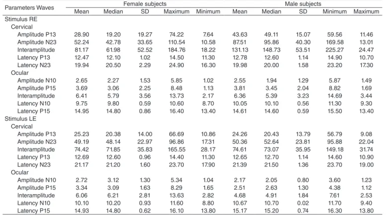
Table 2. Central tendency measurements, dispersion, and position for latency (ms) and amplitude (µV) for the combined ocular and cervical vestibular evoked myogenic potentials for female and male subjects
Parameters Waves Female subjects Male subjects
Mean Median SD Maximum Minimum Mean Median SD Minimum Maximum
Stimulus RE Cervical
Amplitude P13 28.90 19.20 19.27 74.22 7.64 43.63 49.11 15.07 59.56 11.46
Amplitude N23 52.24 42.78 33.65 110.54 10.58 87.51 95.86 40.30 169.58 13.01
Interamplitude 81.17 61.98 52.52 184.76 18.22 131.13 148.73 53.51 225.27 24.47
Latency P13 12.47 12.10 1.02 14.50 11.30 12.78 12.60 1.14 14.90 10.70
Latency N23 19.94 20.50 2.29 24.90 16.30 19.98 20.00 1.58 23.20 17.30
Ocular
Amplitude N10 2.65 2.27 1.53 5.85 1.02 2.55 1.94 1.29 5.87 1.49
Amplitude P15 3.69 3.06 2.25 8.48 1.13 3.81 3.45 2.04 8.82 1.69
Interamplitude 6.41 5.79 3.56 13.73 2.17 6.36 5.39 3.23 14.69 3.44
Latency N10 9.75 9.80 0.59 10.60 8.70 10.05 10.10 0.56 11.30 9.30
Latency P15 14.95 14.80 0.86 16.40 13.40 14.61 14.60 0.59 15.50 13.40
Stimulus LE Cervical
Amplitude P13 25.23 20.38 14.00 66.69 10.86 24.26 20.43 13.79 56.79 9.08
Amplitude N23 49.19 48.14 22.97 96.86 17.31 50.36 52.64 23.81 95.88 22.04
Interamplitude 74.42 71.85 35.83 165.55 28.17 74.61 73.07 35.95 149.18 31.74
Latency P13 12.69 12.60 0.96 14.40 11.30 12.65 12.70 1.14 14.60 10.90
Latency N23 21.17 21.20 1.60 23.70 17.90 21.39 21.50 1.36 23.70 19.00
Ocular
Amplitude N10 2.72 3.12 1.30 5.34 1.04 2.17 2.05 0.80 3.60 1.23
Amplitude P15 3.34 3.09 1.63 8.29 1.65 2.51 2.63 1.30 4.38 1.12
Interamplitude 6.06 6.21 2.81 13.63 2.82 4.68 4.91 1.84 7.61 2.53
Latency N10 10.10 10.20 0.93 11.60 8.80 10.67 10.70 0.02 11.70 9.40
Latency P15 14.93 14.80 0.62 16.10 13.80 15.17 15.20 0.74 16.30 13.80
Caption: SD = standard deviation; RE = right ear; LE = left ear
Table 1. Central tendency measurements, dispersion, and position for latency (ms) and amplitude (µV) for the combined ocular and cervical vestibular evoked myogenic potentials
Caption: SD = standard deviation; RE = right ear; LE = left ear
Parameters waves Mean Median SD Maximum Minimum Stimulus RE
Cervical
Amplitude P13 36.27 41.12 18.57 74.22 7.64 Amplitude N23 69.88 81.54 40.65 169.58 10.58 Interamplitude 106.15 127.70 57.96 225.27 18.22 Latency P13 12.62 21.45 1.08 14.90 10.70 Latency N23 19.96 20.15 1.93 24.90 16.30 Ocular
Amplitude N10 2.60 2.03 1.39 5.87 1.02
Amplitude P15 3.75 3.37 2.11 8.82 1.13
Interamplitude 6.38 5.66 3.35 14.69 2.17
Latency N10 9.90 10.05 0.59 11.30 8.70
Latency P15 14.78 14.75 0.75 16.40 13.40 Stimulus LE
Cervical
Amplitude P13 24.75 20.41 13.66 66.69 9.08 Amplitude N23 49.77 50.39 22.99 96.86 17.31 Interamplitude 74.52 72.46 35.27 163.55 28.17 Latency P13 12.67 12.60 1.04 14.60 10.90 Latency N23 21.28 21.45 1.46 23.70 17.90 Ocular
Amplitude N10 2.45 2.33 1.10 5.34 1.04
Amplitude P15 2.93 2.91 1.44 8.29 1.12
DISCUSSION
The responses obtained in this sample, from normal indi-viduals, demonstrate that it is possible to record combined cer-vical and ocular VEMPs. The response analysis of the VEMP revealed results that are similar to other studies in terms of amplitude and latency values of waves(3,16,17).
When confronting the right and left sides, it was found that there was a difference between the amplitude results for waves P13 and N23. This result is not in coherence with the consulted literature, which did not ind differences between the right and left ears for the amplitude of waves P13 and N23(4-6).
There was also a difference in the amplitude for male sub-jects, between the right and left ears, for wave P13. The average value in amplitude for P13 was higher in the right ear. Among femaleand malesubjects, the differences in the amplitude of waves P13 and N23 were also noted, when the right ear was stimulated. The average value for amplitude in waves P13 and N23 was higher in malesubjects. The differences observed
could be justiied owing to the difference in muscle tone of the sternocleidomastoideus muscle. Once again, there was no difference, for amplitude, between the ears for either gender, in the ocular VEMP.
According to some authors, the amplitude can be inluenced by muscular force, allowing it to alter depending on age and the degree of inclination of the body. Thus, it would not be a reliable parameter for clinical diagnoses regarding the func-tion of the vestibular system(2,16).
On the other hand, studies indicated the importance of monitoring the tension of the sternocleidomastoid muscle during the entire VEMP evaluation, so that the difference between the amplitude could be eliminated and only the sac-cule function actually be assessed. Such presumption is still controversial, seeing that some authors agree and others dis-agree with this discussion(2,3,15,16).
However, there are researchers who agree that the absolute amplitude values should not be utilized in the analysis of the VEMP, because they cannot be reproduced owing to the great intersubject variability, and are dependent on a few factors, Table 3. Comparison between the right and left ear for latency (ms) and the amplitude (µV) for combined ocular and cervical vestibular evoked myogenic potentials
Parameters Stimulus RE Stimulus LE p-value
Mean Median SD Mean Median SD
Cervical
Amplitude P13 36.27 41.12 18.57 24.75 20.41 13.66 0.049a
Amplitude N23 69.88 81.54 40.65 49.77 50.39 22.99 <0.001b
Interamplitude 106.15 127.70 57.96 74.52 72.46 35.27 0.071a
Latency P13 12.62 21.45 1.08 12.67 12.60 1.04 0.888a
Latency N23 19.96 20.15 1.93 21.28 21.45 1.46 0.165b
Ocular
Amplitude N10 2.60 2.03 1.39 2.45 2.33 1.10 0.819a
Amplitude P15 3.75 3.37 2.11 2.93 2.91 1.44 0.158a
Interamplitude 6.38 5.66 3.35 5.37 5.49 2.44 0.367a
Latency N10 9.90 10.05 0.59 10.38 10.30 0.91 0.005b
Latency P15 14.78 14.75 0.75 15.05 15.00 0.68 0.765b
aMann–Whitney test (p≤0.05); bStudent t-test (p≤0.05)
Caption: SD = standard deviation; RE = right ear; LE = left ear
Table 4. Comparison between the right and left ears for latency (ms) and amplitude (µV) for combined ocular and cervical vestibular evoked myogenic potential in male subjects
Parameters Stimulus RE Stimulus LE p-value
Mean Median SD Mean Median SD
Cervical
Amplitude P13 43.63 49.11 15.07 24.26 20.43 13.79 0.008a
Amplitude N23 87.51 95.86 40.30 50.36 52.64 23.81 0.202b
Interamplitude 131.13 148.73 53.51 74.61 73.07 35.95 0.272b
Latency P13 12.78 12.60 1.14 12.65 12.70 1.14 0.995b
Latency N23 19.98 20.00 1.58 21.39 21.50 1.36 0.654b
Ocular
Amplitude N10 2.55 1.94 1.29 2.17 2.05 0.80 0.468a
Amplitude P15 3.81 3.45 2.04 2.51 2.63 1.30 0.054a
Interamplitude 6.36 5.39 3.23 4.68 4.91 1.84 0.152a
Latency N10 10.05 10.10 0.56 10.67 10.70 0.02 0.051a
Latency P15 14.61 14.60 0.59 15.17 15.20 0.74 0.422b
aMann–Whitney test (p≤0.05); bStudent t-test (p≤0.05)
Table 5. Comparison between male and female subjects for latency (ms) and amplitude (µV) for combined ocular and cervical vestibular evoked myogenic potential
Parameters Female subjects Male subjects p-value
Mean Median SD Mean Median SD
Stimulus RE Cervical
Amplitude P13 28.90 19.20 19.27 43.63 49.11 15.07 0.029a
Amplitude N23 52.24 42.78 33.65 87.51 95.86 40.30 0.029a
Interamplitude 81.17 61.98 52.52 131.13 148.73 53.51 0.026a
Latency P13 12.47 12.10 1.02 12.78 12.60 1.14 0.972b
Latency N23 19.94 20.50 2.29 19.98 20.00 1.58 0.089b
Ocular
Amplitude N10 2.65 2.27 1.53 2.55 1.94 1.29 0.852a
Amplitude P15 3.69 3.06 2.25 3.81 3.45 2.04 0.694a
Interamplitude 6.41 5.79 3.56 6.36 5.39 3.23 0.950a
Latency N10 9.75 9.80 0.59 10.05 10.10 0.56 0.560b
Latency P15 14.95 14.80 0.86 14.61 14.60 0.59 0.214b
Stimulus LE Cervical
AmplitudeP13 25.23 20.38 14.00 24.26 20.43 13.79 0.772a
Amplitude N23 49.19 48.14 22.97 50.36 52.64 23.81 0.944b
Interamplitude 74.42 71.85 35.83 74.61 73.07 35.95 0.885a
Latency P13 12.69 12.60 0.96 12.65 12.70 1.14 0.599b
Latency N23 21.17 21.20 1.60 21.39 21.50 1.36 0.439b
Ocular
Amplitude N10 2.72 3.12 1.30 2.17 2.05 0.80 0.330a
Amplitude P15 3.34 3.09 1.63 2.51 2.63 1.30 0.101a
Interamplitude 6.06 6.21 2.81 4.68 4.91 1.84 0.885a
Latency N10 10.10 10.20 0.93 10.67 10.70 0.02 0.740b
Latency P15 14.93 14.80 0.62 15.17 15.20 0.74 0.496b
aMann–Whitney test (p≤0.05); bStudent t-test (p≤0.05) Caption: SD = standard deviation; RE = right ear; LE = left ear
such as the intensity of the stimulus and the level of tonic con-traction of the sternocleidomastoid muscle(5,6,17).
During this test, it was decided that the participants would be told to rest during an average of one minute between each VEMP capture; in other words, there was a break between the stimuli. This was done to avoid tiring out of the individ-ual being tested and, consequently, the fatigue of the sterno-cleidomastoid muscle, because a high rate of discharge can cause exhaustion in the sensory cells and, therefore, habitu-ation of the relex.
In the attempt to make the amplitude a measurable parameter in the VEMP test, some authors suggest using the asymmetry index. This index relects the interaural amplitude difference, measured by the average amplitude of this response. Thus, for the interpersonal comparison of the amplitude of responses, the asymmetry index, not the absolute values of the amplitudes, should be utilized.
In this study, no difference was observed for the asymmetry index. Therefore, this result is coherent with the consulted lit-erature. It is important to emphasize that this index is variable in studies and is considered insigniicant for values between 0 and 47%. However, in this study, the asymmetry index varied between 0 and 69%(16).
The literature described the inluence of the cervical muscu-lar contraction and the intensity of the stimulus over the ampli-tude and latency of the response in the VEMP record, and it was
found that there was a linear relationship between the degree of muscle contraction and the amplitude of the responses, but this variation was not observed in the latency. Thus, the abso-lute latencies are considered useful clinical parameters for the evaluation of the neural conduction, contributing to the auxil-iary diagnosis of neurological pathologies(15,16).
In this study, differences between the latency of the waves were not found. It is important to point out that, in the ocu-lar VEMP, differences were found in the latency of wave N10 between the right and left ears when considering the entire sample. This result is not in coherence with the consulted lit-erature; so, the difference between the ears for latency in wave N10 should be considered with caution(7-9,11,17).
The number of similar studies is reduced, conirming the methodological and logistical dificulty of this type of study. Thus, other studies should be performed with more casuistry and controlling variables that could interfere in the possible results.
Various publications have utilized the VEMPs as a method to diagnose or even contribute to the diagnosis of a range of otoneurological diseases, such as Meniere’s disease, superior semicircular canal dehiscence, vestibular neuronitis, vestibular schwannomas, postintratympanic administration of gentamicin control, and even perilymphaticistulae(1,3,4,6,15).
low cost, and does not cause the patient any discomfort. However, studies for technique standardization are necessary as are those for the sustainability of its utilization as a routine method(2).
CONCLUSION
The combined cervical and ocular VEMP results were con-sistent. The responses generated by the VEMP presented ade-quate morphology, latency, and amplitude.
This study demonstrates the applicability of the protocol of simultaneously recording the cervical and ocular VEMPs. The use of the protocol in the routine of the clinic allows the evaluation of the ipsilateral descending vestibular pathway and the contralateral ascending vestibular pathway. Thus, the evaluation time could be reduced, and, consequently, the time for recording the evoked potential of vestibular origin could also be reduced.
*TRS developed the study and the schedule, did the literature research, the data collection and analysis, the drafting of the article, the submission and proceedings for the article; MARS developed the study and the schedule, analyzed data, edited the article and approved the inal draft; LMR developed the study, edited the article and approved the inal draft.
REFERENCES
1. Kantner C, Gürkov R. Characteristics and clinical applications of ocular vestibular evoked myogenic potentials. Hear Res. 2012;294(1-2):55-63. 2. Rey-Martínez J, Rama-López J, Pérez-Fernández N, Guzmán RBD.
¿Cómo analizar un potencial evocado miogénico vestibular? Aplicación de un método no lineal. Acta Otorrinolaringol Esp. 2011;62(2):126-31. 3. Chiarovano E, Zamith F, Vidal PP, Waele C. Ocular and cervical VEMPs:
a study of 74 patients suffering from peripheral vestibular disorders. Clin Neurophysiol. 2011;122(8):1650-9.
4. Oh SY, Kim JS, Yang TH, Shin BS, Jeong SK. Cervical and ocular vestibular-evoked myogenic potentials in vestibular neuritis: comparison between air- and bone-conducted stimulation. J Neurol. 2013;260(8):2102-9. 5. Park HJ, Lee IS, Shin JE, Lee YJ, Park MS. Frequency-tuning characteristics
of cervical and ocular vestibular evoked myogenic potentials induced by air-conducted tone bursts. Clin Neurophysiol. 2010;121(1):85-9.
6. Manzari L, Tedesco AR, Burgess AM, Curthoys IS. Ocular and cervical vestibular-evoked myogenic potentials to bone conducted vibration in Ménière’s disease during quiescence vs during acute attacks. Clin Neurophysiol. 2010;121(7):1092-101.
7. Todd NP. The origin of the ocular vestibular evoked myogenic potential (OVEMP). Clin Neurophysiol. 2010;121(6):978-80.
8. Rosengren SM, Colebatch JG, Straumann D, Weber KP. Why do oVEMPs become larger when you look up? Explaining the effect of gaze elevation on the ocular vestibular evoked myogenic potential. Clin Neurophysiol. 2013;124(4):785-91.
9. Murofushi T, Nakahara H, Yoshimura E. Assessment of the otolith-ocular reflex using ocular vestibular evoked myogenic potentials in patients with episodic lateral tilt sensation. Neurosci Lett. 2012;515(2):103-6. 10. Iwasaki S, Chihara Y, Smulders YE, Burgess AM, Halmagyi GM, Curthoys
IS, et al. The role of the superior vestibular nerve in generating ocular vestibular-evoked myogenic potentials to bone conducted vibration at Fz. Clin Neurophysiol. 2009;120(3):588-93.
1. Chihara Y, Iwasaki S, Ushio M, Fujimoto C, Kashio A, Kondo K, et al. Ocular vestibular-evoked myogenic potentials (oVEMPs) require extraocular muscles but not facial or cochlear nerve activity. Clin Neurophysiol. 2009;120(3):581-7.
12. Piker EG, Jacobson GP, McCaslin DL, Hood LJ. Normal characteristics of the ocular vestibular evoked myogenic potential. J Am Acad Audiol. 2011;22(4):222-30.
13. Govender S, Rosengren SM, Todd NP, Colebatch JG. Ocular vestibular evoked myogenic potentials produced by impulsive lateral acceleration in unilateral vestibular dysfunction. Clin Neurophysiol. 2011;122(12):2498-504.
14. Schubert MC, Minor LB. Vestibulo-ocular physiology underlying vestibular hypofunction. Phys Therapy. 2004;84(4):373-85.
15. Ribeiro S, Almeida RR, Caovilla HH, Ganança MM. Dos potenciais evocados miogênicos vestibulares nas orelhas comprometida e assintomática na Doença de Ménière unilateral. Rev Bras Otorrinolaringol. 2005;71(1):60-6.
16. Welgampola MS, Myrie OA, Minor LB, Carey JP. Vestibular-evoked myogenic potential thresholds normalize on plugging superior canal dehiscence. Neurology. 2008;70(6):464-72.
17. Chou CH, Wang SJ, Young YH. Feasibility of the simultaneous ocular and cervical vestibular-evoked myogenic potentials in unilateral vestibular hypofunction. Clin Neurophysiol. 2009;120(9):1699-705.
18. Silman S, Silverman CA. Basic audiologic testing. In: Silman S, Silverman CA. Auditory diagnosis: principles and applications. San Diego: Singular Publishing Group; 1997. p. 44-52.