Article/Artigo
INTRODUCTION
Comparison between clinical and ultrasonographic findings in cases of
periportal fibrosis in an endemic area for schistosomiasis mansoni in Brazil
Comparação entre achados clínicos e ultrassonográicos no diagnóstico da ibrose periportal
em uma área endêmica de esquistossomose no Brasil
Aluízio Prata
1, Raiza Ruiz-Guevara
1, Carlos Mauricio de Figueiredo Antunes
2,Carolina Coimbra Marinho
2,
Leonardo Campos Queiroz
2, Izabela Voieta
2and José Roberto Lambertucci
2ABSTACT
Introduction: Abdominal palpation and ultrasound indings among patients from an endemic area for schistosomiasis in Brazil who had been followed up for 27 years were compared.
Methods: In 2004, 411 patients from Brejo do Espírito Santo, in the State of Bahia, were selected for the present investigation ater giving their writen informed consent. Based on clinical data, they were divided into three groups: 41 patients with evidence of liver ibrosis in 2004 (Group 1); 102 patients with evidence of liver ibrosis in the past (1976-1989) but not in 2004 (Group 2); and 268 patients without evidence of liver ibrosis at any time during the 27-year follow-up (Group 3). All of the patients underwent abdominal ultrasound in which the examiner did not know the result from the clinical examination. he data were stored in a database. Results: he prevalence of periportal ibrosis on ultrasound was 82.9%, 56.9% and 13.4% in Groups 1, 2 and 3, respectively. In the presence of hard, nodular liver or prominent let lobe and a hard palpable spleen, ultrasound revealed periportal ibrosis in 70.9%. However, periportal ibrosis was diagnosed using ultrasound in 25.4% of the patients in the absence of clinical evidence of liver involvement. hus, ultrasound diagnosed periportal ibrosis 3.1 times more frequently than clinical examination did. Conclusions: Although clinical examination is important in evaluating morbidity due to Manson’s schistosomiasis in endemic areas, ultrasound is more accurate in diagnosing liver involvement and periportal ibrosis.
Key-words: Schistosomiasis. Abdominal ultrasound. Clinical examination. Periportal ibrosis.
RESUMO
Introdução: Neste estudo, se comparou os achados da palpação abdominal e do ultrassom em pacientes de área endêmica de esquistossomose que foram acompanhados por 27 anos no Brasil. Métodos: Em 2004, 411 pacientes de Brejo do Espírito Santo, no estado da Bahia, após consentimento informado e por escrito foram selecionados para o presente estudo. Baseando-se no exame clínico eles foram divididos em 3 grupos: 41 (Grupo 1) com evidência de ibrose hepática no ano de 2004; 102 (Grupo 2) com evidência de ibrose hepática no passado (1976-1989) mas não em 2004; e 268 (Grupo 3) sem evidência de ibrose hepática em 27 anos de seguimento. Todos foram submetidos a exame ultrassonográico do abdome em que o examinador não sabia o resultado do exame clínico. Os dados foram armazenados em banco de dados. Resultados:
A prevalência de ibrose periportal ao ultrassom foi de 82,9%, 56,9% e 13,4% nos Grupos 1, 2 e 3, respectivamente. Na presença de fígado duro, nodular ou lobo esquerdo proeminente e baço palpável duro, o ultra-som revelou ibrose periportal em 70,9%. Porém, ibrose periportal foi diagnosticada através do ultrassom em 25,4% dos pacientes, na ausência de evidência clínica de envolvimento hepático. Assim, o ultrassom diagnosticou ibrose periportal 3,1 vezes mais frequentemente que o exame clínico. Conclusões: O exame clínico tem importância na avaliação da morbidade da esquistossomose mansônica em áreas endêmicas, mas o ultrassom mostra-se mais preciso quando se pretende diagnosticar o envolvimento hepático e a ibrose periportal.
Palavras-chaves: Esquistossomose. Ultrassom abdominal. Exame clínico. Fibrose periportal.
1. Graduate Course on Tropical Medicine and Infectology, University of Triângulo Mineiro, Uberaba, Minas Gerais, Brazil. 2. Graduate Course on Health Sciences: Infectology and Tropical Medicine, School of Medicine, Federal University of Minas Gerais, Belo Horizonte, MG, Brazil.
Address to: DraRaiza Ruiz-Guevara. Av San Juan Bosco, Ed. Belveder A-62 Altamira, Caracas 1062, Venezuela. Ph: 58-212 2652559, Fax: 58-212 6930454.
e-mail: [email protected]
Received in 11/09/2009
Accepted in 05/03/2010
Periportal ibrosis, irst described by Symmers in 19041, can produce portal hypertension and the
formation of gastroesophageal varices. Rupture of these varices causes digestive hemorrhage, the most frequent and feared complication of hepatosplenic Manson’s schistosomiasis. he hepatosplenic form is the usual clinical counterpart of periportal ibrosis. In clinical-epidemiological studies conducted in endemic areas, morbidity used to be evaluated only on the basis of physical examination of the abdomen. However, there is no consensus about the clinical parameters of the liver and spleen that should be considered in the evaluation. Hepatomegaly and splenomegaly have no uniform deinition. he limit for determining that an increase in liver volume has occurred remains a debatable issue.
Some investigators consider hepatomegaly to be present when the organ exceeds the costal margin by 5cm along the sternal line or by 4cm along the midclavicular line2. Others accept enlargement to be
present if the liver is palpable 2cm below the costal margin3 or simply if the liver is palpable4, without
mentioning whether this organ is palpable at rest or during inspiration. Yet other investigators atribute more importance to liver characteristics such as hard consistency, presence of nodules on the liver surface or prominence of the let lobe5. Regarding the spleen,
palpability would be suicient for some physicians6-8,
whereas others believe that the spleen should be palpable at the costal margin or beyond it9.hus,
the diagnosis of hepatosplenic schistosomiasis varies according to the diferent criteria mentioned.
The routine use of abdominal ultrasound examination has been of great help in studying schistosomiasis because it enables identification of periportal fibrosis10-14. It also provides other
RESULTS METHODS
The present study was designed to compare clinical and abdominal ultrasound indings in order to determine: a) whether clinical alterations suggestive of liver and/or spleen involvement in schistosomiasis are conirmed by ultrasound; b) the association between periportal ibrosis and other ultrasound indings; and c) the agreement between organ sizes measured by clinical examination and by ultrasound.
Study population
A cross-sectional study was conducted in Brejo do Espírito Santo in 2004, a rural community in the municipality of Santa Maria da Vitória, Bahia; this area does not present malaria transmission but is considered hyperendemic for schistosomiasis. A total of 3,766 people have been followed up since 1976 by means of clinical and fecal examinations (Kato-Katz technique)19,and control measures
have been implemented.
Physical examination
he same methodology for clinical evaluation was used on all occasions. he right hepatic lobe was examined on the anterior axillary line and the left lobe on the line through the xiphoid appendix. If it exceeded the costal margin, it was measured in centimeters. When the liver was palpable, it was determined whether its consistency was laccid (lip consistency), slightly hardened (nose tip consistency) or hard (bone consistency), and whether its surface was smooth or nodular. Prominence of the let lobe was deined when this lobe was clearly larger than the right lobe5.he spleen was
palpated and measured under the costal margin and its consistency was determined. We deined splenomegaly as present only when the spleen was palpable below the let costal margin. he following clinical forms were described: hepatointestinal (palpable liver), hepatointestinal with advanced hepatic lesions (at least one of the following: hard, nodular or prominent let liver lobe), hepatosplenic (hepatointestinal with advanced liver lesions plus splenomegaly) and postsurgical hepatosplenic (histologically proven periportal ibrosis conirmed during surgery)9.
Patients with hepatosplenic or hepatointestinal schistosomiasis with advanced hepatic lesions were assumed to have periportal ibrosis and therefore severe clinical disease9.Regarding hepatosplenic
patients who underwent surgery, only those with hard and/or nodular liver and/or prominent let lobe were assumed to have liver ibrosis.
Based on clinical examinations performed by one of us (AP) just before the ield visit of the present study (2004), the patients who were invited to participate in this study were divided into three groups: 41 patients with clinical evidence of liver disease (Group 1); 102 patients with evidence of liver disease in the past (1976-1989) but not in 2004 (Group 2); and 268 patients without evidence of liver disease at any time during the 27-year follow-up (Group 3).
Abdominal ultrasound
he abdominal ultrasound examination was performed using a GE LOGIQ 100 instrument with a convex 3.5 MHz transducer. We used indicators of periportal ibrosis proposed by the Niamey Working Group (Niamey-Belo Horizonte classification)20,21. In
accordance with this protocol, periportal ibrosis was classiied in degrees (incipient, possible, probable, deinite, advanced and
advanced with portal hypertension). he following other parameters were also evaluated: hepatic contours, let lobe hypertrophy, right lobe atrophy, splenomegaly and thickening of the gallbladder wall. he measurements (walls of the intrahepatic portal branches, caliber of the portal vein, let lobe, right lobe and spleen) were adjusted for the patient's height16.
Statistical analysis
he data were analyzed using the SPSS program for Windows (Version 10.0; Copyright® SPSS Inc., 1989-1999). Ater performing
descriptive analysis of normality and homogeneity of variance, the data were analyzed using the Student's t, Pearson and McNemar chi-square tests. Sensitivity and speciicity were estimated. he kappa (k) concordance index was using as an estimate of reproducibility. In all cases, the null hypothesis was rejected when a was less than 5% (p< 0.05).
Etical
All clinical, laboratory and ultrasound examinations were performed following a masked protocol. he study protocol was approved by the Research Ethics Commitees of the University of the Triângulo Mineiro, Federal University of Minas Gerais and the Brazilian National Research Ethics Commitee (CONEP).
A total of 411 patients were studied (199 men and 212 women). he mean ages in years for groups 1, 2 and 3 were: 50.3 ± 12.6, 50.5 ± 12.9 and 39.8 ± 20.5, respectively. Group 3 volunteers were signiicantly younger than the individuals in groups 1 and 2.
Table 1 shows that, from 1976 to 2004, there were marked decreases in the prevalence of S. mansoni infection and in the parasitic load, as well as a signiicant decrease in the proportion of patients with hepatosplenic schistosomiasis.
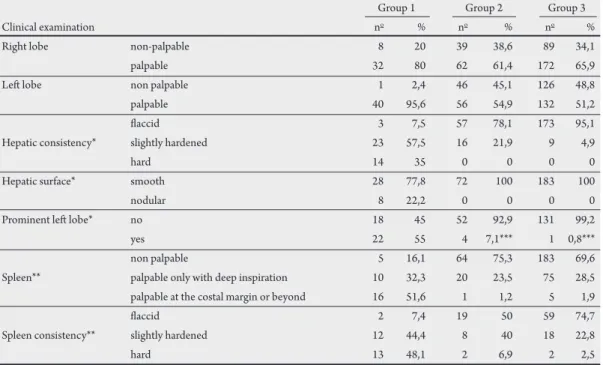
In Group 1, eight (19.5%) patients had hepatointestinal schistosomiasis with advanced liver lesions and 33 (80.5%) had hepatosplenic disease. Among the later, 11 (33.3%) had undergone splenectomy. he clinical indings are summarized in Table 2.
Abdominal ultrasound
Periportal ibrosis was detected in 128 (31.1%) out of the 411 participants. Most of the patients with severe ibrosis were in Group 1 (Figure 1). he mean ages for patients with and without ibrosis were 52 and 34 years respectively (p < 0.01). here was no gender diference.
he liver and spleen sizes of the diferent groups are presented in
Table 3. Ultrasound showed irregular hepatic contours in 11 patients (3%) and gallbladder wall thickening in 114 (29.2%).
Concordance between clinical and abdominal ultrasound examination
here was no relation between palpable right (192/266) or let (153/228) liver lobes and periportal ibrosis on ultrasound. Periportal ibrosis was present in 50 (36.8%) of the 136 patients with a non-palpable right lobe, and in 74 (27.8%) of the 266 with a palpable right liver lobe. Hence, periportal ibrosis was more frequent when the right liver lobe was non-palpable (p = 0.001).
TABLE 1 - Prevalence of schistosomiasis, mean parasitic load (Schistosoma mansoni eggs per gram of feces) and percentage of clinical forms of schistosomiasis in diferent examinations, Brejo do Espírito Santo, Bahia, Brazil.
Year of study
Schistosoma mansoni infection and morbidity 1976 1980 1985 1989 2004
Schistosoma mansoni prevalence (%) 75.2 60.7 44.5 15.2 1.8
infection eggs per gram of feces (mean) 802.5 511.1 255.2 265.3 43.5
hepatointestinal 86.2 67.5 92.3 87.6 94.3
Clinical forms HI* with advanced hepatic lesions 4.0 14.8 1.9 2.0 0.6
hepatosplenic 8.0 10.7 4.3 2.5 1.7
postsurgical hepatosplenic 0.6 1.0 1.1 0.8 2.1
*HI: hepatointestinal.
TABLE 2 - Clinical examination of the study groups, Brejo do Espírito Santo, State of Bahia, Brazil, 2004.
Group 1 Group 2 Group 3
Clinical examination no % no % no %
Right lobe non-palpable 8 20 39 38,6 89 34,1
palpable 32 80 62 61,4 172 65,9
Let lobe non palpable 1 2,4 46 45,1 126 48,8
palpable 40 95,6 56 54,9 132 51,2
laccid 3 7,5 57 78,1 173 95,1
Hepatic consistency* slightly hardened 23 57,5 16 21,9 9 4,9
hard 14 35 0 0 0 0
Hepatic surface* smooth 28 77,8 72 100 183 100
nodular 8 22,2 0 0 0 0
Prominent let lobe* no 18 45 52 92,9 131 99,2
yes 22 55 4 7,1*** 1 0,8***
non palpable 5 16,1 64 75,3 183 69,6
Spleen** palpable only with deep inspiration 10 32,3 20 23,5 75 28,5
palpable at the costal margin or beyond 16 51,6 1 1,2 5 1,9
laccid 2 7,4 19 50 59 74,7
Spleen consistency** slightly hardened 12 44,4 8 40 18 22,8
hard 13 48,1 2 6,9 2 2,5
* Excluding non-palpable patients, ** Excluding splenectomized patients, *** Doubtful.
TABLE 3 - Results from clinical examination and abdominal ultrasound and their relationship with periportal ibrosis and portal hypertension, Brejo do Espírito Santo, State of Bahia, Brazil, 2004.
Fibrosis Portal hypertension
Evaluated parameter N no % no %
right hepatic lobe non-palpable 136 50 36,8* 14 28
let hepatic lobe beyond the costal margin 131 60 45,8* 26 43,3
hardened liver 14 11 78,6* 5 45,4
Clinical examination nodular liver 8 7 87,5* 4 57,1
prominent let hepatic lobe 27 19 70,4* 12 63,1
spleen palpable only with deep inspiration 105 22 22,9* 10 9,5
spleen at the costal margin or beyond 22 16 72,7* 15 93,8*
hardened spleen 17 16 94,1* 14 87,5*
right hepatic lobe atrophy 56 37 66,1* 17 46
Abdominal ultrasound let hepatic lobe enlargement 167 47 28,1 21 44,7
splenomegaly 38 23 60,5* 20 87*
irregular liver contours 11 11 100* 5 45,5
thickening of the gallbladder wall 114 100 87,7* 37 37
*Statistically signiicant.
this inding was more apparent in Group 1. Eleven (78.6%) out of the 14 patients with a hardened liver, 32 (66.7%) out of the 46 patients with a slightly hardened liver and 46 (19.7%) out of the 233 patients with a laccid liver had periportal ibrosis on ultrasound. Periportal ibrosis was also found in seven (87.5%) out of the eight patients with nodular liver and in 78 (27.6%) out of the 283 patients with smooth liver surface. Periportal ibrosis was detected in 19 (70.4%) out of the 27 patients with a prominent let lobe and in 107 (28.5%) out of the 375 patients without a prominent let lobe. All these diferences were statistically signiicant (p=0.001). hese associations were not observed with portal hypertension.
Periportal ibrosis was present in 61 (24.2%) out of the 252 patients with a non-palpable spleen, and in 24 (22.9%) of the 105 patients with a spleen that was only palpable during deep inspiration. Interestingly, 16 (72.7%) out of the 22 patients presenting enlarged spleen had periportal ibrosis (p<0.004) and portal hypertension (p<0.004). A hardened spleen was also associated with periportal ibrosis (16 out of 17 patients) and portal hypertension (14 out of 17 patients) (p<0.001).
In Group 1, 34/41 (82.9%) showed periportal fibrosis on ultrasound, and among them, 23 (67.7%) had portal hypertension. In Group 2, 58/102 (56.9%) had periportal ibrosis, and 18 (31%) had portal hypertension. In Group 3, 36/269 (13.4%) had periportal ibrosis and three (8.3%) had portal hypertension (Figure 1). Groups 2 and 3 were pooled together and compared with Group 1, and this showed that periportal ibrosis was signiicantly higher in Group 1 (p<0.001).
Comparison between other ultrasound findings and periportal ibrosis
hirty-seven (66.1%) out of the 56 patients with right liver lobe atrophy had periportal ibrosis, as opposed to 89 (25.5%) out of the 349 patients with normal right liver lobe (p < 0.0001). his was not observed for the let liver lobe.
Twenty-three (60.5%) out of the 38 patients with splenomegaly diagnosed using ultrasound had periportal ibrosis, as opposed to 84 (24.1%) out of the 348 without splenomegaly (p < 0.001). Splenomegaly was also associated with portal hypertension (Table 3).
Irregular hepatic contours and thickening of the gallbladder wall were associated with periportal ibrosis (Table 3). A good agreement was found between gallbladder wall thickening and periportal ibrosis (kappa = 0.8).
Comparison of liver and spleen size determined on clinical examination and ultrasound
Twenty-eight (50.9%) out of the 55 patients who had right liver lobe atrophy on ultrasound had a palpable liver. Conversely, 50 (30.5%) out of the 164 patients with an enlarged let lobe on ultrasound had a non-palpable let liver lobe.
Among the 125 patients with a palpable spleen, ultrasound conirmed the presence of splenomegaly in 31 (24.8%). On the other hand, six (15.8%) out of the 38 patients with splenomegaly on ultrasound had a non-palpable spleen.
DISCUSSION
Palpable right liver lobe and palpable spleen are not good indicators of periportal ibrosis in ield-based work. Among the cases of a prominent, hard nodular palpable liver, ultrasound revealed periportal ibrosis in 70.9%. It is worth noting that periportal ibrosis occurred in 25.4% of the patients without clinical evidence of liver involvement. In addition, ultrasound diagnosed periportal ibrosis 3.1 times more frequently than clinical examination would suggest. Even in the cases with severe periportal ibrosis on ultrasound, with or without portal hypertension, ultrasound diagnosed 2.1 times more cases than clinical examination did.
Regarding the size of the right liver lobe, there was more ibrosis when this lobe was non-palpable and even more when the lobe was found to be atrophic by ultrasound, as has been previously reported2, 11.
With reference to the size of the let hepatic lobe, Houston et al22
observed that hepatomegaly showed high specificity (94%), but low (28%) sensitivity as a fibrosis marker. It is common knowledge that the liver size decreases with increasing degree of ibrosis15, 22-24. Doehring-Schwedtsfeger et al25 found no relationship
FINANCIAL SUPPORT CONFLICT OF INTEREST
he authors declare that there is no conlict of interest.
REFERENCES
FAPEMIG, CNPq and Federal University of Triângulo Mineiro, Brasil
lobe was also prominent. hus, we do not think that increased let lobe size alone is a good clinical marker of hepatic ibrosis. When analyzing let lobe hypertrophy on ultrasound, Cerri et al11 and
Kardorf et al2 detected correlation with portal ibrosis, a inding that
was not conirmed in our study.
A distinction should be made between small and large spleen sizes. Only in individuals with large spleens has a relationship with liver ibrosis been found. hus, in endemic areas, the importance of small palpable spleens is uncertain; it may or may not be related to schistosomiasis (initial stages of the disease). Other causes of splenomegaly should be investigated, as reported by Lambertucci et al26.
In our study, we also detected a relationship between clinical splenomegaly and the WHO ibrosis paterns25,27-32. In addition,
patients with severe periportal ibrosis without splenomegaly were also found30,33.Splenomegaly diagnosed using ultrasound was related
both to the presence of the WHO paterns and of periportal ibrosis, as already described11,20.
he liver was found by ultrasound to have irregular contours in individuals who had current or past clinical evidence suggestive of periportal ibrosis. However, the absence of irregular contours did not rule out periportal ibrosis. heir presence, as well as gallbladder wall thickening, correlated with periportal ibrosis but not with portal hypertension. his inding has been reported before 2,14,20,25. In one
study, the inding of gallbladder wall thickening was also suggestive of non-reversal of liver ibrosis31.
No agreement was found between ultrasound and clinical examination regarding organ size, as already reported34. One
advantage of the Niamey–Belo Horizonte protocol is the adjustment of the measurements to the patients’ height21. Nonetheless, we
occasionally still detected periportal thickening or dilatation of the portal vein in individuals with no qualitative signs of ibrosis on ultrasound (WHO paterns).
Although ultrasound is superior to clinical examination for detection of periportal fibrosis, it does not allow evaluation of important data such as liver and spleen consistency. On the other hand, the subjectivity of clinical examination was unexpectedly demonstrated in the present study: the prevalence of hepatointestinal schistosomiasis with advanced liver disease increased from 4% in 1976 to 14.8% in 1980 and that of hepatosplenic schistosomiasis from 8% to 10.7% (Table 1). Ater a careful investigation, it was noted that a diferent physician was responsible for the clinical examination in the study area in 1980.
Brejo do Espírito Santo has not been a hyperendemic area for schistosomiasis since the implementation of control measures, which started in 1976. he current clinical and ultrasound indings, many of them slight and some in a regression phase, represent the sequelae from lesions that previously were severe. hese were established in the past when S. mansoni transmission was high. he implementation of efective measures has changed the disease proile in this area, along with the emergence of hepatosplenic schistosomiasis. However, it is important to emphasize that severe disease persists even ater achieving a good control of transmission31.
In conclusion, although clinical examination is still important in evaluating morbidity due to Manson’s schistosomiasis in endemic areas, ultrasound is more accurate in diagnosing liver involvement and periportal thickening.
1. Symmers WSC. Note on a new form of liver cirrhosis due to the presence of the ova of Bilharzia haematobia. J Pathol Bacterol 1904;9: 237-239.
2. Kardorf R, Gabone RM, Mugashe C, Obiga D, Ramarokoto CE, Mahlert C, et al. Schistosoma mansoni-related morbidity on Ukerewe Island, Tanzania: clinical, ultrasonographical and biochemical parameters. Trop Med Intern Hlth 1997;2: 230-239.
3. Gryseels B. he morbidity of schistosomiasis mansoni in the Rusizi (Burundi). Trans R Soc Trop Med Hyg 1988;82:582-587.
4. Kong A, Verlé P, Dieng A, Talla I, Rouquet PH. Clinical investigations of a population recently infected with Schistosoma mansoni (Richard-Toll, Senegal). Trop Med Intern Hlth 1996;1:191-198.
5. Prata A, Bina JA. Development of the hepatosplenic form of schistosomiasis. Gaz Méd Bahia 1968;68:49-60.
6. Gersparcher-Lara R, Pinto-Silva R, Serufo JC, Rayes AAM, Drummond SC, Lambertucci JR. Splenic palpation for the evaluation of morbidity due to
Schistosoma mansoni. Mem Inst Oswaldo Cruz 1998;93: 245-248.
7. Kloetzel K. Critério usado para incluir um paciente na forma hepatesplênica da esquistossomose. In: Program and abstracts of the II Simpósio sobre Esquistossomose. Salvador, Bahia, Brazil; 1970. p. 179.
8. Lambertucci JR, Serufo JC, Gerspacher-Lara R, Rayes AAM, Teixeira R, Nobre V, Antunes CMF. Schistosoma mansoni: assessment of morbidity before and ater control. Acta Tropica 2000;77:101-109.
9. Prata A. Como caracterizar a forma hepatesplênica da esquistossomose
In: Program and abstracts of the II Simpósio sobre Esquistossomose. Salvador, Bahia, Brazil; 1970. p. 179-184.
10. Abdel-Wahab MF, Esmat G, Farrag A, El-Boraey Y, Strickland GT. Ultrasonographic prediction of esophageal varices in schistosomiasis mansoni. Am J Gastroenterol 1993;88: 560-563.
11. Cerri GG, Alves VAF, Magalhães A. Hepatosplenic schistosomiasis mansoni: ultrasound manifestations. Radiology 1984;153: 777-780.
12. Gersparcher-Lara R, Pinto-Silva R, Rayes AAM, Drumond SC, Lambertucci JR. Ultrasonography of periportal ibrosis in schistosomiasis mansoni in Brazil. Trans R Soc Trop Med Hyg 1997;91: 307-309.
13. Homeida MA, Abdel-Gadir AF, Cheever AW, Bennet JL, Arbab BMO, Ibrahium SZ, et al. Diagnosis of pathologically conirmed Symmers´ periportal ibrosis by ultrasonography: a prospective blinded study. Am J Trop Med Hyg 1988;38:86-91.
14. Pinto-Silva A, Abrantes WL, Antunes CMF, Lambertucci JR. Sonographic features of portal hypertension in schistosomiasis mansoni. Rev Inst Med Trop S Paulo 1994;36: 353-361.
15. Abdel-Wahab MF, Esmat G, Farrag A, El-Boraey YA, Strickland GT. Grading of hepatic schistosomiasis by the use of ultrasonography. Am J Trop Med Hyg 1992;46: 403-408.
16. Yazdanpanah Y, Thomas AK, Kardorff R, Talla I, Sow S, Niang M, et al. Organometric investigation of the spleen and liver by ultrasound in Schistosoma mansoni endemic and non endemic villages in Senegal. Am J Trop Med Hyg 1997;57: 245-249.
17. Lambertucci JR, dos Santos Silva LC, Andrade LM, de Queiroz LC, Carvalho VT, Voieta I, et al. Imaging techniques in the evaluation of morbidity in schistosomiasis mansoni. Acta Trop 2008;108:209-217.
19. Katz N, Chaves A, Pellegrino J. A simple device for quantitative stool thick-smear technique in schistosomiasis mansoni. Rev Inst Med Trop S Paulo 1972;14: 397-400.
20. Richter J, Coutinho Domingues AL, Barata CH, Prata A, Lambertucci JR. Report of the Second Satellite Symposium on ultrasound in schistosomiasis. Mem Inst Oswaldo Cruz 2001;96 (suppl 1):151-156.
21. Richter J, Hatz C, Campagne G, Bergquist NR, Jenkins JM editors. Ultrasound in schistosomiasis. A practical guide to standardized use of ultrasonography for the assessment of schistosomiasis-related morbidity. 2000, Geneva, Switzerland; 2000. World Health Organization, TDR/STR/SCH/00.1.
22. Houston S, Munjoma M, Kanyimo K, Davidson RN, Flowerdew G. Use of ultrasound in a study of schistosomal periportal ibrosis in rural Zimbabwe. Acta Trop1993;53:51-58.
23. Abdel-Wahab MF, Esmat G, Milad M, Abdel-Razek S, Strickland GT. Characteristic sonographic patern of schistosomal hepatic ibrosis. Am J Trop Med Hyg 1989;40:72-76.
24. Richter J, Silva-Monteiro E, Moreira-Braz R, Abdalla M, Abdel-Rahim IM, Fano U, et al. Sonographic organometry in Brazilian and Sudanese patients with hepatosplenic schistosomiasis mansoni and its relation to the risk of bleeding from esophageal varices. Acta Trop 1992;51:281-290.
25. Doehring-Schwedtfeger E, Abdel-Rahim IM, Ali QM, Elsheik M, Schlake J, Kardorf R, et al. Ultrasonographical investigation of periportal ibrosis in children with Schistosoma mansoni infection: evaluation of morbidity. Am J Trop Med Hyg 1990;42:581-586.
26. Lambertucci JR. Schistosoma mansoni: pathological and clinical aspects. In: Jordan P, Webbe G, editors. Human Schistosomiasis, Cab International, Wallingford, UK; 1993. p. 195-225.
27. Gersparcher-Lara R, Pinto-Silva R, Rayes AAM, Drumond SC, Lambertucci JR. Ultrasonography of periportal ibrosis in schistosomiasis mansoni in Brazil. Trans R Soc Trop Med Hyg 1997;91:307-309.
28. Hofmann N, Esterre P, Ravaolimalala VA, Ehrich JHH, Doehring E. Morbidity of schistosomiasis mansoni in the highlands of Madagascar and comparison of current sonographical classiication systems. Trans R Soc Trop Med Hyg 2001;95: 623-629.
29. Homeida MA, Ahmed S, Defalla AA, Suliman S, El Tom J, Nash T, et al. Morbidity associated with Schistosoma mansoni infections as determined by ultrasound: a study in Gezira, Sudan. Am J Trop Med Hyg 1988;39:196-201.
30. Lambertucci JR, Cota GF, Pinto-Silva RA, Serufo JC, Gerspacher-Lara R, Drummond SC, et al. Hepatosplenic schistosomiasis: a combined clinical and sonographic deinition. Mem Inst Oswaldo Cruz 2001;96 (suppl 1):147-150. 31. Cota GF, Pinto-Silva A, Antunes CM, Lambertucci JR. Ultrasound and clinical
investigation of hepatosplenic schistosomiasis: evaluation of splenomegaly and liver ibrosis four years ater mass chemotherapy with oxamniquine. Am J Trop Med Hyg 2006;74:103-107.
32. Lambertucci JR, Silva LC, Andrade LM, de Queiroz LC, Pinto-Silva A. Magnetic resonance imaging and ultrasound in hepatosplenic schistosomiasis mansoni. Rev Soc Bras Med Trop 2004;37:333-337.
33. Prata A, Andrade ZA. Fibrose hepática de Symmers sem esplenomegalia. O Hospital 1963;63:609-623.