Agenesis of maxillary lateral incisor in an
Angle Class II, Division 1 malocclusion patient
Guilherme Thiesen1
How to cite this article: Thiesen G. Agenesis of maxillary lateral incisor in an Angle Class II, Division 1 malocclusion patient. Dental Press J Orthod. 2015 Sept-Oct;20(5):108-17.
DOI: http://dx.doi.org/10.1590/2177-6709.20.5.108-117.bbo
Submitted: June 06, 2015 - Revised and accepted: August 17, 2015
Contact address: Guilherme Thiesen
Av. Madre Benvenuta, 1285 Santa Mônica 88035-001 - Florianópolis / SC Brazil - Email: [email protected]
» The author reports no commercial, proprietary or financial interest in the prod-ucts or companies described in this article.
1 Professor of Orthodontics, Universidade do Sul de Santa Catarina (UNISUL),
Florianópolis, Santa Catarina, Brazil. Certified by the Brazilian Board of Orthodontics and Dentofacial Orthopedics (BBO).
* Clinical case report approved by the Brazilian Board of Orthodontics and
Dentofacial Orthopedics (BBO).
» Patient displayed in this article previously approved the use of their facial and in-traoral photographs.
INTRODUCTION
A 26-year and 5-month-old male patient sought orthodontic treatment in good general health and with occasional smoking habit. His chief complaint was “im-paired esthetics in the anterior region.” His dental history reported trauma of tooth #11 sufered ive years before, when he was subject to endodontic treatment. The pa-tient presented with dark discoloration of the clinical
crown. He had also been subject to esthetic rehabilita-tion of tooth #23 with dental composite and stripping, and presented with adequate oral health. Functionally, the patient presented lateral disocclusion through molar guidance on the let side. Mouth opening and closure movements were performed without deviation, with the temporomandibular joint free of any symptoms.
DOI: http://dx.doi.org/10.1590/2177-6709.20.5.108-117.bbo
The present case report describes the orthodontic treatment of a patient with agenesis of maxillary left lateral incisor and Angle Class II, Division 1 malocclusion. The patient also presented with maxillary midline deviation and inclina-tion of the occlusal plane in the anterior region. Treatment objectives were: correcinclina-tion of sagittal relainclina-tionship between the maxilla and the mandible; correction of midline deviation, so as to cause maxillary and mandibular midlines to coincide; correction of overbite and leveling of the occlusal plane, so as to create ideal conditions for esthetic rehabili-tation of anterior teeth. This case was presented to the Brazilian Board of Orthodontics and Dentofacial Orthope-dics (BBO) as a requirement for the title of certified by the BBO.
Keywords:Anodontia. Angle Class II malocclusion. Corrective Orthodontics.
O presente caso clínico relata o tratamento ortodôntico de um paciente portador de agenesia do incisivo lateral supe-rior esquerdo e má oclusão de Classe II 1a divisão de Angle. Apresentava, ainda, desvio da linha média superior e
incli-nação do plano oclusal na região anterior. Os objetivos do tratamento foram adequar a relação sagital entre as arcadas, proporcionar a coincidência entre as linhas médias superior e inferior, corrigir a sobremordida e nivelar o plano oclusal, criando condições apropriadas para a reabilitação estética dos dentes anteriores. Este caso foi apresentado à Diretoria do Board Brasileiro de Ortodontia e Ortopedia Facial (BBO) como parte dos requisitos para a obtenção do título de Diplomado pelo BBO.
DIAGNOSIS
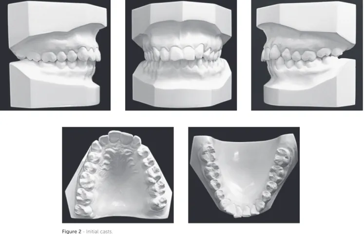
As shown in Figure 1, facial analysis revealed a convex proile. The lower third of the face was balanced and as-sociated with a concave proile (UL – S-Line = -2 mm, LL – S-Line = -2 mm), there was passive lip seal, normal nasolabial angle and acute mentolabial angle. Smile analysis revealed maxillary midline deviation to the let, with dis-tinct inclination of the occlusal plane in the anterior region. Dental analysis (Figs 1, 2) revealed Angle Class II, Divi-sion 1 maloccluDivi-sion, mild curve of Spee, 3-mm overbite and 3.5-mm overjet. There was tooth-bone discrepancy of -1.5 mm in the maxilla and -3 mm in the mandible, in addition to absence of tooth #22.
Panoramic radiograph (Fig 3) revealed that all permanent teeth were present, except for tooth #22. Maxillary third molars were unerupted, whereas mandibular third molars were proclined and impacted at the mandibular ramus. A more thorough analysis performed by means of anterior periapical radiograph evinced mild shortening of tooth #11 root which had been endodontically treated. All remaining incisors had normal root and bone trabeculae contours.
Figure 2 - Initial casts.
Figure 3 - Initial panoramic (A) and periapical radiographs of maxillary (B) and mandibular (C) incisors.
A C
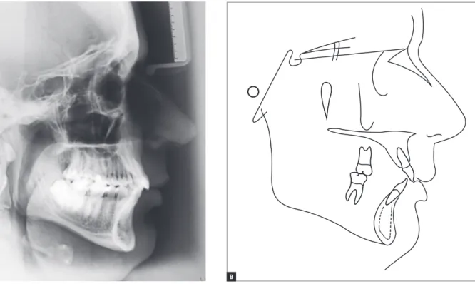
maxilla. Vertical pattern analysis revealed decreased angular measurements: SN-GoGn = 27°, FMA = 18° and Y-axis = 56.5°. The patient also presented with discreet labial proclination and protrusion of maxil-lary incisors (1.NA = 24° and 1-NA = 5 mm) asso-ciated with signiicant labial proclination and protru-sion of mandibular incisors (IMPA = 107°, 1.NB = 34°, 1-NB = 7.5 mm and 1-APo = 3.5 mm).
TREATMENT PLAN
The main treatment objectives were: correction of sag-ittal relationship between the maxilla and the mandible; correction of midline deviation, so as to cause maxillary and mandibular midlines to coincide with the facial midline; correction of overbite and leveling of the occlusal plane in the anterior region, so as to create the ideal conditions for esthetic rehabilitation of anterior teeth. Thus, treatment planning included extraction of tooth #14, with distaliza-tion of anterior teeth on the right side. The inal goal was to achieve Class II molar relationship on both sides, and Class I canine relationship on the right side. On the let side, the irst premolar would replace the canine, so as to occlude with teeth #33 and #34. Treatment planning also
maxillary irst molars and mandibular irst and second mo-lars, and bonding to the remaining teeth. In the maxilla, enameloplasty and 1-mm interproximal reduction were planned for tooth #23, so as to adjust its shape and achieve lateral guidance. The procedures were followed by extrac-tion of tooth #14. In the mandible, 1.5-mm interproximal reduction was planned for all anterior teeth, so as to aid crowding correction and prevent potential proclination of teeth during alignment and leveling.
Subsequently, space closure of the extraction site would be performed by means of sliding mechanics, associated with correction of the lower curve of Spee achieved with rectangular archwire with reversed curve and torque control in the anterior region. Dur-ing space closure, a cantilever would be installed on the right side of the maxilla for intrusion of anterior teeth and leveling of the occlusal plane. The moment resulting from the use of a cantilever on tooth #16 would aid anchorage control. Intermaxillary elastics would be used for anchorage control. Correction of midline deviation was also planned.
TREATMENT PROGRESS Figure 4 - Initial lateral cephalogram (A) and cephalometric tracing (B).
Figure 6 - Intraoral photographs after space closure with a cantilever and intermaxillary elastics concur-rently used on the right side.
alignment and leveling of maxillary and mandibular teeth were performed with superelastic nickel-titanium 0.012-in, 0.014-in and 0.016-in wires followed by stainless steel 0.016-in, 0.018-in and 0.020-in wires. At this phase, a stainless steel 0.019 x 0.026-in archwire was installed in the maxilla with a hook placed distally to tooth #12, with a superelastic nickel-titanium closed loop from tooth #12 to #16, under 300 g of force, for space closure by sliding me-chanics (Fig 5). As for the mandible, a stainless steel 0.019 x 0.025-in archwire was manufactured with reverse curve of Spee and buccal root torque to control incisors procli-nation in the anterior region.
As planned, during space closure after extraction of tooth #14, a cantilever was placed on the right side of the maxilla for intrusion of anterior teeth and lev-eling of the occlusal plane. At space closure onset, in-termaxillary Class III elastics were used to enhance molar intercuspation on both sides and control buc-cal proclination of mandibular incisors. By the end of
the space closure phase, Class III elastics remained in use on the right side, whereas Class II elastics were the choice for the left side. This was done so in order to enhance intercuspation of the premolar replacing the canine on the left side and to cause maxillary and mandibular midlines to coincide (Fig 6).
Once the aforementioned procedures had been carried out, stainless steel 0.019 x 0.025-in rectangular coordinate archwires were installed under ideal torque. Treatment in-ishing was performed with 0.018-in archwires associated with single bends, whenever necessary, and intermaxillary elastics 1/8-in in diameter for intercuspation.
Once the active phase of treatment was concluded, the full ixed appliance was removed and a ixed retainer was bonded to mandibular teeth, from canine to canine. A wraparound removable retainer was used in the max-illa. The patient was referred for esthetic treatment, so as to have esthetic rehabilitation of dark teeth, also in need of reshaping, carried out with dental composite.
RESULTS
As revealed by inal examinations (Figs 7 to 10), pa-tient’s facial proile remained without signiicant altera-tions ater treatment completion. Upper and lower lips remained practically unchanged, with only a minor al-teration in the upper lip (UL – S-line went from -2 mm to -1 mm). Improvements in the mentolabial angle were also noted, with preservation of passive lip seal. Smile analysis revealed signiicant changes in smile harmony as a result of correction of occlusal plane inclination.
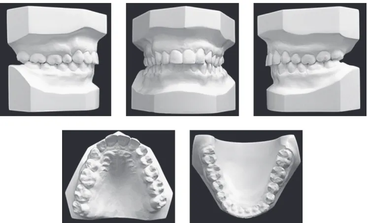
Class II molar relationship was satisfactorily achieved on both sides, in addition to Class I canine relationship on the right side and adequate intercuspation on the let side,
with maxillary irst premolar occluding at canine position. Additionally, the canine replacing the lateral incisor was reshaped and rendered favorably esthetic. Adequate overjet and overbite were also achieved, with satisfactory relation-ship between the maxilla and the mandible in both vertical and horizontal directions. In terms of function, balanced occlusion was achieved during protrusion and lateral guid-ance movements on both right and let sides.
Figure 8 - Final casts.
Figure 9 - Final panoramic (A) and periapical radiographs of maxillary (B) and mandibular (C) incisors.
A C
Figure 10 - Final lateral cephalogram (A) and cephalometric tracing (B).
Figure 11 - Total (A) and partial (B) cephalometric superimpositions of initial (black) and final (red) tracings.
A B
relationship remained stable, with SNA, SNB and ANB angles and Wits value unchanged. Facial height also remained stable, with SN-GoGn, FMA and Y-axis angles unchanged.
Cephalometric superimpositions of initial and inal tracings (Fig 11) revealed unchanged vertical dimensions as well as unchanged maxilla and mandible. Facial proile underwent minimal changes, with minor opening of the mentolabial angle. Maxillary partial superimposition re-vealed that incisors underwent mild retraction and lingual tipping, whereas the right molar was signiicantly mesi-ally tipped without extrusion. Mandibular superimposi-tion revealed that incisors underwent mild retracsuperimposi-tion and lingual tipping, whereas molars remained unchanged.
FINAL CONSIDERATIONS
Epidemiological studies1-6 found a prevalence of
agenesis of maxillary lateral incisors varying from 1% to 3%, with genetics most likely representing the ma-jor etiological factor.7 In Caucasians, maxillary lateral
incisors represent approximately 20% of missing teeth.8
Such an alteration is viewed as an extremely complex issue to be addressed by Orthodontics. There is an in-creasing need for therapy capable of solving this prob-lem, since this condition has a highly negative impact on facial aesthetics due to lack of continuity resulting from absence of lateral incisors in the maxilla.9-11
As for the case presented herein, it is worth noting the importance of thorough reshaping of the let maxil-lary canine performed at treatment inishing. The pro-cedure was carried out with a view to rendering this tooth similar to the lateral incisor and achieving group disocclusion on the same side, since the irst premolar
replaced the canine, both in position and function.12
In the past, some authors13 used to consider canine
Class I relationship as key to periodontal health and sat-isfactory occlusion. However, from the 1950s on, the procedures of canine lateral guidance and mesialization of the irst premolar became rather popular, with some studies yielding great results, also in the long term.8,14,15,16
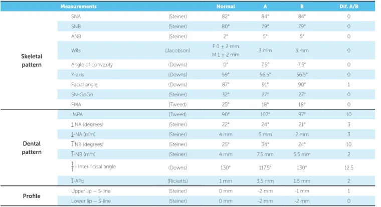
Table 1 - Initial (A) and final (B) cephalometric values.
Measurements Normal A B Dif. A/B
Skeletal pattern
SNA (Steiner) 82° 84° 84° 0
SNB (Steiner) 80° 79° 79° 0
ANB (Steiner) 2° 5° 5° 0
Wits (Jacobson) F 0 ± 2 mm
M 1 ± 2 mm 3 mm 3 mm 0
Angle of convexity (Downs) 0° 7.5° 7.5° 0
Y-axis (Downs) 59° 56.5° 56.5° 0
Facial angle (Downs) 87° 91° 90° 1
SN-GoGn (Steiner) 32° 27° 27° 0
FMA (Tweed) 25° 18° 18° 0
Dental pattern
IMPA (Tweed) 90° 107° 97° 10
1.NA (degrees) (Steiner) 22° 24° 21° 3
1-NA (mm) (Steiner) 4 mm 5 mm 2 mm 3
1.NB (degrees) (Steiner) 25° 34° 24° 10
1-NB (mm) (Steiner) 4 mm 7.5 mm 5.5 mm 2
1
1- Interincisal angle (Downs) 130° 117.5° 130° 12.5
1-APo (Ricketts) 1 mm 3.5 mm 1.5 mm 2
Profile Upper lip — S-line (Steiner) 0 mm -2 mm -1 mm 1
1. Fujita Y, Hidaka A, Nishida I, Morikawa K, Hashiguchi D, Maki K. Developmental anomalies of permanent lateral incisors in young patients. J Clin Pediatr Dent. 2009;33(3):211-5.
2. Gomes RR, Fonseca JA, Paula LM, Faber J, Azevedo AC. Prevalence
of hypodontia in orthodontic patients in Brasilia, Brazil. Eur J Orthod. 2010;32(3):302-6.
3. Nordgarden H, Jensen JL, Storhaug K. Reported prevalence of
congenitally missing teeth in two Norwegian countries. Community Dent Health. 2002;19(4):258-61.
4. Pinho T, Tavares P, Maciel P, Pollmann C. Developmental absence of
maxillary lateral incisors in the Portuguese population. Eur J Orthod. 2005;27(5):443-9
5. Prskalo K, Zjaca K, Skaric‘-Juric‘ T, Nikolic‘ I, Anic‘-Milosevic‘ S, Lauc T. The prevalence of lateral incisor hypodontia and canine impaction in Croatian population. Coll Antropol. 2008;32(4):1105-9.
6. Silva Meza R. Radiographic assessment of congenitally missing teeth in orthodontic patients. Int J Paediatr Dent. 2003;13(2):112-6.
7. Garib DG, Alencar BM, Ferreira FV, Ozawa TO. Associated dental
anomalies: the orthodontist decoding the genetics which regulates the dental development disturbances. Dental Press J Orthod. 2010;15(2):138-57.
8. Robertsson S, Mohlin B. The congenitally missing upper lateral incisor. A retrospective study of orthodontic space closure versus restorative treatment. Eur J Orthod. 2000;22(6):697-710.
9. Araújo EA, Oliveira DD, Araújo MT. Diagnostic protocol in cases of congenitally missing maxillary lateral incisors. World J Orthod. 2006;7(4):376-88.
10. Rizzatto SMD, Thiesen G, Rego MVNN, Marchioro EM. A extração de incisivos permanentes com inalidade ortodôntica. Rev Clín Ortod Dental Press. 2004;3(2):73-87.
11. Zachrisson BU. Improving the esthetic outcome of canine substitution for missing maxillary lateral incisors. World J Orthod. 2007;8(1):72-9. 12. Kokich VO, Kinzer GA. Managing congenitally missing lateral incisors.
Part I: Canine substitution. J Esthet Restor Dent. 2005;17(1):5-10. 13. Angle EH. Treatment of malocclusion of the teeth. 7th ed. Philadelphia:
SS White manufacturing Co.; 1907.
14. Dueled E, Gotfredsen K, Damsgaard MT, Hede B. Professional and patient based evaluation of oral rehabilitation in patients with tooth agenesis. Clin Oral Implants Res. 2009;20(7):729-36.
15. Holm U. Problems of the closing of spaces and compensatory extraction in agenesis of upper lateral incisors. Fortschr Kieferorthop. 1971;32(2):233-47.
16. Thodarson A, Zachrisson BU, Mjor IA. Remodeling of canines to the shape of lateral incisors by grinding: a long-term clinical and radiographic evaluation. Am J Orthod Dentofacial Orthop. 1991;100(2):123-32. 17. Andrade DC, Loureiro CA, Araújo VE, Riera R, Atallah AN. Treatment
for agenesis of maxillary lateral incisors: a systematic review. Orthod Craniofac Res. 2013;16(3):129-36.
REFERENCES Thus, replacing the lateral incisor by a canine instead
of opening space for prosthetic rehabilitation proves to be advantageous, since it yields satisfactory esthetic out-comes, does not induce functional problems to arise at the temporomandibular joint and allows periodontal health conditions to be better maintained, when com-pared to implant-prosthetic rehabiliation cases.8,14,15,16
Furthermore, although canines might not end up with satisfactory color and shape in some cases, recontour-ing procedures associated with bleachrecontour-ing and composite resin restoration provides great esthetic results.10,11,12,17
In the present case, only one maxillary right premo-lar was extracted with a view to correcting maxilpremo-lary midline deviation and counterbalancing Class II mo-lar relationship with the least esthetic damage possible, while causing minimal changes to patient’s lower face. Extracting three premolars (teeth #14, 34 and 44) with a view to counterbalancing agenesis at the upper let hemiarch would invariably lead to retraction of man-dibular incisors with potential deterioration of patient’s facial proile. Another possibility would be the distaliza-tion of posterior teeth on the right side, which would aim at achieving Class I molar relationship on the same side. Nevertheless, this alternative would render the use of Class III elastics to control the position of mandibular incisors impossible, in addition to extending treatment time and requiring extraction of tooth #18 with a view to opening space in the tuberosity for molar distaliza-tion on the same side.