A descriptive study of primary pterygium
surgery with fibrin glue
Estudo descritivo de cirurgia de pterígio
primário com adesivo de fibrina
Carla Christina de Lima Pereira
1, Sarah Apolonio Vieira
2, Edivania Pereira Leite
3, Gabriela Alves de Lima Felix
4,
Jamili Anbar Torquato
51Preceptor at the Residency Programme in Ophthalmology, Federal University of Paraiba.
22nd-year Resident Physician at the Residency Programme in Ophthalmology, Federal University of Paraiba. 3Preceptor at the Residency Programme in Ophthalmology, Federal University of Paraiba.
41st-year Resident Physician at the Residency Programme in Ophthalmology, Federal University of Paraiba. 5Professor at the Master’s Programme in Healthcare Science, Cruzeiro do Sul University.
The authors declare no conflict of interest.
Received for publication: 16/7/2012 - Accepted for publication: 20/4/2013
RESUMO
Objetivo: Avaliar a técnica cirúrgica de exérese de pterígio primário com adesivo de fibrina, quanto à sintomatologia e dados epidemiológicos. Métodos: Ensaio clínico prospectivo, não-controlado e aleatório com 30 pacientes do Hospital Universitário Lauro Wanderley, concordantes com o termo de consentimento livre e esclarecido, aprovado pelo Comitê de Ética em Pesquisa. As cirurgias foram avaliadas nos 1º, 7º e 21º pós-operatório (PO). Resultados: Dos 30 pacientes operados, 16 (53,3%) eram do sexo feminino, com idade variando de 21 a 67 anos (42,2). Quanto à graduação do pterígio (1 a 3): grau 1-10 (33,3%), grau 2-10 (33,3%) e grau 3-10 (33,3%), sendo 16 (53,3%) no olho direito e 14 (46,7%) no olho esquerdo. 56,7% dos pacientes afirmaram intensa exposição solar durante a vida e possuíam casos semelhantes na família. No transoperatório, houve queixas de dor (43,3%) e sensação de corpo estranho (46,7%). O tempo cirúrgico variou de 11 a 32 minutos (17,7). As queixas no 1º, 7º e 21º PO, respectivamen-te, foram: dor (60%, 26,6% e 6,66%), hiperemia (93,3%, 66,6% e 36,6%), sensação de corpo estranho (53,3%, 46,6% e 20%), epífora (83,3%, 43,3% e 6,66%), secreção (33,3%, 36,6% e 6,66%), ardência (53,3%, 36,6% e 16,6%) e olho seco (6,66%, 26,6% e 23,3%). 43,3% afirmaram estar muito satisfeitos com a cirurgia e 63,8% consideraram o aspecto estético excelente. Três pacientes (10%) não compareceram ao 7 DPO e 5 (16,6%) ao 21 DPO. Não houve perda do enxerto nos casos estudados. Conclusão: O transplante autólogo de conjuntiva é atualmente a alternativa mais eficaz para o tratamento do pterígio. Associado ao uso do adesivo de fibrina, que é composto de fibrinogênio e trombina e tem a vantagem de ser totalmente absorvível, apresenta vantagens, dentre elas a redução do tempo cirúrgico. Neste estudo, corroborando com estudos recentes, o tempo cirúrgico médio foi 19,05 (±6,1) minutos e houve alta incidência de familiares portadores de pterígio e exposição aos raios ultravioleta. A queixa de olho seco aumentou progressivamente após a cirurgia, revelando uma possível alteração do ritmo do piscar. Pouco abordada na literatura, a sintomatologia avaliada neste estudo, mostrou que as queixas leves transoperatórias e acentuadas no 1º DPO, regrediram a partir do 7º DPO, justificando a maior satisfação dos pacientes e o excelente aspecto estético referido com o uso desta técnica.
Descritores: Pterígio; Transplante autólogo; Espuma de fibrina; Epidemiologia; Sintomas locais
ABSTRACT
Objective: To evaluate the surgical technique for excision of primary pterygium with fibrin glue, as the symptoms and epidemiological data. Methods: Clinical prospective non-controlled and randomized study with 30 patients of the Hospital Universitário Lauro Wanderley, consistent with the term informed consent, approved by the Ethics in Research. The surgeries were evaluated at 1, 7 and 21 postoperatively (PO). Results: Of the 30 operated patients, 16 (53,3%) were female, aged 21-67 years (42.2). As for the degrees of pterygium (1-3): grau 1-10 (33,3%), grade 2-16 (33,3%) and grade 3-10 (33,3%), 16 (53,3%) in the right eye and 16 (44.4%) in the left eye. 56,7% of patients had intense sun exposure during their lifetime and cases in the family. In the trans-operative period, there were complaints of pain (43,3%) and foreign body sensation (46,7%). Surgical time ranged from 11 to 32 minutes (17.7). The complaints in the first, seventh and 21 PO, respectively, were pain (60%, 26,6% e 6,66%), redness (93,3%, 66,6% e 36,6%), foreign body sensation (53,3%, 46,6% e 20%), epiphora (83,3%, 43,3% e 6,66%), discharge (33,3%, 36,6% e 6,66%),burning (53,3%, 36,6% e 16,6%) and dry eye (6,66%, 26,6% e 23,3%). 43,3% reported being very satisfied with the surgery and 63.8% considered excellent aesthetic appearance. 3 (10%) patients did not attend the 7 DPO and 5(16,6%),at 21 DPO. There was no graft loss in the cases studied. Conclusion: Autologous conjunctiva is currently the most effective alternative for the treatment of pterygium. Associated with the use of fibrin adhesive, which is composed of fibrinogen and thrombin and has the advantage of being totally absorbable, has advantages, among them a reduction in surgical time. In this study, corroborating recent studies, the mean operative time was 19.05 (± 6.1) minutes and there was a high incidence of family pterygium patients and UV exposure. Complaints of dry eye increased progressively after surgery, revealing a possible change in blink rate. Rarely addressed in the literature, the symptoms assessed in this study, showed that complaints Intraoperative mild and marked the first POD, regressed from the 7th POD, justifying the higher patient satisfaction and excellent aesthetics that using this technique.
INTRODUCTION
P
terygium excision associated with autologous conjunctival grafting is currently the most effective method to treat pterygium due to its low rates of recurrence(1). Sutures can be used in the procedure, usually polyglactin (Vycril™) or nylon 10.0. However, pterygium excision with sutures often cau-ses postoperative discomfort, as despite the procedure’s high rates of success, the use of sutures increases tissue manipulation, requiring constant repositioning for adequate fixation. Furthermore, using a needle and suture produces increased tissue trauma, thus increasing the incidence of granuloma. In such situations, loose sutures need to be removed, thus increasing the chance of infection and, more importantly, the surgical time(2).In order to minimise the adverse effects of sutures, a compound with excellent adhesiveness and functionality has been developed: the fibrinogen adhesive or biological glue, used as an alternative to skin sutures(3). Fibrin glue mimics the final reactions of the coagulation cascade, resulting in a firm fibrin clot resulting from the polymerisation of fibrinogen by thrombin. Thus, it aims to provide adhesion between tissues during surgical procedures as an alternative to sutures. Additionally, it provides several benefits in terms of tissue regeneration, reducing surgical time, the friction and tension applied to tissues, tissue necrosis, inflammation, the risk of infection, and the formation of foreign body granuloma, thus providing greater patient comfort(4,5). However, the surgeon needs to be experienced in the technique and has to handle the glue swiftly due to its fast precipitation(6). In ophthalmology, fibrinogen adhesives have been tested in various surgical procedures, such as: amniotic membrane fixation and autologous conjunctival grafting for pterygium, limbal transplantation, lamellar keratoplasty, reconstruction of the ocular surface, glaucoma surgery, cataract surgery, phacoemulsification with scleral tunnel, corneal perforations, oculoplastic surgery, conjunctival surgery, radial keratotomy, lamellar grafting, penetrating keratoplasty, refractive surgery, and even surgery for superficial burns(5,7).
Thus, we aimed to conduct a prospective analysis of patients submitted to pterygium surgery using fibrin glue for autologous conjunctival graft fixation, assessing factors such as: surgical time, symptoms, and intra- and postoperative visual acuity; patient satisfaction and postoperative aesthetic appearance; the need for symptom-relieving medication; and the time needed to readapt to daily activities. This surgical technique is still a matter of debate due to the effective and well-established use of sutures; however, many ophthalmologists are still unfamiliar with the multiple benefits of fibrinogen adhesives(8).
METHODS
Prospective, descriptive, randomised clinical trial comparing subgroups of patients. The study participants were 30 patients treated at the Ophthalmology Clinic of University Hospital Lauro Wanderley (HULW). The patients gave their free and informed consent to undergo pterygium excision with autologous conjunctival grafting using fibrin glue. The study was approved by HULW’s Research Ethics Committee under protocol CEP/ HULW 439/11). This project is part of an ongoing controlled comparative study whose early results are presented here.
The study’s inclusion criteria were: symptomatic patients with primary medial pterygium grade 1 (up to 2 mm), 2 (2 to 4 mm) or 3 (more than 4 mm) who voluntarily opted for surgical treatment. Exclusion criteria were: associated corneal or surface disorders, poor general health, and mental or physical inability to collaborate with the study.
Patient charts included the following information: date, name, age, sex, group, eye, start and finish time of the procedure; symptoms reported during surgery (pain, foreign body sensation, other); postoperative complaints (pain, redness, foreign body sensation, epiphora, discharge, burning, and dry eye); postoperative discomfort rated from 0 to 10 on the Visual Analogue Scale (VAS); patient satisfaction on postoperative (PO) days 1, 7 and 21, rated as very poor, poor, fair, good, and excellent; pre- and postoperative visual acuity (PRE VA, POST VA); aesthetic appearance on PO day 21; and rehabilitation time to perform daily activities. The VAS measures the intensity of ocular discomfort and is an important tool to reliably asses the patient’s condition during treatment: the patient is asked to rate their degree of ocular discomfort, with 0 being no discomfort and 10 the maximum tolerable level of discomfort.
All procedures were performed by the same surgeon. After preparation of the surgical field, asepsis and topical anaesthesia with 1% proparacaine eye drops, the operative time was measured and the following surgical steps were performed: infiltration with 2% lidocaine in the body of the pterygium; detachment of the pterygium head with a cold blade number 15; dissection and excision of the pterygium body from the limbus with conjunctival scissors; and excision of Tenon’s capsule, avoiding cauterisation whenever possible. The ipsilateral upper conjunctival graft was marked and dissected, being 2 mm larger in size than the excised conjunctiva. The two components of fibrin glue were then applied separately, with fibrinogen on the base of the sclera and thrombin on the graft’s inner surface. Then, within 1 minute, the graft was placed on the desired position, the donor limbus was positioned on the host limbus, the conjunctival epithelium was exposed, and excess glue was removed.
Postoperative assessments were conducted by an independent, previously-calibrated observer. After surgery, an occlusive dressing with an ointment of tobramycin plus dexamethasone was applied. After removal, patients were prescribed gatifloxacin plus prednisolone acetate (Zypred™, Allergan) eye drops, 1 drop every 6 hours until PO day 21, with lubricating eye drops (Optive™, Allergan) every 6 hours or more if necessary.
Table 1
Assessing the normality of quantitative variables using the Shapiro-Wilk Test
Variable p-Value
Age 0,391
Time 0,004
PRE VA 0,011
POST VA 0,003
VAS PO 1 0,041
VAS PO 7 0,003
VAS PO 21 0,000
Significant differences between the three grades of pterygium were found in the variable “surgical time” (p=0.033). The average surgical times of grades 1, 2, and 3 were 14.8, 17.8, and 20.5 minutes, respectively. A significant difference was found between grades 1 and 3, with longer surgical times for grade 3 (p=0.019).
As regards intraoperative symptoms, 43.3% of patients reported pain, 46.6%, reported foreign body sensation, and 23.3% reported burning. No statistically-significant differences were found between the study groups regarding the frequency of
RESULTS
The study sample consisted of 30 participants. Mean age was 45.2 years (SD=12.8), ranging from 22 to 67 years, with a predominance of females (53.3%). Participants were separated into groups 1, 2 and 3 according to the grade of pterygium, with 10 patients in each group. Sixteen (53.3%) patients had pterygium in the right eye and 14 (46.7%) in the left eye. As regards their personal background, 56.7% of patients reported a history of intense exposure to sunlight, and 56.7% of patients had a family history of pterygium.
The Shapiro-Wilk test was used to verify if the data to be analysed were normally distributed. In other words, if the p-value in the table below is <0.05, then the data have a normal distribution (Table 1).
VAS: Visual Analogue Scale for ocular discomfort; PRE VA: Preoperative visual acuity; POST VA: Postoperative visual acuity; PO: Post-operative day.
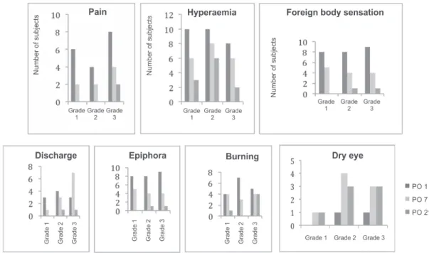
Figure 1: Symptoms on postoperative days 1, 7 and 21, per study group.
postoperative symptoms, except for the variable “discharge” on PO day 7 (p=0.048). A higher frequency of discharge was observed in subjects with pterygium grade 3. A gradual and progressive regression of pain, redness, foreign body sensation, burning, and epiphora was observed on PO days 1, 7, and 21 (Figure 1). However, groups 2 and 3 showed an increase in complaints of dry eye over time.
To compare PRE VA vs. POST VA and VAS, as the data were not normally distributed, we used the nonparametric Wilcoxon and Friedman tests (PRE VA vs. POST VA) followed by the Wilcoxon test (VAS PO 1 vs. PO 7 vs. PO 21). Statistically-significant differences were found between PRE VA and POST VA for the sample as a whole (overall p=0.005) and more specifically for pterygium grade 3 (p=0.027). An improvement in POST VA in comparison with PRE VA was observed.
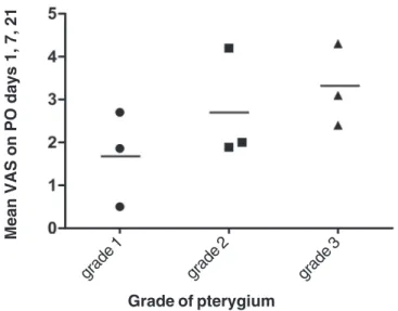
Figure 2 shows the analysis of the degree ocular discomfort according to VAS using the Wilcoxon test, where pterygium gra-des 1, 2 and 3 were assessed on PO days 1, 7, and 21. For pterygium
Figura 3: Aesthetic appearance preoperatively and on postoperative day 21.
DISCUSSION
Compared to other adhesives such as cyanoacrylate, fibrin glue has the advantage of being fully absorbable; it is therefore the first choice for use in biological tissues(9). Commercially-available fibrin adhesives consist of two compounds that, when mixed, promote the formation of a fibrin clot. The first compound is a concentrate of human plasma rich in fibrinogen, diluted in an aprotinin solution which increases the duration of adhesive action by slowing down fibrinolysis. The second is a solution of thrombin and calcium chloride. A direct precursor of insoluble fibrin, fibrinogen is the fundamental structure of blood clots. This fibrin polymer results from the activation of fibrinogen by the proteolytic enzyme thrombin, which breaks down fibrinogen into two fibrinopeptides, A and B, corresponding to the fibrin monomer. The preparation of autologous tissue adhesives using fresh plasma is already done in some blood banks(8,10). Fibrin glue reduces surgical time, making it an excellent choice for ante-rior segment surgery and especially for pterygium surgery, which used to be a feared procedure due to postoperative discomfort(11) with symptoms such as burning, foreign body sensation, and dry eye, amongst others(12-14).
Despite the positive results described here, this study was limited due to the absence of a control group, with results being compared to data from the literature only. Also, the comparison of results was done between the different grades of pterygium; consequently, the symptoms were more severe among patients
Figura 2: Comparison of Visual Analogue Scale scores on PO days 1, 7 and 21 in subjects with different grades of pterygium.
With regard to the use of lubricating eye drops, 37% of patients reported that they did not need to use them, 35% needed to use them a few times, and 28% used them several times a day (more than 3 times). Also, 62.5% of patients reported that they resumed their usual activities within the first 15 PO days.
Patient satisfaction was rated as very poor, poor, fair, good, and excellent. A total of 70% of patients rated their satisfaction as good or excellent on PO day 1, 77.7% on day 7, and 96% on day 21. Aesthetic appearance was also rated as very poor, poor, fair, good, and excellent and was assessed on PO days 7 and 21, with 55.5% of patients rating their appearance as good or excellent on PO day 7 and 80% on PO day 21.
with higher grades. Thus, the superiority of this technique over others cannot be guaranteed.
This study shows the influence of the environment on the formation of pterygium, with patients reporting intense exposure to sunlight and a family history of pterygium. The highly variable age of participants in this study shows that pterygium affects various age groups, causing discomfort and dissatisfaction(8,10).
This study also showed that the use of fibrin glue significantly reduced surgical time (17.7 minutes on average) when compared to sutures while minimising symptoms, in agreement with other studies found in the literature(8).
During the procedure, patients reported pain, foreign body sensation and, to a lesser degree, burning; however, the complaints were mild and most patients did not present any symptoms, feeling relatively comfortable during the procedure. Still, due to shortage of data in the literature, a comparison could not be made.
Most postoperative symptoms were reported on PO day 1 and decreased progressively thereafter. Patients were questioned about pain, foreign body sensation, hyperaemia, and epiphora; these symptoms were found to be present, but gradually decreased towards PO day 21, in accordance with the literature as regards the procedure’s efficacy and mild symptoms(10). However, there were cases of dry eye sensation, probably because the removal of pterygium causes corneal erosion and a reduced surface area of intact conjunctiva, where the mucin in the tear film is produced. Also, the irregularities caused by its removal interfere affect surface integrity, impairing lubrication and thus contributing to an increased blink rate(12). The presence of discharge appears to be linked to these dry eye-inducing factors.
The significant improvement in postoperative visual acuity observed in this study is related to the advanced grades of pterygium which block the visual axis; this effect is reduced postoperatively with proper epithelial healing(13,14).
Even though symptoms are subjective, the VAS is an important tool to globally assess postoperative ocular discomfort. We found statistically-significant differences in VAS scores between the grades of pterygium and during the postoperative period, highlighting a satisfactory recovery with this surgical technique(13).
In our study, 37% of patients felt no need for lubricating eye drops, and 35% required them only a few times, indicating greater ocular comfort. On PO day 21, almost all patients (96%) showed high levels of satisfaction with the procedure, and 80% rated their postoperative aesthetic appearance as good or excellent. These results support the use of fibrin glue over sutures, as by preventing the need to remove sutures postoperatively, fibrin glue causes less postoperative discomfort and pain (e.g. Figure 3). No studies were found in the literature that assess these variables.
The topics covered in this study are part of an ongoing project which will follow-up participants for a period of 3 months using biomicroscopy and will perform a comparison with surgical procedures that used nylon 10.0 or vycril 8.0 sutures. The results of this project will be published upon its conclusion.
Mean VAS on PO days 1, 7, 21
grade 1 grade 2 grade 3
CONCLUSION
O uso da cola de fibrina, como opção terapêutica do pterígio primário, mostra resultados satisfatórios quanto aos sintomas per e pós-operatórios e benefícios na estética, proporcionando satisfação aos pacientes já nos primeiros dias após a cirurgia, assegurando ser uma técnica rápida e eficaz.
REFERENCES
1. Farid M, Pirnazar JR. Pterygium recurrence after excision with conjunctival autograft: a comparison of fibrin tissue adhesive to absorbable sutures. Cornea. 2009;28(1):43-5.
2. Viveiros MM, Schellini SA, Rogato S, Rainho C, Padovani CR. Análise do cultivo de fibroblastos de pterígios primários e recidivados e da cápsula de Tenon normal. Arq Bras Oftalmol. 2006;69(1):57-62.
3. Cronkite EP, Lozner EL, Deaver JM. Use of thrombin and fi-brinogen in skin grafting. Preliminary report. JAMA. 1944;124(14):976-8.
4. Brown AL, Nantz FA. The use of fibrin coagulum fixation in ocular surgery; in retinal detachment. Trans Am Acad Ophthalmol Otolaryngol. 1949;54:126-30.
5. Pizzol MF, Roggia MF, Kwitko S, Marinho DR, Rymer S. Utilização de adesivo de fibrina em cirurgias oftalmológicas. Arq Bras Oftalmol. 2009;72(3):308-12.
6. Nieuwendaal CP, van der Meulen IJ, Mourits M, Lapid-Gortzak R. Long-term follow-up of pterygium surgery using a conjuncti-val autograft and Tissucol. Cornea. 2011;30(1):34-6.
7. Panda A, Kumar S, Kumar A, Bansal R, Bhartiya S. Fibrin glue in ophthalmology. Indian J Ophthalmol. 2009;57(5):371-9. Comment in Indian J Ophthalmol. 2010;58(2):176.
8. Hall RC, Logan AJ, Wells AP. Comparison of fibrin glue with sutures for pterygium excision surgery with conjunctival autografts. Clin Experiment Ophthalmol. 2009;37(6):584-9. 9. Coral-Ghanem R, Oliveira RF, Furlanetto E, Ghanem MA,
Ghanem VC. Transplante autólogo de conjuntiva com uso de cola de fibrina em pterígios primários. Arq Bras Oftalmol. 2010;73(4):350-3
10. Karalezli A, Kucukerdonmez C, Akova YA, Altan-Yaycioglu R, Borazan M. Fibrin glue versus sutures for conjunctival autografting in pterygium surgery: a prospective comparative study. Br J Ophthalmol. 2008;92(9):1206-10.
11. Adamis AP, Starck T, Kenyon DR. The management of ptery-gium. Ophthalmol Clin North Am. 1990;3(4):611-23.
12. Takahagi RU, Gonçalves F, Yamamoto RK, Viveiros MM, Schellini SA, Padovani CR. Ritmo de piscar em portadores de pterígio antes e após a exérese. Arq Bras Oftalmol. 2008;71(3):381-4. 13. Rubin MR, Dantas PE, Nishiwaki-Dantas MC, Felberg S.
Eficácia do adesivo tecidual de fibrina na fixação de enxerto conjuntival autógeno em cirurgias de pterígio primário. Arq Bras Oftalmol. 2011;74(2):123-6.