SECONDARY BILATERAL SYNCHRONY DUE TO
FRONTO-MESIAL LESIONS
AN INVASIVE RECORDING STUDY
ARTHUR CUKIERT*, CASSIO FORSTER**, JOSE AUGUSTO BURATINI**, VIVIANE BORGES FERREIRA**, GARY GRONICH***
ABSTRACT - Frontal lobe epilepsies may present difficulties in focus localization in the pre-operative work-up for epilepsy surgery. This is specially true in patients with normal MRIs. We report on a 16 years-old girl that started with seizures by the age of 8 years. They were brief nocturnal episodes with automatisms such as bicycling and boxing. Seizure frequency ranged from 4-10 per night. Scalp EEG showed few right frontal convexity spiking and intense secondary bilateral syncrhony (SBS). High resolution MRI directed to the frontal lobes was normal. Ictal SPECT suggested a right fronto-lateral focus. Ictal video-EEG showed no focal onset. She was submitted to invasive recordings after subdural plates implantation. Electrodes covered all the frontal convexity and mesial surface bilaterally. Ictal recordings disclosed stereotyped seizures starting from the right mesial frontal. Using a high-resolution tool to measure intra and interhemispheric latencies, the timing and direction of seizure spread from the right fronto-mesial region were studied. Motor strip mapping was performed by means of electrical stimulation. She was submitted to a right frontal lobe resection, 1.5 cm ahead of the motor strip and has been seizure free since surgery (8 months). Pathological examination found a 4 mm area of cortical dysplasia. Invasive studies are needed to allow adequate localization in patients with non-localizatory non-invasive work-up and may lead to excellent results in relation to seizures after surgery.
KEY WORDS: frontal lobe epilepsy, surgery, sub-dural electrodes.
Sincronia bilateral secundária devido a lesões mesiais: estudo com registros invasivos
RESUMO - Podem ocorrer dificuldades na localização de focos nas epilepsias frontais, principalmente nos pacientes que possuem ressonância magnética (RM) normal. Relatamos o caso de uma paciente de 16 anos de idade que começou a apresentar crises convulsivas aos 8 anos. As crises eram breves episódios noturnos que incluíam automatismos tais como pedalar e boxear. A frequência das crises era 4-10 por noite. O eletrencefálograma (EEG) mostrou raras descargas na convexidade frontal direita e intensa sincronia bilateral secundária não localizatória. A RM foi normal e o SPECT ictal sugeriu um foco frontal lateral direito. Realizou-se implante com eletrodos subdurais cobrindo a convexidade frontal e a superfície mesial bilateralmente. Registros ictais mostraram o início das crises na superfície fronto-mesial direita. As latências intra e inter-hemisféricas foram medidas com aparelhagem de alta resolução. O mapeamento da área motora foi realizado através de estimulação elétrica. A paciente foi submetida a ressecção frontal 1,5 cm anterior à área motora. Está sem crises desde a cirurgia há 8 meses. O estudo anátomo-patológico mostrou uma área de 4 mm de displasia cortical. Estudos invasivos são necessários para permitir a localização adequada para o tratamento cirúrgico das epilepsias em pacientes com focos frontais e RM normais. A despeito da maior complexidade, os resultados quanto às crises podem ser excelentes após o tratamento cirúrgico.
PALAVRAS-CHAVE: lobo frontal, epilepsia, cirurgia, eletrodos subdurais.
Serviço de Neurologia e Neurocirurgia (SN) do Hospital Brigadeiro, São Paulo & Laboratório de Neurofisiologia Clínica (LNF): *Chefe do SN; **Médico do SN; ***Diretor do LNF. Aceite: 6-maio-1999.
are probably the responsible for these events. Magnetic resonance image (MRI), video-EEG monitoring and ictal SPECT4 have been used to enhance our ability to define frontal lobe foci but
many patients still require invasive intracranial recordings in the pre-operative evaluation work-up5.
This is specially true for patients with normal MRI scans.
Frontal lobe resections are the second more frequently performed procedure in epilepsy surgery, surpassed only by temporal lobe resections. Frontal cortical resections are usually larger than those performed in the temporal lobe6. Not only the frontal lobe is bigger than the temporal lobe but
cortical generators seem to be more widespread in the former. There is also no such an amplifier and concentrating relay as the hippocampus in the frontal lobe. Frontal lobe resections are usually limited by the pre-central motor cortex. Intra- or pre-operative mapping of the motor strip is mandatory.
Stereotyped clinical epileptic syndromes have been described over the last years7. Mesial
frontal epilepsy typically presents with frequent nocturnal brief seizures which include hypermotor activity and automatisms such as boxing and bicycling. This paper reports a patient with clinically suspected frontomesial epilepsy in whom non-invasive work-up showed non-localizatory SBS and who was submitted to invasive studies and surgery.
CASE REPORT
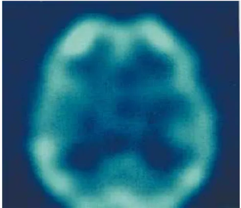
EJT, a 16 years-old girl started with seizures by the age of 8 years. More then 90% of the seizures occurred during sleep. They were brief (5-10 sec) episodes during which there was a sudden appearance of automatisms such as bicycling and boxing, without interruption of sleep. There was no head deviation or any lateralized motor phenomenon. Seizure frequency ranged 4-10 per night. She had been treated with high doses of carbamazepine, phenytoin, phenobarbital, valproate and lamotrigine without significant improval. Scalp EEG showed few right frontal convexity spiking but SBS prevailing in the frontal regions was the main EEG picture. High resolution MRI directed to the frontal lobes was normal. Ictal SPECT suggested a right fronto-lateral focus (Fig 1). Video-EEG monitoring confirmed the family’s description of the seizures. Eight seizures were recorded and no focal onset could be determined. All seizures began with a sentinel spike over both frontal lobes followed by a brief period of background desynchronization and a non-lateralized SBS afterwards.
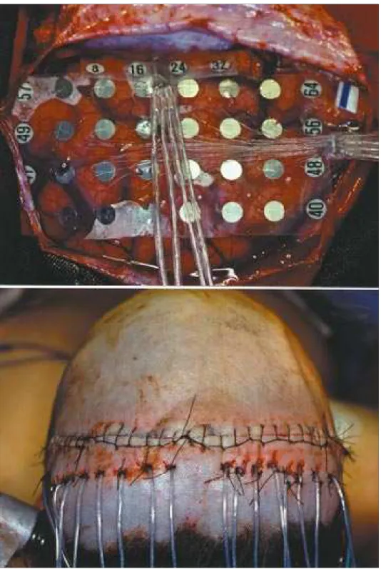
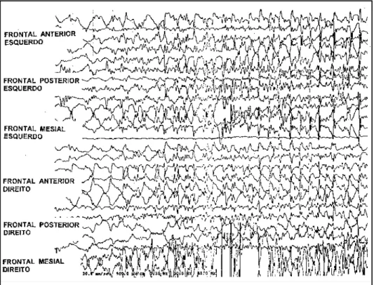
She was submitted to invasive recordings after subdural plates implantation. Electrodes covered all the frontal convexity and mesial surface bilaterally (Fig 2). Inter-ictal recordings showed SBS and focal spiking over the parasagittal and mesial regions of the right frontal lobe. lctal recordings disclosed stereotyped seizures starting from the right mesial frontal cortex, just rostral to the supplementary motor area (Fig 3). Using a highresolution tool to measure intra- and interhemispheric latencies, the timing and direction of seizure spread from the right fronto-mesial region were studied (Fig 4). Motor strip mapping was performed by means of electrical stimulation.
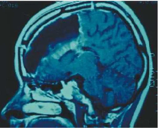
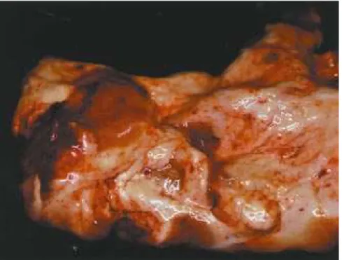
She was submitted to a right frontal lobe resection, 1.5 cm rostral to the motor strip (Fig 5). She experienced a transient left hemiparesis which subsided over the following 2 weeks . She has been seizure free since surgery (8 months). Pathological examination found a 4 mm area of cortical dysplasia in the pre-supplementary cortical region (Fig 6). Careful review of the pre-operative MRI directed to the area where the lesion was found was again negative. There was no neuropsychological morbidity.
A
B
C
D
Fig 4. A, B, C, D: Diagrams showing the pattern of seizure spread beginning in the right mesial region (4 different seizures). Numbers in parenthesis represent msec after seizure start.
DISCUSSION
Frontal lobe epilepsies still represent a challenge to epileptologists in terms of electroclinical localization and surgical treatment. Nevertheless, in the presence of a highly suspected frontal lobe focus, aggressive work-up may lead to focus localization.
This patient presented with poor localizatory findings in the non-invasive work-up. Both interictal EEG and ictal SPECT suggested a focus in the right frontal convexity and not a mesial focus. Non-localizatory SBS over both frontal lobes was the main EEG feature. Invasive EEG made it possible to adequately localize a single focus and to study the neurophysiologic characteristics of seizure spread in this patient8.
The frontal lobe resection performed was much bigger than the lesion found on pathology. The extent of resection was aimed to cover both the non-invasive and invasive neurophysiological data9. Small frontal lobe resections are usually associated with poor outcome in relation to seizures10.
Interhemispheric latency of seizure spread is compatible with known human transcallosal latencies11.
As usual, motor strip mapping through the use of sub-dural plates can be easily achieved.
Despite the continuous development of the non-invasive methods for focus localization, there are still some patients who would need invasive recordings in the pre-operative work-up12. Even in
these more challenging patients, adequate work-up can lead to focus localization and good seizure outcome after surgery.
REFERENCES
1. Cukiert A, Gronich G, Marino R Jr. Secondary bilateral synchrony associated to a parasagittal tumor: case report. Arq Neuropsiquiatr 1991:49:333-337.
2. Cukiert A . The role of the cortex and the corpus callosum in the pathophysiology of the secondary bilateral synchrony in the cat (Abstr). Arq Neuropsiquiatr 1997;55:159-160.
3. So NK. Mesial frontal epilepsy. Epilepsia 1998;39(Suppl 4): S49-S61.
4. Laich E, Kuzniecky R, Mountz J, et al. Supplementar sensorimotor area epilepsy: seizure localization, cortical propagation and subcortical activation pathways usign ictal SPECT. Brain 1997;120:855-864.
5. Munari C. Giallonardo AT, Brunet P, Broglin D, Bancaud J. Stereotactic investigations in frontal lobe epilepsies. Acta Neurochir 1989;39(Suppl):46:9-12.
6. Bleasel A, Comair Y, Luders HO. Surgical ablations of the mesial frontal lobe in humans. Adv Neurol 1996;70:217-235. 7. Waterman K, Purves SJ, Kosaka B, Strauss E, Wada JA. An epileptic syndrome caused by mesial frontal lobe seizure foci.
Neurology 1987;37:577-582.
8. Toczek MT, Morrell MJ, Risinger MW, Shuer L. Intracranial ictal recordings in mesial frontal lobe epilepsy. J Clin Neurophysiol 1997;14:499-506.
9. Cukiert A, Olivier A, Andermann F. Post-traumatic frontal lobe epilepsy with structural changes: excellent results after cortical resection. Can J Neurol Sci 1996;23:114-117.
10. Cukiert A, Olivier A, Andermann F. The role of frontal lobe resections combined to callosal sections in the treatment of secundary generalized epilepsy. In Reeves AG, Roberts DW. Advances in behavioral biology 45: Epilepsy and the corpus callosum 2, New York: Plenum Press, 1995:201-208.
11. Buser P, Bancaud J, Chauvel P. Callosal transfer between mesial frontal areas in frontal lobe epilepsies. Adv Neurol 1992;57:589-604.