Noninvasive method to assess the electrical brain activity from rats
Método não invasivo para avaliar a atividade elétrica cerebral de ratos
Rosana FerrariI Aldo Ivan Cespedes ArceI Mariza Pires de MeloI Ernane Jose Xavier CostaI*
ISSN 0103-8478
ABSTRACT
This research presents a noninvasive method for the acquisition of brain electrical signal in rat. Was used an electroencephalography (EEG) system developed for bovine and adapted to rats. The bipolar electrode system (needle electrodes) was glued on the surface of the head of the animal without surgical procedures and the other electrode was glued to the tail, as ground. The EEG activity was sampled at 120Hz for an hour. The accuracy and precision of the EEG measurement was performed using Fourier analysis and signal energy. For this, the digital signal was divided into sections successive of 3 seconds and was decomposed into four frequency bands: delta (0.3 to 4Hz), theta (4-8Hz), alpha (8-12Hz) and beta (12-30Hz) and energy (μV2) of the series of time fi ltered were calculated. The method allowed the acquisition of non-invasive electrical brain signals in conscious rats and their frequency patterns were in agreement with previous studies that used surgical procedures to acquire EEG in rats. This system showed accuracy and precision and will allow further studies on behavior and to investigate the action of drugs on the central nervous system in rats without surgical procedures.
Key words: biological signals, digital signal processing, central nervous system, EEG.
RESUMO
Este trabalho apresenta um método não invasivo para a aquisição de sinal elétrico cerebral em ratos. Utilizou-se um sistema de eletroencefalografi a EEG desenvolvido para bovino e adaptado para ratos. O sistema bipolar de eléctrodos (eléctrodos de agulha) foi colado na superfície da cabeça do animal, sem procedimentos cirúrgicos e outro eletrodo foi colado na cauda, como terra. A atividade do EEG foi amostrada em 120Hz durante uma hora. Foi calculada a exatidão e a precisão da medida de EEG usando análise de Fourier e energia do sinal. Para isso, o sinal digital foi dividido em trechos sucessivos de 3 segundos, decomposto em quatro faixas de frequência: delta (0,3-4Hz), teta
(4-8Hz), alfa (8-12Hz) e beta (12-30Hz) e a energia (μV2) das
séries de tempo fi ltradas foram calculadas. O método não invasivo permitiu adquirir sinais elétricos cerebrais em ratos conscientes e seus padrões de frequências foram de acordo com trabalhos anteriores, que utilizaram procedimentos cirúrgicos para adquirir EEG em ratos. Esse sistema apresentou exatidão e precisão e permitirá novos estudos sobre comportamento e para investigar a ação de drogas no sistema nervoso central sem procedimentos cirúrgicos em ratos.
Palavras-chave: sinais biológicos, processamento de sinal digital, sistema nervoso central, EEG.
INTRODUCTION
The monitoring of brain electrical activity represented by the electroencephalography (EEG) enables temporal analysis of neurophysiologic processes. EEG pattern can have its frequency pattern and its temporal dynamics changed in several situations. Thus, EEG has been used for different purposes, as studies the action of substances in central nervous system (RADEK et al., 2004; IHMSEN et al., 2008); to monitoring and studying the behavior of animals (ARCE et al., 2009), as a method of sensory analysis of foods (HASHIDA et al., 2005) and tables of epilepsy (KOTAGAL & YARDI, 2008).
EEG collection methods require the use of electrodes that are transducers that transform ionic signals generate in brain during the neurotransmission into electric signals. The major studies of EEG signal
in rats are placement with invasive methods. In these cases, intracranial electrodes (needle) are placed within a stereotactic surgical (DIMPFEL, 2005). The bregma is used as a reference point to determine the stereotactic coordinates to attach the electrodes (BLASIAK et al., 2010). Thus, surgical procedures are requiring to place the electrodes and prophylaxis, anesthesia and a post-surgical interval are necessary (LAPRAY et al., 2008). Therefore, the improvement of methods for recording and processing electrical signals to the brain without any kind of interference is extremely relevant, as showed in this work. Electrodes placed just on the surface of the animals’ head, such as in humans, compel a higher sensibility of the brain electrical signals collector equipment, in order to provide a good signal quality, measured by the signal-to-noise ratio.
There are several different mathematical models and digital signal processing techniques that enables to remove more information of EEG that their representation in the form of a time series.Such processing methods include the Fourier analysis (SILVA et al., 2005), time-frequency (MARCHANT, 2003), Wavelets (DAUBECHIE, 1992). Commonly, the EEG amplitude is analyzed by studies from different situations. However, we calculated the accuracy and precision of the EEG measure by using Fourier analysis and signal energy, which is proportional to the square of amplitude (PROAKIS & MANOLAKIS, 2006). Thus, this study it is demonstrated that is possible to acquire high quality electroencephalography signals, using a non-invasive approach, in conscious rats.
MATERIAL E METHODS
Animals
Six male Wistar rats weighing 235g±15g (about 2 months old) were obtained from the Faculty of Animal Science and Food Engineering (University of São Paulo, Pirassununga, SP). The rats were maintained at 23oC under a cycle of 12h light: 12h
darkness and were housed in individual cages arranged in order to diminish the environmental effects on the measured parameters. The animals received water and commercial food to rodent without restriction.
Electrodes and placement
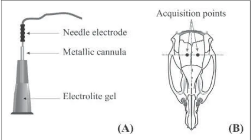
The needle electrode (Bio-Logic Systems Corp) was attached into the conical guide with 2cm-long (Figure 1A), made from a stainless steel hypodermic needle of 0.80x25 (BD PrecisionGlide®).
The guide was fi lled with conductive gel (Carbogel Ltda.), and was glued with synthetic resin (Scotch Bond, 3M®) on the skin’s surface of the animal’s head.
The electrode placement was made by selecting four imaginary quadrants in an animal (Figure 1B), and each one was fi xed on each side of the head (bipolar register) after trichotomy. One needle electrode was glued with adhesive tape on the tail, as ground. After all of the electrodes were in place, they were connected to the amplifi er (module one of EEG equipment). The EEG activity was recorded with 120Hz of sampling frequency immediately after the electrodes placement for approximately 3 hours in awake freely moving rats. All experiments were performed at the same time of day (10:00 to 12:00 am).
EEG Equipment
The EEG equipment was developed by SILVA et al. (2005) and ARCE et al. (2009) for bovine and adapted for rat by FERRARI (2008) and was built following the security and quality patterns for medical equipment. The system consisted of two modules: module one that amplifi es and transmits the EEG signal (telemetric system) and module two, which receives and makes the connection with a computer. The dimensions of module one were 2.5x7.0x11cm and the weight was 180g. This module has three basic functions: signal amplifi cation and storage; digital conversion and digital micro-processed communication system control, and, digital data transmission. Module two has the same size and weight attributes, and is responsible for the digital signal reception, sent by module one. Therefore, it can be considered a radio base station. The interface between the user and the radio-base point is made using software that connects with module two using an Application Program Interface (API), for Windows®, generating EEG graphics. All
of the information is stored in a database. The fi gure 2 shows the schematic diagram of the EEG collections.
Data analysis
The EEG recorded were analyzed by using digital signal processing techniques implemented in Matlab® software (MathWorks, Inc., MA, USA). In
order to measure the system accuracy by comparing with literature results, the digital signal collected was analyzed by using Fourier transform (FT). The system precision was measured by the following procedure:
fi rst of all, the signal was divided in successive epochs of 3 second of EEG artifact-free by visual inspection.
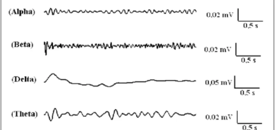
After, for each epoch, the signal was decomposed into four frequency bands: delta (0.3-4Hz), theta (4-8Hz), alpha (8-12Hz) and beta (12-30Hz), using four order elliptic fi lters resulting in more than 240 epochs to be processed. Finally, in each epochs, the energy (μV2) of the time series
fi ltered in specifi cs band frequencies (delta, theta, alpha and beta) was calculated and expressed as mean±S.E.M, analyzed by ANOVA followed by Tukey test, considering level of P<0.05. The precision of one measurement was defi ned by comparing it with the mean of N measurement as described by NORTHROP (2005).
RESULTS AND DISCUSSION
The time domain of EEG signals recorded are shown in fi gure 3A and it is clear by visual inspection that the amplitude is qualitatively comparable with those described in the literature (BO, 2003; LAPRAY et al., 2008), with amplitudes that ranged from 1-25μV, considering that these amplitudes differ due the invasive electrode placement as described in the literature (LAPRAY et al., 2008). The frequency domain of EEG signals recorded is shown in fi gure 3B. It is observed, in this fi gure, that there is a prevalence of frequencies ranging from 1Hz to 40Hz, in agreement with previous literature reports (LAPRAY et al., 2008). This result can be used to assess the accuracy of the system developed by comparing quantitatively with those described in the literature. The result of EEG epoch decomposed in the band frequencies delta (0.3-4Hz), theta (4-8Hz), alpha (8-12Hz) and beta (12-30Hz) is shown in
fi gure 4. This EEG decomposition was in accordance to observe by others authors (DRINGENBERG &
DIAVOLITSIS, 2002). To assess the precision of the system the energy (μV2) of the more than 240 epochs
of the time series fi ltered in specifi cs band frequencies was calculated. The energy value (μV2) for these
band frequencies was: 0.2211±0.0076 (delta), 0.1059±0.0052 (theta), 0.0971±0.0034 (alpha) and 0.1009±0.0039 (beta) and there was no statistical difference between the animals in each frequency band showing that the system precision was achieved.
Due to the non-invasive placement, the position of the electrodes produces changes in the signal-to-noise ratio of the EEG signal. To verify the effect of electrode placement position and the signal-to-noise ratio, different collection points had been tested, and no signifi cant signal-to-noise ratio differences were observed. In this regard, certain factors must be considered. The skull anatomy of the rat allows for the dissipation of electrical currents generated in the brain to the head’s surface, but this interference was not as signifi cant as has been observed in other species such as cattle (SILVA et al.,
2005). The dimensions of the non-invasive electrode placement system were small enough to allow the animal to move freely and did not cause apparent discomfort that can promote changes in the behavior of the animals. One differential EEG channel (bipolar register) was suffi cient for capturing these electrical currents favoring the animal handling.
There are several techniques to assess changes in brain activity such as positron emission tomography (PET), magnetic resonance imaging (fMRI), measures of brain magnetic activity (Magnetoencephalography - MEG) and measures of brain electrical activity (EEG). The advantage of monitoring brain electrical activity by EEG is mainly in the time resolution, low cost of procedures and the possibility of using mathematical tools to remove a greater amount of information of the signal obtained. Thus, a simple method for acquiring non-invasive EEG for rats was developed in this work and allowed the capture of animals awake EEG, without the need for surgical procedures for implantation of Figure 3 -Pattern of the electrical signal recorded by non-invasive electrodes. (A) EEG signal of 3 seconds;
(B) Spectral power density for these stretched under the control condition. Spectrum was calculated using the Welch method with an overlay of 50%. The result shown has a confi dence interval of 95% (central line).
electrodes and using only one channel. Furthermore, main advantage of the system presented herein is the lack of the experimental stressors such as pain or discomfort that are normally produced by implanted electrodes in the skull in accordance with the ethical aspects to animal science. The signal-to-noise ratio was improved by using the digital signal processing techniques, like digital fi lters implemented in the Matlab® environment.
CONCLUSION
The results shown herein demonstrate that it is possible to acquire and extract information from brain electrical activity using a non-invasive approach in conscious rats by monitoring the EEG activity with accuracy and precision. The EEG patterns observed were similar to the invasive methodology already documented in the literature.
ACKNOWLEDGMENTS
The authors gratefully acknowledge the generous support of the National Council for Scientifi c and Technological Development (CNPq, Proc. 300416/2009-1) for the research described in this work.
ETHICS COMMITTEE AND BIOSECURITY
All experimental procedures were done in accordance of guidelines established by the Brazilian College for Animal Experimentation (Cobea) and approved by Animal Ethics Committee of the Faculty of Animal Science and Food Engineering, University of São Paulo (Protocol n.100904).
REFERENCES
ARCE, A.I.C. et al. Monitorización de rebaños através de redes de sensores inalámbricos. Archivos de Zootecnia (Universidad de Córdoba), v.58, p.253-263, 2009. Available from: <http://scielo. isciii.es/pdf/azoo/v58n222/art10.pdf>. Accessed: Abr. 04, 2012. doi: 10.4321/S0004-05922009000200010.
BLASIAK, T. et al. A new approach to detection of the bregma point on the rat skull. Journal of Neuroscience Methods, v.185, p.199-203, 2010. Available from: <http://www.sciencedirect.com/ science/article/pii/S0165027009005317>. Accessed: Abr. 04, 2012. doi: 10.1016/j.jneumeth.2009.09.022.
BO, P. et al. Quantifi ed EEG analysis monitoring in a novel model of general anaesthesia in rats. Brain Research Protocols, v.11, p.155-161, 2003. Available from: <http://www.sciencedirect.com/ science/article/pii/S1385299X03000424>. Accessed: Abr. 04, 2012. doi: 10.1016/S1385-299X(03)00042-4.
DAUBECHIE, I. Ten lectures on “wavelet”s. Philadelphia: CBMS Lectures notes series - SIAM, 1992. 353p.
DIMPFEL, W. Pharmacological modulation of cholinergic brain activity and its refl ection in special EEG frequency ranges from
various brain areas in the freely moving rat (Tele-Stereo-EEG).
European Neuropsychopharmacology, v.15, p.673-682, 2005. Available from: <http://www.sciencedirect.com/science/article/pii/ S0924977X05000817>. Accessed: Abr. 04, 2012. doi: 10.1016/j. euroneuro.2005.03.006.
DRINGENBERG, H.C.; DIAVOLITSIS, P. Eletroencephalographic activation by fl uoxetine in rats: role of 5-HT1A receptors and enhancement of concurrent acetylcholinesterase inhibitor treatment.
Neuropharmacology, v.4, p.154-161, 2002. Available from: <http:// www.sciencedirect.com/science/article/pii/S0028390801001642>. Accessed: Abr. 04, 2012. doi: 10.1016/S0028-3908(01)00164-2. FERRARI, R. Effects of indole-3-acetic acid (IAA) administration on metabolism and electroencephalic parameters on rats. 2008. 108f. Thesis (Ph.D. in Animal Science) - College of Animal Science and Food Engineering. University of São Paulo, SP.
HASHIDA, J.C. et al. EEG pattern discrimination between salty and sweet taste using adaptative Gabor transform. Neurocomputing, v.68, p.251-257, 2005. Available from: <http://www.sciencedirect. com/science/article/pii/S0925231205001190>. Accessed: Abr. 04, 2012. doi: 10.1016/j.neucom.2005.04.004.
IHMSEN, H. et al. Concentration-effect relations, prediction probabilities (Pk) and signal-to-noise ratios of different electroencephalographic parameters during administration of desclurane, isofl urane, and sevofl urane in rats. Anesthesiology, v.108, p.276-285, 2008. Available from: <http://journals.lww.com/ anesthesiology/pages/articleviewer.aspx?year=2008&issue=0200 0&article=00016&type=abstract>. Accessed: Abr. 04, 2012. doi: 10.1097/01.anes.0000300074.04200.b1.
KOTAGAL, P.; YARDI, N. The relationship between sleep and epilepsy. Seminars in Pediatric Neurology, v.15, p.42-49, 2008. LAPRAY, D. et al. A novel miniature telemetric system for recording EEG activity in freely moving rats. Journal of Neuroscience Methods, v.168, p.119-126, 2008. Available from: <http://www.physiologie.uni-mainz.de/physio/luhmann/146.pdf>. Accessed: Abr. 04, 2012. doi:10.1016/j.jneumeth.2007.09.029. MARCHANT, B.P. Time-frequency analysis for biosystems engineering. Biosystems Engineering, v.85, n.3, p.261-281, 2003. Available from: <http://www.sciencedirect.com/science/ article/pii/S1537511003000631>. Accessed: Abr. 04, 2012. doi: 10.1016/S1537-5110(03)00063-1.
NORTHROP, R.B. Introduction to instrumentation and measurements. 2.ed. CRC Press, 2005. 743p.
PROAKIS, J.G.; MANOLAKIS, D.G. Digital signal processing: principles, algorithms, and applications. 4.ed. New Delhi: Prentice-Hall; 2006. 1004p.
RADEK, R.J. et al. The adenosine kinase inhibitor ABT-702 augments EEG slow waves in rats. Brain Research, v.1026, p.74-83, 2004. Available from: <http://www.sciencedirect.com/science/ article/pii/S0006899304013058>. Accessed: Abr. 04, 2012. doi:10.1016/j.brainres.2004.08.011.