Arq Neuropsiquiatr 2002;60(2-B):478-480
PROGRESSION OF AN ARTERIAL
INFUNDIBULUM TO ANEURYSM
Case report
Carolina Martins
1, Mladen Macanovic
2, Isabel Eugenia Costa e Silva
1,
Fatima Griz
1, Hildo R. C. Azevedo-Filho
1ABSTRACT - In this case an aneurysm of the right posterior communicating artery developed 11 months after an infundibular dilation of this artery had been angiographycally and surgically demonstrated. In the best of the authors’ knowledge, there are only eleven such cases reported in the literature. This report brings about diagnostic and therapeutic questions regarding arterial infundibula and the need of a better understanding of those lesions.
KEY WORDS: aneurysm, arterial infundibulum, bilateral supratentorial aneurysms, infundibular dilation.
Progressão de infundíbulo arterial a aneurisma: relato de caso Progressão de infundíbulo arterial a aneurisma: relato de caso Progressão de infundíbulo arterial a aneurisma: relato de caso Progressão de infundíbulo arterial a aneurisma: relato de caso Progressão de infundíbulo arterial a aneurisma: relato de caso
RESUMO - Relatamos a transformação de uma dilatação infundibular da origem da artéria comunicante posterior em aneurisma, após onze meses do diagnóstico angiográfico e cirúrgico. Apenas onze casos semelhantes foram descritos na literatura internacional. Relatos como esse trazem à tona os problemas diagnósticos e terapêuticos representados por essas lesões e suscitam a necessidade do melhor entendimento das dilatações infundibulares encontradas nas artérias cerebrais.
PALAVRAS-CHAVE: aneurismas, aneurismas bilaterais, dilatação infundibular, infundíbulo.
Department of Neurosurgery, Hospital da Restauração, Recife PE, Brazil: 1Neurosurgeon; 2Medical Student, Imperial College, UK.
Received 7 April 2001, received in final form 12 December 2001. Accepted 9 January 2002.
Dra. Carolina Martins - Rua Deputado Pedro Pires Ferreira 325/1601 - 52050-480 Recife PE - Brasil. E-mail: [email protected]
Infundibula (IFs) are funnel-shaped symmetrical enlargements of the origin of cerebral arteries. Most frequently they affect the origin of the posterior com-municating artery (PComA) at its junction with the internal carotid artery (ICA), and are considered as normal anatomical variants devoid of pathogenic significance. Some authors do not agree with this statement and consider an IF a “pre-aneurysmal” le-sion1-4. This belief rests on the increasing incidence of infundibular widening with age and on the histo-logical demonstration of changes in some IFs simi-lar to that characteristic of saccusimi-lar aneurysms1.
We report the progression of an IF to aneurysm, with subsequent rupture, in a hypertensive patient previously operated of multiple and bilateral aneurysms, through a unilateral craniotomy.
CASE
A 53-year-old caucasian woman was admitted on March 30, 1998, complaining of a sudden bitemporal hea-dache, started 12 hours before admission. She had no
loss of consciousness or vomiting. She scored 15 on GCE, but had a marked stiff neck. A CT performed on admission revealed widespread subarachnoid haemorrhage (SAH), mainly in the left Sylvian fissure. Five days later, the patient developed a complete III nerve palsy on the left side. An-giography revealed aneurysms on the origin of the left PComA, on the bifurcation of the middle cerebral artery and ICA bifurcation on the right side and an infundibular origin of the right PComA (Fig 1). She was submitted to a left pterional craniotomy and microsurgical clipping of the lesions in both sides (unilateral craniotomy to bilateral aneurysms) (Fig 2). The origin of the right PComA was explored and the presence of the IF confirmed. The posto-perative period was unremarkable and the control angio-gram performed on May 15, 1998 revealed no aneurysms. The follow up visits during the next nine months confirmed a good control of the mild hypertension (diagnosed dur-ing admission), no evidence of aortic coarctation, policystic disease or inflammatory connective tissue disease, and a partial recovery of the left third nerve palsy - the residual deficit only seen during the upward gaze (deficit of the superior division of the oculomotor nerve).
complai-Arq Neuropsiquiatr 2002;60(2-B) 479
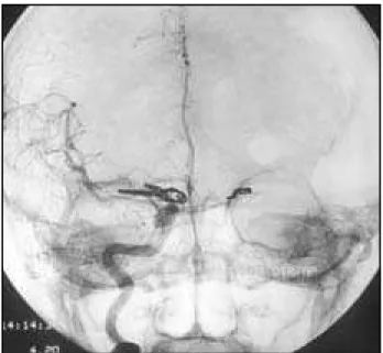
Fig 2. Postoperative angiogram (AP) showing clips occluding aneurysms on the left PComA, right ICA and MCA, through an unilateral craniotomy.
Fig 1. Diagnostic angiogram (profile) showing infundibular origin of right PComA.
Fig 3. Diagnostic angiogram (profile) showing progression from infundibulum to aneurysm eleven month after the first angiogram (Fig 1).
ning of a “headache just like the other time”. This event had taken place one week before and had been initially neglected by the patient until her eye became involved. On admission there was a heavy stiff neck and a 24-hour history of right ptosis. The CT performed at that time (nine days after the beginning of the complaint) failed to dem-onstrate HSA. The angiography disclosed an aneurysm at the origin of the right PComA (Fig 3). A right pterional craniotomy was performed and the aneurysm clipped. The microdissection on the right Sylvian fissure was accom-plished uneventfully, but there were hard adherences on the lamina terminalis and right carotid cisterns. A control angiogram obtained on July 4, 1999 demonstrated com-plete occlusion of the lesion.
DISCUSSION
IFs are apparent on 7 to 25% of otherwise nor-mal angiograms, and the incidence seems to be grea-ter in cases of familial or multiple aneurysms3. Angio-graphically, IFs are triangular shaped lesions, which base does not measure more than 3 mm and the branch artery arises from its apex2. An IF thus should appear as a symmetric bulge without a neck in mar-ked contrast to an intracranial aneurysm, which bul-ges, asymmetrically from a well-defined neck3. Al-though this guidelines help in most cases, differen-tiation of aneurysms from infundibular dilations of the PComA remains a difficult radiological prob-lem1,2,5, especially when considering patients with subarachnoid haemorrhage and no other angiogra-phic abnormality or patients with multiple
aneu-rysms. According to Marshmann et al.2, it may be
unwise to report “negative” angiographic results in cases of SAH in which IFs are present.
480 Arq Neuropsiquiatr 2002;60(2-B)
Our patient had one hemodynamic factor remo-ved (treated hypertension) but remains to be under-stood if the effects of the presence of multiple an-eurysms, or the results of their clipping could have contributed to the progression of the IF to aneu-rysm. This question is even more important when considering the time of progression in this patient: eleven months, in contrast with a mean period of seven years as described in literature1-4.
Considering the pre-aneurysmal role that IF could play, some authors have advocated IF wrapping at craniotomy in precisely such cases3. Following this view imply that, added to the other benefits provided by the contralateral approach, this patient might have been spared of additional trouble. The question however, is not that simple: the co-operative study findings indicating that no signs of rupture were found in any bulging less than 3mm in size suggest that this procedure could be excessive3,6. A systematic follow up, with angiography, especially in young patients who have IFs and multiple aneurysms is the recommendation of many authors1,3,7. These reports, however, do not suggest any specific follow up times.
Based on the experience with this case, the litera-ture data, the risks of the angiography itself and costs, the management of patients harbouring in-fundibular bulges at the Department of Neurosur-gery at Hospital da Restauração include a careful watch of the cases and, in those patients in whom a high risk of progression would be foreseen, an an-nually based angiography is recommended. In cen-ters where computer tomographic angiography is available, this relatively non-invasive method, which provides detailed information about vascular lesions’ shape and direction as well as its relationship with adjacent structures8 may be used, with an even smal-ler interval, to follow these patients. It should be stated, though, that this method is still being perfec-ted and few reports of its use to infundibular dila-tions have been made9.
Considering patients with multiple and bilateral aneurysms, which account for 32% of all patients
treated in our department annually10, a single cran-iotomy has the advantage of avoiding a second ap-proach and anaesthesia, and reducing the risks of rebleeding11, especially when an acute surgery is not possible. This technique, however, is not recom-mended for all the patients. The individual anatomy and clinical setting are the factors that have a major influence on the sucess12. In our case the unilateral approach gave us an unexpected advantage, as say, not only the possibility of clipping all aneurysms through the same operative approach, but above all, the opportunity of inspecting the origin of the right PcomA and being able to confirm the progression of IF to aneurysm, co-validating, thus, this report.
REFERENCES
1. Stuntz JT, Ojemann GA, Alvord EC. Radiographic and histologic dem-onstration of an aneurysm developing of the infundibulum of the pos-terior communicating artery: case report. J Neurosurg 1970:33:591-595. 2. Archer CR, Silbert S. Infundibula may be clinically significant.
Neuroradiology, 1978;15:247-251.
3. Marshman LAG, Ward PJ, Walter PH, Dossetor RS. The progression of an infundibulum to aneurysm formation and rupture: case report and literature review. Neurosurgery 1981;43:1445-1449.
4. Trassi S, Vincent LM, Zingesser LH. Development of aneurysm from infundibulum of posterior communicating artery with documentation of prior hemorrhage. AJNR 1981;2:368-370.
5. Endo S, Furuichi S, Takaba M, Hirashima Y, Nishijima M, Takaku A. Clinical study of enlarged infundibular dilation of the origin of the posterior communicating artery. J Neurosurg 1995;83:421-425. 6. Locksley HB: Report on the co-operative study of intracranial
aneu-rysms and subarachnoid haemorrhage: section V-part II. Natural his-tory of subarachnoid haemorrhage, intracranial aneurysms and arte-riovenous malformations-based on 6368 cases in the co-operative study. J Neurosurgery 1966;25:321-368.
7. Itakura T, Fuminori O, Nakai E, Fujii T, Hayashi S, Komai N. Bilateral aneurysm formation developing from junctional dilation (infundibu-lum) of the posterior communicating arteries: case report. J Neurosurg 1983;58:117-119.
8. Sekhar LN, Kalia KK, Yonas H, et al. Cranial base approaches to in-tracranial aneurysms in the subarachnoid space. Neurosurgery 1994; 3:472-483.
9. Ng SH, Wong HF, Ko SF, et al. CT angiography of intracranial aneu-rysms: advantages and pitfalls. Eur J Radiol 1997;25:14-19.
10. Martins C, Lay J. Estudo das hemorragias aracnoideas aneurismáticas: revisão da literatura e análise dos casos atendidos no Hospital da Restauração no ano 2000. Monografia de Conclusão da Residência Médica em Neurocirurgia. Recife, 2001.
11. Oliveira E, Tedeschi H, Siqueira M, et al. Anatomical and technical aspects of the contralateral approach for multiple aneurysms. Acta Neurochir (Wien) 1996;138:1-11.