PB 83
Obesity and periodontal disease in diabetic pregnant women
Obesidade e doença periodontal em gestantes diabéticas
Ana Chapper* Artur Munch**
Camila Schermann***
Carolina Carraro Piacentini*** Maria Thereza Martins Fasolo*
ABSTRACT: This cross-sectional study investigated the impact of pregestational overweight and obesity on peri-odontal status of patients with gestational diabetes mellitus (GDM). Sixty pregnant women with gestational diabe-tes mellitus (GDM) were recruited for the study. According to the pregestational body mass index (BMI), patients were classified into 3 groups: normal, overweight or obese. The periodontal assessment parameters were the presence of gingival bleeding (GB) and bleeding on probing (BOP) per tooth. Clinical attachment loss (CAL) was assessed per tooth and classified according to following values: 1) absence of attachment loss; 2) between 1 and 2 mm, 3) between 3 and 5 mm; and 4) CAL ≥ 6 mm. The means of individual percentage of teeth with GB and BOP and the means of the individual classified values of CAL were compared through ANOVA. Differences between the groups were established through post hoc Bonferroni test for multiple comparisons (p < 0.05). The analysis revealed significant differences between the normal group and the obese group considering GB (52.76% ± 27.99% and 78.85% ± 27.44%, respectively) and CAL (2.21 ± 0.41 and 2.61 ± 0.54, respectively). Although an increase was found in BOP as the BMI increased (ranging from 55.65% to 75.31%), no statistically significant differences were found among the groups. Patients with GDM and pregestational obesity had significantly more gingivitis and peri-odontal attachment loss that those with normal pregestational BMI. Periperi-odontal treatment should be considered in the establishment of future recommendations for metabolic control for this special group of patients.
DESCRIPTORS: Diabetes, gestational; Obesity; Gingivitis; Periodontitis; Oral manifestations.
RESUMO: O objetivo do presente estudo foi examinar o efeito da massa corporal prévia à gestação (IMC - índice de massa corpórea) sobre o periodonto de pacientes com diabete mellitus gestacional (DMG). A amostra constituiu-se de 60 gestantes classificadas em 3 grupos segundo o IMC: normal, sobrepeso ou obeso. Os parâmetros de avalia-ção periodontal foram sangramento gengival (SG), sangramento à sondagem (SS) e perda de inseravalia-ção clínica perio-dontal (PI) categorizada de acordo com os seguintes valores: 1) correspondente à ausência de perda de inserção; 2) PI entre 1 e 2 mm, 3) PI entre 3 e 5 mm; e 4) PI ≥ 6 mm. Médias das porcentagens dos dentes com SG e SS e as médias dos valores categorizados, por dente, da perda de inserção foram comparadas por meio do teste ANOVA e as diferenças entre os grupos foram estabelecidas por meio do teste post hoc de Bonferroni para comparações múltiplas (p < 0,05). Diferenças estatisticamente significantes foram identificadas entre o grupo normal e obeso com relação às médias percentuais da presença de SG (52,76 ± 27,99% e 78,85 ± 27,44%, respectivamente) e às médias da categorização da PI (2,21 ± 0,41 mm e 2,61 ± 0,54 mm, respectivamente). Embora tenha se observado aumento no percentual médio de SS à medida do incremento do IMC, não foram observadas diferenças estatis-ticamente significantes entre os grupos, e a variação foi de 55,65% a 75,31%. Pacientes com DMG e obesidade pré-gestacional apresentaram significativamente mais gengivite e perda de inserção periodontal que aquelas com IMC pré-gestacional normal. O tratamento periodontal deve ser considerado na determinação de futuras recomen-dações de controle metabólico para esse grupo especial de pacientes.
DESCRITORES: Diabetes mellitus gestacional; Obesidade; Gengivite; Periodontite; Manifestações bucais.
INTRODUCTION
Gestational diabetes mellitus (GDM) is a chronic systemic disease characterized by changes in glucose, lipid and protein metabolism. Obesity has been considered a risk factor for GDM along with age older than 25 years and family history of diabetes mellitus14. In the past decades, obesity
has been considered one of the main public health problems. Its prevalence has increased, particu-larly in the female population. Data obtained in 1996 in Brazil showed that 10.2% of women of childbearing age (20 to 49 years) were obese6.
* Masters in Periodontology; ***Graduate Students – School of Dentistry, Lutheran University of Brazil.
84 85
84 85
The influence of systemic conditions on the oral environment, especially on dental gingival tis-sues, has been shown. Pregnancy-associated gin-givitis, for instance, is inflammatory changes found in pregnant women gingival tissues which happen due to the microbial challenge of the dental biofilm when facing the increased circulating hormone lev-els22. Also, an increased risk of periodontal break-down is observed in diabetic patients in whom the condition has not been properly controlled17,29. As well as pregnancy-associated gingivitis, increased gingival inflammation can be found in diabetic patients who present the same amount of bacte-rial plaque than non-diabetic control patients4,16. In addition, recent epidemiological studies have shown a significant correlation between obesity and increased risk of periodontitis3,28.
Considering their impact on systemic health, the understanding of periodontal diseases as local-ized entities has been questioned. Researches have shown that periodontal infections can adversely affect blood glucose levels in diabetes, increase the risk of cardiovascular diseases and negatively influence pregnancy outcomes like the increased risk of premature delivery10,20. In 2002, Yuli et al.30 described the following oral manifestations in two patients with GDM: candidiasis, angular cheilitis, gingivitis and periodontitis30. Though concern over women’s oral health has been voiced22,27, specific studies relating GDM patients and oral health are not frequently observed in literature.
Therefore, the purpose of this study was to investigate the possible relation between pregesta-tional body mass index and periodontal status of patients diagnosed with gestational diabetes.
MATERIALS AND METHODS
This cross-sectional study was carried out at the Gestational Diabetes Outpatient Unit at Fêmi-na Hospital (Conceição Hospital Group) in Porto Alegre, Brazil. The convenience sample consisted of gestational diabetes mellitus (GDM) patients who, after a physician appointment in the hospital, were guided to keep control of their diabetic state by the nutritional service. Sixty non-smoking GDM women were included in the sample.
The study was in accordance with Resolution 196/96 of Brazilian National Health Committee and its amendments, and with the Helsinki Decla-ration of 1975 as revised in 1983. This study was approved by the Ethics Committee of Conceição Hospital Group (protocol 44/2001). Patients
re-ceived a description of the study and provided a written informed consent.
Body mass index (BMI) was calculated using patients’ height from the hospital’s protocols and pregestational self-reported weight. The obtained result for each patient was classified as normal, overweight or obese. The BMI reference values are 18.5 – 24.9 kg/m2, 25.0 – 29.9 kg/m2 and ≥ 30 kg/ m2, respectively18. The glucose tolerance test (75 g Oral Glucose Tolerance Test - OGTT) was used for establishing the diagnosis of GDM24. In order to discard previous diabetes mellitus, measurements of glycated protein (GHb - glycated hemoglobin) were used for monitoring glycemic control24.
Periodontal examination included assessment of gingival bleeding (GB)1, bleeding on probing (BOP)5 and clinical attachment loss (CAL)3. As-sessments of GB and BOP were carried out by two trained examiners, but only one trained and cali-brated examiner performed CAL measurements. The CAL Kappa value obtained during a calibration period previous to the study utilizing 79 periodon-tal sites was 0.79.
GB and BOP exams were used to assess the presence of inflammatory signs linked to the pres-ence of supra- and subgingival bacterial biofilm, respectively. Exams were considered positive for any given tooth if there was bleeding on one or more surfaces. CAL measurements were performed to the nearest 1 mm using a Williams periodontal probe (Newmar, São Paulo, Brazil). Six sites per tooth were assessed, but only the greatest meas-ure was taken into account. After that, the CAL measure observed in each tooth was classified ac-cording to the following values: 1) corresponding to absence of attachment loss; 2) if attachment loss was between 1 and 2 mm, 3) if attachment loss was between 3 and 5 mm; and 4) if the loss was greater than or equal to 6 mm. Patients with periodontal needs were referred for periodontal treatment.
84 85
84 85
RESULTS
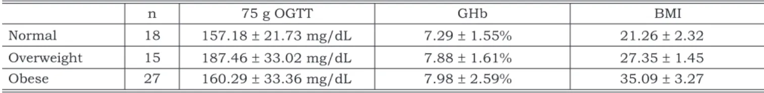
Mean age of the 60 patients included in the study was 32.75 ± 6.01 (19-45 years old) and the pregnancy time ranged from 16 to 40 weeks (32.98 ± 5.01). Laboratorial assays and BMI values, according to the groups, are listed in Table 1. The mean values of 75 g OGTT were 157.18 ± 21.73 mg/ dL in the normal group, 187.46 ± 33.02 mg/dL in the overweight group and 160.29 ± 33.36 mg/dL in the obese group. GHb means ranged from 7.29% to 7.98%. The BMI results for normal, overweight and obese groups were 21.26 ± 2.32, 27.35 ± 1.45 and 35.09 ± 3.27, respectively.
The results of the clinical periodontal assess-ments are shown in Table 2. Significant differ-ences were found only between the normal and obese groups considering GB (52.76 ± 27.99% and 78.85 ± 27.44%, respectively), and CAL (2.21 ± 0.41 mm and 2.61 ± 0.54 mm, respec-tively). Although an increase in BOP was found when comparing the normal and obese groups, no significant differences were found among groups. The percentage means of BOP ranged from 55.65% to 75.31%. The medians and their respective per-centiles (25-75) of attachment loss classification in the normal, overweight and obese groups were 2 (2-2), 2 (2-3) and 3 (2-3), respectively. Two patients presented attachment loss median equal to four:
one with BMI of 38.90 kg/m2 and another with BMI of 26.20 kg/m2.
DISCUSSION
In this study, patients who presented preges-tational obesity showed significantly more gingi-vitis and attachment loss than pregnant women with normal pregestational BMI.
Diabetes mellitus, smoking and certain micro-organisms in the dental biofilm are considered risk factors of periodontal breakdown25. Although other periodontitis risk indicators have been studied (for instance: stress, osteoporosis and genetic factors), few analytical epidemiological studies have been conducted establishing the relationship between obesity and periodontitis and using the clinical periodontal attachment loss parameter as the outcome factor3,28. Vecchia et al.28 (2003), using the criteria “30% of the teeth or more presenting CAL ≥ 5 mm” to determine the presence of peri-odontitis found that obese women had 1.65 more chances of having periodontitis after adjustment for age and smoking (Confidence Interval 1.04-2.64)28. When smoking was not considered, obese women had 2.28 times more chances of having periodontitis (Confidence Interval 1.05-4.93). The authors excluded diabetic patients from the study. The biological plausibility of this association may
TABLE 1 - Means and standard deviation of 75 g oral glucose tolerance test (OGTT), glycated hemoglobin assay (GHb) and body mass index (BMI) of 60 pregnant women distributed according to normal, overweight and obese groups.
n 75 g OGTT GHb BMI
Normal 18 157.18 ± 21.73 mg/dL 7.29 ± 1.55% 21.26 ± 2.32 Overweight 15 187.46 ± 33.02 mg/dL 7.88 ± 1.61% 27.35 ± 1.45 Obese 27 160.29 ± 33.36 mg/dL 7.98 ± 2.59% 35.09 ± 3.27
TABLE 2 - Clinical parameters of 60 gestational diabetes mellitus patients distributed according to body mass in-dex (BMI) groups.
n Number of Teeth* GB BOP CAL CAL**
Normal 18 24.44 ± 6.43 52.76 ± 27.99a 55.65 ± 27.65 2.21 ± 0.41a 2 (2-2)
Overweight 15 25.27 ± 5.24 65.64 ± 23.31 71.47 ± 20.35 2.40 ± 0.53 2 (2-3) Obese 27 26.25 ± 5.10 78.85 ± 27.44a 75.31 ± 30.33 2.61 ± 0.54a 3 (2-3)
p value 0.556 0.008 0.062 0.032
86 87
86 87
be explained by the increased levels of pro-inflam-matory mediators found in obese patients19,23.
Our study sample consisted of 60 patients with GDM, with 45% presenting pregestational obesity. In the period before delivery, obese preg-nant women present a larger number of pre-exist-ing complications. The prevalence of gestational diabetes mellitus ranges from 14 to 39.4% in obese patients, while in normal weight pregnant women it ranges from 1.85 and 4.3% (p < 0.05)7,12. They also may develop more medical changes when compared to normal weight controls: an increased risk of hypertension, anemia, perinatal mortality and urinary track infection.
Gingivitis is the most frequent oral manifesta-tion associated with pregnancy. Its occurrence is reported to range from 30 to 100%4. In this study, a statistically significant difference was found in GB between the normal and the obese group. Vec-chia et al.28 (2003), when assessing the average percentage of teeth with gingival bleeding in 386 non-pregnant women according to the BMI catego-ries, found that GB ranged from 64.1 to 69.2%, without differences between normal, overweight and obese women groups28. The differences found among studies could be related to changes found in the protection periodontium that take place after the second month of pregnancy. Evidence sug-gests that the levels of sexual hormones change the composition of the biofilm, as well as influence vascular, cellular and immune responses of the inflammatory process4. And although it results predominantly just in gingivitis, an increased risk of periodontitis in these patients should not be ignored. However, no differences were found in the prevalence of periodontitis when pregnant and non-pregnant women were compared8,12.
Periodontal clinical attachment loss is con-sidered the most adequate indicator of destruc-tive periodontal disease9. The CAL observed in the study sample revealed that loss of periodontal attachment in at least one of the tooth surfaces is frequent. Unlike diabetes mellitus, hormone changes found in pregnancy do not represent an increased risk for the onset and progression of support periodontium destruction8,13. Also, to es-tablish the diagnosis of GDM, it has to be shown that carbohydrate metabolism was completely nor-mal before pregnancy26. Considering these facts, it is interesting to investigate factors that could account for the oral manifestations found in ges-tational diabetes patients.
When examining the relationship between periodontal disease and obesity, Al-Zahrani et al.3 (2003) defined periodontal disease using CAL
greater than or equal to 3 mm associated with probing depth greater than or equal to 4 mm3. In the present study, the values of tooth attachment loss in patients of the obese group ranged from 3 to 5 mm, while in the normal weight group it ranged from 1 to 2 mm. Losses found in the normal weight group can be linked only to gingival reces-sion, which can be related to bacterial plaque or not. Although probing depth, like BOP, is an in-flammatory parameter commonly used in studies, it is limited when determining the presence and severity of periodontal disease1,21.
In this study, no differences were found among the groups considering the presence of BOP, a pa-rameter that shows inflammation associated with the presence of bacterial plaque in the subgingi-val environment, although a trend of percentage increase with the increase of BMI could be found. This could be explained by the limits of the study sample size. Also, this could be due to the con-founding factor associated with the presence of bleeding of the marginal gingiva21. New studies in this group of patients with separate description of parameters for free and proximal surfaces could better elucidate the patients’ hygiene pattern. Ad-ditionally, the analysis of BOP after control of su-pragingival plaque could better define the sites with true subgingival periodontal disease activity. For now, it is clear that in the studied group there is a significant history of periodontal support loss and gingivitis.
As a whole, the results of this study showed that clinicians should assess the periodontal status of gestational diabetes patients with pregestational obesity history. It has been shown that periodontal infections can change the metabolic-endocrine sta-tus of the host, leading to difficulties in the control of blood sugar levels, which can contribute to in-sulin-resistance, hyperglycemia and complications in the metabolic control of diabetes mellitus2,11,15. Moreover, periodontal treatment improves diabetes control2. Therefore, periodontal infection control could be one additional important tool used in the metabolic control of gestational diabetes.
CONCLUSION
86 87
86 87
REFERENCES
1. Ainamo J, Bay I. Problems and proposals for recording gingivitis and plaque. Int Dent J 1975;25(4):229-35. 2. Aldridge JP, Lester V, Watts TLP, Collins A, Viberti G,
Wilson RF. Single-blind studies of the effects of improved periodontal health on metabolic control in Type 1 diabetes mellitus. J Clin Periodontol 1995;22:271-5.
3. Al-Zahrani MS, Bissada NF, Borawskit EA. Obesity and periodontal disease in young, middle-aged, and older adults. J Periodontol 2003;74(5):610-5.
4. Amar S, Chung KM. Influence of hormonal variation on the periodontium in women. Periodontol 2000 1994;6:79:87. 5. Badersten A, Nilvéus R, Egelberg J. Scores of plaque,
bleed-ing, suppuration and probing depth to predict probing at-tachment loss. 5 years of observation following nonsurgical periodontal therapy. J Clin Periodontol 1990;17:102-7. 6. BEMFAM. Pesquisa Nacional sobre Demografia e Saúde.
Rio de Janeiro; 1997.
7. Bianco AT, Smilen SW, Davis Y, Lopez S, Lapinski R, Lock-wood CJ. Pregnancy outcome and weight gain recommen-dations for the morbidly obese woman. Obstet Gynecol 1998,91:97-102.
8. Brabin BJ. Epidemiology of infection in pregnancy. Rev Infect Dis 1985;7:579-603.
9. Genco RJ. Current view of risk factors for periodontal dis-ease. J Periodontol 1996,60(10):1041-9.
10. Genco R, Glurich I, Haraszthy V, Zambon J, DeNar-din E. Overview of risk factors for periodontal disease and implications for diabetes and cardiovascular disease. Com-pend Contin Educ Dent 1998;19(1):40-5 [spec issue]. 11. Grossi SG, Genco RJ. Periodontal disease and
dia-betes mellitus: a two-way relationship. Ann Periodontol 1998;3(1):51-61.
12. Johnson JW, Longmate JA, Frentzen B. Excessive maternal weight and pregnancy outcome. Am J Obstet Gynecol 1992;167:353-70.
13. Jonsson R, Howland BE, Bowden GHW. Relationships between periodontal health, salivary steroids, and Bacte-roides intermedius in males, pregnant and non-pregnant women. J Dent Res 1988;67:1062-9.
14. Jovanovic L, Pettin DJ. Diabetes mellitus gestacional. JAMA Brasil 2003;7(2):1446-8.
15. Kornmann KS, Page RC, Tonetti MS. The host re-sponse to the microbial challenge in periodontitis: assem-bling the players. Periodontol 2000 1997;14:33-53. 16. Lalla E, Lamster IB, Drury S, Caifeng F, Schmidt AM.
Hyperglycemia, glycoxidation and receptors for advanced glycation endproducts: potential mechanisms underlying diabetic complications, including diabetes-associated peri-odontitis. Periodontol 2000 2000;23:50-62.
17. Mattews DC. The relationship between diabetes and periodontal disease. J Can Dent Assoc 2002;68(3):161-4.
18. National Institutes of Health. Clinical guidelines on the identification, evaluation, and treatment of overweight and obesity in adults – The evidence report. Obes Res 1998;6(Suppl):51S-209S.
19. Nishmura F, Murayama Y. Periodontal inflammation and insulin resistance – lessons from obesity. J Dent Res 2001;80(8):1690-4.
20. Offenbacher S, Beck JD. Periodontitis: a potential risk factor for spontaneous preterm birth. Compend Contin Educ Dent 1998;19(1):32-9 [spec issue].
21. Oppermann RV, Azevedo L, Chapper A, Cartão VV, Susin C. Marginal bleeding influence in the treatment of periodontitis [abstract online]. J Dent Res 2002. Available from: URL: http://iadr.confex.com/iadr/2002SanDiego/ techprogram/abstract_20732.htm.
22. Otomo-Corgel J, Steinberg BJ. Medicina periodontal e a mulher como paciente. In: Rose LF, Genco RJ, Meadley B, Cohen DW. Medicina Periodontal. São Paulo: Santos; 2002. p. 151-66.
23. Perlstein MI, Bissada NF. Influence of obesity and hypertension on the severity of periodontitis in rats. Oral Surg Oral Med Oral Pathol 1977;43(5):707-19.
24. Sacks DB, Bruns DE, Goldstein DE, Maclaren NK, McDonald JM, Parrott M. Guidelines and recommendations for laboratory analysis in the diagnosis and management of diabetes mellitus. Clin Chem 2002;48(3):436-72. 25. Salvi GE, Lawrence H, Offenbacher S, Beck JD.
Influ-ence of risk factors on the pathogenesis of periodontitis. Periodontol 2000 1997;14:173-201.
26. Simpson R, Kast S. Management of gestational dia-betes with a conservative insulin protocol. Med J Aust 2000;172(11):537-40.
27. Steinberg BJ. Saúde Bucal das Mulheres. Compên-dio de Educação Continuada em Odontologia: mulheres e odontologia 2001;22(1):8-14.
28. Vecchia CFD, Susin C, Oppermann RV, Rösing CK, Albandar J. Sobrepeso e obesidade como indicadores de risco à periodontite [resumo Pc399]. Pesqui Odontol Bras 2003;17(Supl 2):259.
29. Yalda B, Offenbacher S, Collins JG. Diabetes as a modifier of periodontal disease expression. Periodontol 2000 1994;6:37-49.
30. Yuli M, Andreína M, Yuraima P. Manifestaciones bucales de la diabetes mellitus gestacional - presentación de dos casos y revisión de la literatura. Acta Odontol Venez [article online] 2002 [cited 2003 Dec 25]. Available from: URL: http://www. actaodontologica.com/40_2_2002/32.asp.