1 – PhD in Medical Sciences - Assistant – Heart Institute - HCFMUSP 2 - Livre-Docente - InCor - HCFMUSP
3 - Livre-Docente - InCor - HCFMUSP 4 - PhD in Medical Sciences - InCor - HCFMUSP 5 - Cardiovascular Surgery resident - InCor - HCFMUSP 6 - PhD in Medical Sciences - InCor - HCFMUSP 7 - Livre-Docente - InCor - HCFMUSP 8 - Professor Emeritus - InCor - HCFMUSP
9 - Titular Professor of Cardiology - InCor - HCFMUSP 10 - Livre-Docente and Regent Professor - InCor - HCFMUSP Work performed in the Heart Institute of the Medicine School, University de São Paulo, São Paulo, SP.
Correspondence address:
Fábio Antonio Gaiotto. Rua Capote Valente, 361 Ap 121 - São Paulo, SP - CEP: 05409-001. Tel 11-3898-3023 / 9375-8386.
E-mail: [email protected]
Fabio Antonio GAIOTTO1, Luiz Boro PUIG2, Charles MADY3, Fábio FERNANDES4, Carlos Eduardo TOSSUNIAM5, Miriam Magalhães PARDI6, Luis A. O. DALLAN7, Sérgio Almeida de OLIVEIRA8, José F. RAMIRES9, Pablo M. A. POMERANTZEFF10
Article received in April 26th, 2006 Article accepted in December 26th, 2006
RBCCV 44205-870
Substituição da valva mitral com tração dos músculos papilares em pacientes com miocardiopatia
dilatada
Mitral valve replacement with chordae tendineae
preservation, traction and fixation in end-stage
dilated cardiomyopathy
Abstract
Objective: This study aimed at evaluating results of mitral
valve replacement using a new technique of complete chordae tendineae adjustment for left ventricular remodeling.
Methods: Twenty end-stage idiopathic dilated
cardiomyopathy patients with severe functional mitral valve regurgitation underwent mitral valve replacement. Seventeen (85%) were in Functional Class IV. Both anterior and posterior leaflets of the mitral valve were divided to obtain 4 pillars of chordae tendineae. These were displaced with traction toward the left atrium and anchored between the
mitral annulus and a valvular prosthesis. To evaluate the left ventricular remodeling, Doppler echocardiography was performed. For statistical analysis, variance analysis and Friedman’s test were employed.
Results: Two (10%) early deaths occurred. Kaplan-Meyer
Alternatives to heart transplantation have been proposed with the aim of increasing and improving the quantity and quality of life of these patients. Bolling et al. [5] demonstrated satisfactory results with the surgical approach to secondary mitral valve insufficiency with mitral valve repair using a flexible ring. Buffolo et al. [6] demonstrated good outcomes with mitral valve replacement and fixation of groups of chordae tendineae related to the anterior cuspid in the commissural regions.
For patients with secondary mitral valve insufficiency due to advanced dilated myocardiopathy, Puig et al. [7], in 2000, proposed mitral valve replacement with chordae tendineae traction and anchoring, aiming at ventricular remodeling. A 5-year evaluation of the technique is presented in this work.
METHOD
The patients included in this study were those in advanced stage dilated myocardiopathy with three or more hospitalizations for decompensated heart failure, under maximum medicinal treatment, Functional Class III or IV, accentuated systolic dysfunction (ejection fraction of less than 30% using the Teicholz method) and accentuated secondary mitral valve insufficiency. Patients with Chagasic disease, ischemic or gestational myocardiopathy were INTRODUCTION
Secondary mitral valve insufficiency is commonly present in advanced stage dilated myocardiopathy. The changes in the left ventricle geometry, with an alteration from a truncated cone to a spherical format, cause deformities in the subvalvar apparatus with detachment of the papillary muscle base. There are also mitral ring dilatation and transvalvar pressure alterations that, together with the subvalvar alterations cause secondary mitral valve insufficiency. The association of dilated myocardiopathy with secondary mitral valve insufficiency causes refractivity to clinical treatment and high mortality, mainly in Functional Class III and IV patients (NYHA). Survival is less certain under these conditions: 59% with absence of secondary mitral valve insufficiency over 32 months and 17% with accentuated insufficiency [1].
Heart transplantation is the best treatment method for advanced heart failure, with good survival rates over the first 5 years, but, there are insufficient donors thereby limiting its use. In the United States between 6000 and 8000 patients per year join the waiting list for transplants. The number of donors is close to 2500 per year [2]. Mortality on the waiting list is high (40% to 50%). Many patients leave the waiting list or do not enter it due to contraindications for heart transplantation.
of the end diastolic diameter (p=0.038), end systolic diameter (p=0.008), end systolic volume (p=0.029) and end diastolic volume (p=0.009). No statistical differences were noted in the systolic volume. Comparing pre-operative and third- and six-month follow-ups, the Friedman test showed no statistical differences for all studied variables. Variance analyses between pre, three-month and final evaluations showed no significant differences.
Conclusion: This technique of mitral valve replacement
improved the left ventricle ejection fraction and decreased the end diastolic and systolic diameters and the end systolic diastolic volumes up to the third month of follow-up. From then on the variables stabilized.
Descriptors: Mitral valve, surgery. Heart failure,
congestive. Cardiomyopathy, dilated. Heart valve prosthesis.
Resumo
Objetivo: Avaliar a geometria e a função do ventrículo
esquerdo (VE) após a troca mitral com tração e fixação dos papilares, em portadores de insuficiência cardíaca terminal com insuficiência mitral secundária.
Método: Dos 20 pacientes avaliados, 70% eram homens,
com idade média de 50,2 anos e 55% recebiam inotrópicos. A fração de ejeção (FEVE) foi menor que 30% em todos; 85%
estavam em classe funcional (CF) IV. Dezoito receberam próteses de pericárdio bovino e dois, mecânicas. Os períodos considerados foram: 3, 6, 12 e 18 meses. As variáveis consideradas: volume sistólico do VE (VS), a FEVE, os diâmetros sistólico e diastólico finais (DSF e DDF) e os volumes sistólico e diastólico finais (VSF e VDF). No estudo estatístico, empregou-se da análise de variância (AV) e o teste de Friedmann (F). A sobrevida foi aferida pelo método de Kaplan-Meyer.
Resultados: Dois (10%) faleceram no período imediato. A
sobrevida no primeiro ano foi de 85%, no segundo, 44%, no terceiro, 44%, no quarto, 44% e no quinto, 44%. A comparação entre pré e 3 meses, empregando-se a AV, não revelou alteração significativa para o VS (p=0,086). Houve acréscimo da FEVE (p=0,008) e decréscimo do DDF (p=0,038); do DSF (p=0,008); do VDF (p=0,029) e do VSF (p=0,009). Os momentos pré, 3 e 6 meses, com o teste F, não revelaram alterações. Entre os momentos pré, 3 meses e final, empregando-se a AV, não houve significância.
Conclusão: Há melhora da FEVE, dos VDF, VSF, DDF e
DSF; até o terceiro mês. A partir de então, as variáveis permanecem estáveis.
Descritores: Valva mitral, cirurgia. Insuficiência cardíaca
excluded from this study. All patients were informed of the nature of the study and the protocol followed the norms of the institutional.
From July 2000 to December 2003, 20 patients were consecutively submitted to surgery in the Heart Institute of the Hospital das Clínicas of the Medicine School, University of São Paulo. Preoperative clinical data are shown in Table 1.
atrial roof and interatrial septum, enabling ample exposure of the valve and subvalvar apparatus [8].
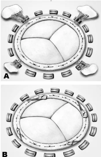
The chordae tendineae are divided into four groups, starting with the isolation of the anterior cuspid section in the middle of its free portion and continuing up to the mitral ring, extending both sides up to the commissures. The chordae tendineae of the posterior cuspid are also divided into two groups, as the chordae are thicker (Figure 1).
Variable Age Male: female
Systemic arterial hypertension Diabetes mellitus
Previous valvuloplasty Ejection fraction Systolic volume End-systolic volume End-diastolic volume End-systolic diameter End-diastolic diameter NYHA Functional Class III
IV
Cardiogenic shock
Number of hospitalizations on the ward Number of hospitalizations in the ICU Cardiac cachexia
value 50.2 ± 9.0 years
14:6 7 (35%) 2 (10%) 1 (5%) 23.3% ± 4.1% 73.6 mL ± 20 mL
246 mL ± 73 mL 320 mL ± 87 mL 6.83 cm ± 0.92 cm 7.68 cm ± 0.98 cm
3 (15%) 17 (85%)
11(55%) 5.45 ± 3.1 2.4 ± 1.27 12 (60%) Table 1. Preoperative data
Fig. 1 – A: Mitral subvalvar apparatus anatomy. B: Mitral valve anatomy: incision lines
Transthoracic echocardiographic evaluations were performed using Sonos 5500 Phillips Medical Systems (2000, Hannover, MA, USA) and HDI 3500 (1996, Seattle, WA, USA) devices. The degree of mitral vale insufficiency was measured and to assess remodeling, the left ventricular systolic volume (SV), end diastolic and systolic volumes (EDV and ESV) and end diastolic and systolic diameters (EDD and ESD) were chosen.
In the postoperative follow-up, all patients were accompanied for between 1 and 60 months, with a mean of 23.2 months. The clinical conditions of the patients were evaluated at three-monthly intervals.
Surgical technique
Patients were operated on by sternotomy using normothermic cardiopulmonary bypasses. Myocardial protection was achieved with intermittent anterograde blood cardioplegia with coronary reperfusion by aortic declamping with the aim of reducing the overall ischemia time.
Access, in all cases, was by the right atrium, the left
of forces. Excess of chordae and cuspid that remain in the left atrium are fixed on the prosthetic ring. The prosthesis is implanted using “U-shaped” sutures (Figure 2).
RESULTS
Immediate
Eighteen bovine pericardial bioprostheses were implanted and two patients received St Jude mechanical prostheses. Under-sized prostheses were chosen aiming at remodeling of the left ventricular base (sizes 27 in 2; 29 in 16; 31 in 1 and 33 in 1). The DeVega procedure was performed on the tricuspid valve in 12 (60%) patients. The cardiopulmonary bypass time varied from 80 to 180 minutes (105 ± 23 minutes) and aortic clamping time varied from 30 to 74 minutes (46 ± 12 minutes). All patients received endovenous inotropic support in the postoperative period. Intra-aortic balloons were used in four (20%) patients.
Two (10%) patients suffered early deaths, one 18 days after the procedure due to bronchopneumonia and the other 25 days after the procedure with multiple organ failure.
Late
All the patients were followed up and the functional class, after 48 months of follow up, had improved (p=0.013 – McNemar test) with four (40%) patients in Class I, three (30%) in Class II and three (30%) in Class III (Figure 3).
Fig. 2 –A: Implanted bioprosthesis with papillary traction. B: Concluded operation with the removal of excess chordae
Fig. 3 - Functional Class at 48 months of follow-up
Statistical analysis
Statistical analysis was structured with the objective of understanding the temporal behavior of the selected variables. To compare the variables in the preoperative periods and at 3 months, analysis of variance for repeated measures was utilized. For the study of the preoperative period and at 3 and 6 months, Friedmann’s nonparametric test was chosen. In the comparison of pre-, 3-month and final evaluations, analysis of variance for repeated measures was again used. The functional class was analysed by McNemar’s test, at the end of 48 months of follow up. The number of hospital readmissions was evaluated using the Wilcoxon’s test and survival by the Kaplan-Meyer method.
Evaluation of survival demonstrated rates of 85% at the end of first year, 44%, for the second, 44% for the third, 44% for the fourth and 44% for the fifth year (Figure 4).
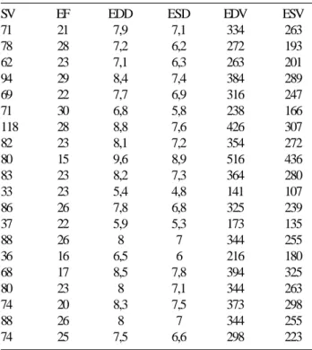
Table 2. Preoperative echocardiography variable
SV – systolic volume in mL; EF – left ventricular ejection fraction; EDD – end-diastolic diameter in cm; ESD – end-systolic diameter in cm; EDV – end-diastolic volume in mL; ESV – end-systolic volume in mL
SV 71 78 62 94 69 71 118 82 80 83 33 86 37 88 36 68 80 74 88 74 EF 21 28 23 29 22 30 28 23 15 23 23 26 22 26 16 17 23 20 26 25 EDD 7,9 7,2 7,1 8,4 7,7 6,8 8,8 8,1 9,6 8,2 5,4 7,8 5,9 8 6,5 8,5 8 8,3 8 7,5 ESD 7,1 6,2 6,3 7,4 6,9 5,8 7,6 7,2 8,9 7,3 4,8 6,8 5,3 7 6 7,8 7,1 7,5 7 6,6 EDV 334 272 263 384 316 238 426 354 516 364 141 325 173 344 216 394 344 373 344 298 ESV 263 193 201 289 247 166 307 272 436 280 107 239 135 255 180 325 263 298 255 223
3rd month 6th month Last
evaluation LVEF (%) 29±6.9 26.8±7.7 29±11 SV (ml) 81±27 66±24 77±27 EDV (ml) 279±70 247±69 270±112 ESV (ml) 198±59 179±58 192±102 EDD (cm) 7.2±0.8 6.8±0.8 7.0±1.1 ESD (cm) 6.2±0.7 5.9±0.7 6.0±1.2 Table 3. Echocardiography variable in the late postoperative
Fig. 4 - Actuarial curve - Kaplan-Meier method
Echocardiography (Tables 2 & 3) Preoperative period
The ejection fraction (LVEF) varied from 15% to 30%, with a mean of 23 ± 4%. The mean SV was 73 ± 20 mL ranging from 36 to 97 mL. The mean ESV was 246 ± 87 mL varying from 135 to 436 mL and the mean EDV was 320 ± 73 mL ranging from 173 to 516 mL. The mean DDE was 7.68 ± 0.98 cm varying from 5.9 to 9.6 cm and the mean ESD was 6.38 ± 0.92 cm varying from 5.3 to 8.9 cm.
Study of preoperative period and 3 months of follow-up
Analysis of variance for repeated measures showed improvement in the LVEF (p=0.008) and a reduction in the EDD (p=0.038), ESD (p=0.008), EDV (p=0.029) and ESV (p=0.009). There was no significant change in the SV.
Study of preoperative period and 3 and 6 months of follow-up
The Friedman’s nonparametric test did not show significant changes for any of the variables analysed.
Study of preoperative, 3-month and final evaluations
The Analysis of variance for repeated measures did not demonstrate significant changes for variables analysed.
DISCUSSION
Heart failure is frequently observed in end-stage myocardiopathies, with the 1-year survival rate estimated at 50% for patients in Functional Class IV [9,10]; a value that is surely higher when patients with secondary mitral valve insufficiency and those that use inotropic support for prolonged periods are considered. Barreto & Ramires [11] demonstrated that heart failure is responsible for two-thirds of cardiovascular procedures. The surgical treatment may be performed in patients who do not respond to clinical treatment.
number of donors and the high mortality on the waiting list makes the procedure unviable on a large scale [12]. Many patients are contraindicated for transplantation and so no option exists. Some alternatives have been proposed and studied for these patients: cardiomyoplasty [13], coronary artery bypass grafting surgery for ischemic patients [14], partial left ventriculectomy [15], ventricular resynchronization [16], valve repair [5,17] and mitral valve replacement [6,7]. In some cases, a combination of techniques may benefit the patient.
Secondary mitral valve insufficiency is a factor of worst prognosis in end-stage heart failure [1,18]. The geometric changes of the left ventricle and dilatation of the mitral annulus cause mitral valve insufficiency without structural alterations of the mitral valve cuspids. Spherical dilatation of the left ventricle is a factor of worst prognosis [19]. The size of the ventricular cavity increases, the papillary muscles migrate in posterolateral and apical directions, the heart base dilates and secondary mitral valve insufficiency results. Due to the similarity of mechanisms, tricuspid valve insufficiency is frequently associated with mitral valve insufficiency. Mitral valve insufficiency causes volume overload of the affected left ventricle with the cycle being perpetuated until the death of the patient. Correction of mitral valve insufficiency may break this cycle and delay the natural history of the phenomenon.
Mitral valve repair is attractive, to preserve all the natural tissue of the patient. With a reduction of the mitral ring, mitral valve insufficiency may be eliminated. There is a much better ventricular result as the volume overload is treated and the diastolic volume reduces. The amount of regurgitation is also diminished and the patient has a clinical improvement.
Residual mitral valve insufficiency is common with mitral valve repair worsening in the follow-up period [5,20]. Mitral valve replacement does not allow relapse but replacing it with a prosthesis is inappropriate taking into account the life expectation of these patients [6]. Lillehei et al. [21], in 1964, reported high mortality rates after mitral valve replacement without repair of the subvalvar apparatus. On preserving the continuity of the mitral ring with the left ventricle, the mortality rate decreased from 34% to 17%. Later, Miki et al. [22] suggested preserving the posterior cuspid and splitting up the chordae tendineae of the anterior cuspid, anchoring them on the commissures. Buffolo et al. [6] also proposed this strategy for patients with dilated myocardiopathy, believing that the elliptical format of the heart could be reestablished.
The papillary muscles play an important role in ventricular contraction. With them fixed onto the mitral ring, they bring the anteromedial and posterolateral walls closer together during systole and cause an alteration in the shape
of the heart: a tendency of a spherical shape during diastole and elliptical during systole [23].
The proposed technique removes any excess of chordae tendineae and maintains them under tension in respect to the left atrium thereby helping to reduce the longitudinal axis of the left ventricle and returning it to an elliptical shape. Adequate single-plane traction with anchoring of the chordae tendineae at four points on the ventricular ring may cause a physiological spread of the forces that act on the ventricular wall. This dissipation of the forces will be transmitted to the ventricular wall during diastole, which, with a reduction of volume overload, may result in an improved systolic performance and promote remodeling of the left ventricle. The echocardiographic data suggest that this improvement is seen up to the third month, with maintenance afterwards. Echocardiographic data also show a reduction in the volume and diameters, as expected by the proposed mechanism. There is even improvement in the functional class, reduction in the number of patients returning to hospital and better survival.
The myocardial protection method utilized, cardioplegia with intermittent reperfusion, may help myocardial recovery in the postoperative period. The greater the ischemia time, the better the myocardial recovery will be and, undoubtedly, the option to use the access route described by Guiraudon et al. [8] facilitated this strategy.
Some patients present with tricuspid regurgitation associated to secondary mitral valve insufficiency due to the dilatation of the heart base. We believe that tricuspid repair is essential and must always be considered. Radovanovic et al. [24] suggested repair of the mitral and tricuspid insufficiency at the first decompensation of heart insufficiency. One patient in this series presented right-sided heart failure due to accentuated tricuspid insufficiency and was submitted to tricuspid bioprosthesis implantation showing a good evolution in the postoperative period. The patient was discharged from hospital on the 20th
postoperative day in Functional Class II.
Additional studies must be performed to find the best moment to perform mitral and tricuspid repair [25]. The patients may improve with clinical treatment, but the moment for mitral valve repair must always be remembered. Regurgitation is less harmful when the myocardium is healthy, but when there is myocardiopathy it may be catastrophic and a dilatation cycle may be activated. There is no ventricular reserve to adapt to secondary mitral valve insufficiency. Early indication may improve survival and functional class.
REFERENCES
1. Blondheim DS, Jacobs LE, Kotler MN, Costacurta GA, Parry WR. Dilated cardiomyopathy with mitral regurgitation: decreased survival despite a low frequency of left ventricular thrombus. Am Heart J. 1991;122(3 Pt 1):763-71.
2. Zaroff JG, Rosengard BR, Armstrong WF, Babcock WD, D`Alessandro A, Dec GW, et al. Consensus conference report: maximizing use of organs recovered from the cadaver donor: cardiac recommendations, March 28-29, 2002, Crystal City VA. Circulation. 2001;106(7):836-41.
3. Aaronson KD, Mancini DM. Mortality remains high for outpatient transplant candidates with prolonged (>6 months) waiting list time. J Am Coll Cardiol. 1999;33(5):1189-95.
4. Stevenson LW, Fowler MB, Schroeder JS, Stevenson WG, Dracup KA, Fond V. Poor survival of patients with idiopathic cardiomyopathy considered too well for transplantation. Am J Med. 1987;83(5):871-6.
5. Bolling SF, Pagani FD, Deeb GM, Bach DS. Intermediate-term outcome of mitral reconstruction in cardiomyopathy. J Thorac Cardiovasc Surg. 1998;115(2):381-8.
6. Buffolo E, Paula IAM, Palma H, Rodrigues JNB. Nova abordagem cirúrgica para o tratamento de pacientes em insuficiência cardíaca refratária com miocardiopatia dilatada e insuficiência mitral secundária. Arq Bras Cardiol. 2000;74(2):129-34.
7. Puig LB, Gaiotto FA, Pardi MM, Bacal F, Mady C, Fernandes F, et al. Mitral valve replacement and remodeling of the left ventricle in dilated cardiomyopathy with mitral regurgitation: initial results. Arq Bras Cardiol. 2002;78(2):224-9.
8. Guiraudon GM, Ofiesh JG, Kaushik R. Extended vertical transatrial septal approach to the mitral valve. Ann Thorac Surg. 1991;52(5):1058-62.
9. Keogh AM, Freund J, Baron DW, Hickie JB. Timing of cardiac transplantation in idiopathic dilated cardiomyopathy. Am J Cardiol. 1988;61(6):418-22.
10. Rankin JS, Feneley MP, Hickey MS, Muhlbaier LH, Wechsler AS, Floyd RD, et al. A clinical comparison of mitral valve repair versus replacement in ischemic mitral regurgitation. J Thorac Cardiovasc Surg. 1988;95(2):165-77.
11. Barreto ACP, Ramires JAF. Insuficiência cardíaca: um problema de saúde pública. Rev Bras Cardiol 2000;2:142-7.
12. Evans RW, Orians CE, Ascher NL. The potential supply of organ donors: an assessment of the efficacy of organ procurement efforts in the United States. JAMA 1992;267(2):239-46.
13. Carpentier A, Chachques JC, Acar C, Relland J, Mihaileanu S, Bensasson D, et al. Dynamic cardiomyoplasty at seven years. J Thorac Cardiovasc Surg. 1993;106(1):42-54.
14. Dreyfus GD, Duboc D, Blasco A, Vigoni F, Dubois C, Bunodaty D, et al. Myocardial viability assessment in ischemic cardiomyopathy: benefits of coronary revascularization. Ann Thorac Surg. 1994;57(6):1402-8.
15. Batista RJ, Santos JL, Takeshita N, Bocchino L, Lima PN, Cunha MA, et al. Partial left ventriculectomy to improve left ventricular function in end-stage heart disease. J Card Surg. 1996;11(2):96-8.
16. Wilensky RL, Yndelman P, Cohen AI, Fletcher RD, Atkinson J, Virmani R, et al. Serial eletrocardiographic changes in idiopathic cardiomyopathy confirmed at necropsy. Am J Cardiol. 1988;62(4):276-83.
17. Cohn LH, Kowalker W, Bhatia S, DiSesa VJ, St John-Sutton M, Shemin RJ, et al. Comparative morbidity of mitral valve repair versus replacement for mitral regurgitation with and without coronary artery disease. Ann Thorac Surg. 1988;45(3):284-90.
18. Bolling SF. Mitral reconstruction in cardiomyopathy. J Heart Valve Dis. 2002;11(suppl 1):S26-S31.
19. Juilliere Y, Danchin N, Briancon S, Khalife K, Ethevenot G, Balaud A, et al. Dilated cardiomyopathy: long-term follow-up and predictors of survival. Int J Cardiol. 1988;21(3):269-77.
20. Calafiore AM, Gallina S, Di Mauro M, Gaeta F, Iaco AL, D'Alessandro S, et al. Mitral valve procedure in dilated cardiomyopathy: repair or replacement? Ann Thorac Surg. 2001;71(4):1146-53.
21. Lillehei CW, Levy MJ, Bonnabeau RC Jr. Mitral valve replacement with preservation of papillary muscles and chordae tendineae. J Thorac Cardiovasc Surg. 1964;47:532-43.
22. Miki S, Kusuhara K, Ueda Y, Komeda M, Ohkita Y, Tahata T. Mitral valve replacement with preservation chordae tendineae and papillary muscles. Ann Thorac Surg. 1988;45(1):28-34.
23. Rushmer RF, Finlayson BL, Nash AA. Movements of the mitral valve. Circ Res. 1956;4(3):337-42.
24. Radovanovic N, Mihajlovic B, Selestiansky J, Torbica V, Mijatou MK, Popou M. Reductive annuloplasty of double orifices in patients with primary dilated cardiomyopathy. Ann Thorac Surg. 2002;73(3):751-5.