Regulates Glutamate-Dependent Oxidative
Phosphorylation on Demand
Frank Norbert Gellerich1*, Zemfira Gizatullina2, Odeta Arandarcikaite3, Doreen Jerzembek1, Stefan Vielhaber2, Enn Seppet4, Frank Striggow1
1KeyNeurotek Pharmaceuticals AG, ZENIT Technology Park, Magdeburg, Germany,2Department of Neurology, Otto von Guericke University Magdeburg, Magdeburg, Germany,3Institute for Biomedical Research, Kaunas University of Medicine, Kaunas, Lithuania,4Department of Pathophysiology, Centre of Molecular and Clinical Medicine, University of Tartu, Tartu, Estonia
Abstract
We present unexpected and novel results revealing that glutamate-dependent oxidative phosphorylation (OXPHOS) of brain mitochondria is exclusively and efficiently activated by extramitochondrial Ca2+in physiological concentration ranges
(S0.5= 360 nM Ca2+). This regulation was not affected by RR, an inhibitor of the mitochondrial Ca2+ uniporter. Active
respiration is regulated by glutamate supply to mitochondria via aralar, a mitochondrial glutamate/aspartate carrier with regulatory Ca2+-binding sites in the mitochondrial intermembrane space providing full access to cytosolic Ca2+. At
micromolar concentrations, Ca2+ can also enter the intramitochondrial matrix and activate specific dehydrogenases.
However, the latter mechanism is less efficient than extramitochondrial Ca2+regulation of respiration/OXPHOS via aralar.
These results imply a new mode of glutamate-dependent OXPHOS regulation as a demand-driven regulation of mitochondrial function. This regulation involves the mitochondrial glutamate/aspartate carrier aralar which controls mitochondrial substrate supply according to the level of extramitochondrial Ca2+.
Citation:Gellerich FN, Gizatullina Z, Arandarcikaite O, Jerzembek D, Vielhaber S, et al. (2009) Extramitochondrial Ca2+
in the Nanomolar Range Regulates Glutamate-Dependent Oxidative Phosphorylation on Demand. PLoS ONE 4(12): e8181. doi:10.1371/journal.pone.0008181
Editor:Mark R. Cookson, National Institutes of Health, United States of America
ReceivedApril 28, 2009;AcceptedOctober 30, 2009;PublishedDecember 9, 2009
Copyright:ß2009 Gellerich et al. This is an open-access article distributed under the terms of the Creative Commons Attribution License, which permits unrestricted use, distribution, and reproduction in any medium, provided the original author and source are credited.
Funding:This work was supported by the European Huntington network, the DFG (Ge 664/11-2), the German Federal Ministry of Economics and Technology, grant No. IWO72052 (MitoscreenTest), Estonian Ministry of Education and Research (SF0180114As08) and Estonian Science Foundation (grants No. 7117 and 7823). The funders had no role in study design, data collection and analysis, decision to publish, or preparation of the manuscript.
Competing Interests:Frank Gellerich, Zemfira Gizatullina, Doreen Jerzembeck and Frank Striggow are employees of Keyneurotek Pharmaceuticals AG, a privately held biotechnology company.
* E-mail: [email protected]
Introduction
It has been assumed that ADP formed by ATP-consuming enzymes activates OXPHOS [1]. However, cytosolic ADP of the heart muscle is only insignificantly increasedin vivoduring elevated
work loads [2,3]. Therefore, two hypotheses have been proposed, (i) the dynamic compartmentation of ADP, assuming that necessary ADP augmentations occur exclusively within the mitochondrial intermembrane space [4,5] and (ii) the stimulation of OXPHOS due to Ca2+
influx into the mitochondrial matrix via Ca2+uniporter, followed by the activation of distinct
intramito-chondrial dehydrogenases [6,7]. Some authors also assume a Ca2+
stimulation of F0F1-ATP synthase [8,9]. However, both scenarios comply only partially with thein vivofindings outlined above [10].
Recent data suggest that the activity of the malate aspartate shuttle (MAS), including glutamate/aspartate carriers as aralar, is activated by extramitochondrial Ca2+ (S
0.5= 324 nM) [11–13]. The N-terminal regulatory Ca2+-binding site of aralar is located
within the mitochondrial intermembrane space [11–13] where it can interact with Ca2+
passing through porin pores of the outer membrane. Aralar supplies OXPHOS with glutamate, a key mitochondrial substrate. In this study, we addressed the question whether OXPHOS can be directly activated by extramitochon-drial Ca2+
and if so, whether aralar is involved in this regulation.
Results
First, we investigated the influence of Ca2+ on OXPHOS of
isolated rat brain mitochondria in a medium containing 150 nM free Ca2+
(Ca2+
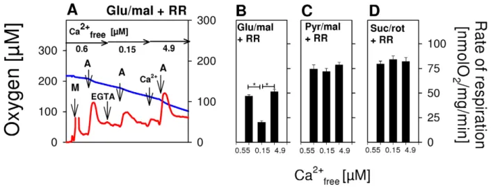
free), corresponding to basal levels of cytosolic Ca2+ under physiological conditions [14]. ADP was added so as to fully activate phosphorylation-related respiration (state 3). Using glutamate/malate as substrate, a relatively low state 3glu/malwas obtained (Fig. 1A,B). However, state 3glu/mal nearly doubled immediately after a pulse addition of 4.9mM Ca2+
free(Fig. 1A,B). This Ca2+
activation was not limited by the mitochondrial capacity of OXPHOS, but rather was due to its efficacy in metabolizing glutamate, as succinate conspicuously enhanced respiration above the level of state 3glu/mal. With pyruvate/malate (Fig. 1C), state 3pyr/malsignificantly exceeded state 3glu/mal(Fig. 1A,B). However, added Ca2+
did not augment state 3pyr/mal, whereas added succinate did (Fig. 1C). Fig. 1D demonstrates that there was also no Ca2+
effect on complex II-dependent state 3sucwith succinate/ rotenone. Overall, these results show that Ca2+
activation of OXPHOS in isolated brain mitochondria is a glutamate-specific phenomenon. The next series of experiments revealed that RR, an inhibitor of the mitochondrial Ca2+uniporter [15], is not able to
modulate Ca2+
RR concentrations (250 nM) in order to avoid possible unspecific RR effects. Nevertheless, even in the presence of up to 5mM RR, extramitochondrial Ca2+
-induced stimulation of state 3glu/malwas detectable (Data not shown).
Next, we investigated the kinetics of Ca2+
activation (Fig. 1E,F). Ca2+
was increased in steps. Increments of Ca2+
-induced state 3glu/mal were plotted against fluorimetrically measured Ca2+ (Fig. 1F) in order to determine the half-activation constant (S0.5) and the extent of Ca2+
stimulation (S0.5= 356639 nM Ca2+free, Vmax= 8665 nmol O2/mg/min). Neither parameter was affected by RR (S0.5= 306635 nM Ca2+free, Vmax= 8868 nmol O2/mg/ min). Thus, Ca2+influx into the mitochondrial matrix appears not
to be required for state 3glu/mal stimulation and, hence, Ca2+ activation must be an extramitochondrial effect.
To exclude furthermore artificial Ca2+
effects due to potential interactions of digitonin with mitochondrial membranes, we also varied the digitonin concentration used during the preparation of mitochondria to permeabilize synaptosomal membranes. Omitting digitonin did not cause any significant changes in the extent of extramitochondrial Ca2+ activation of state 3
glu/mal (Fig. 1G) compared with control mitochondria prepared with digitonin (Fig. 1G). Thus, digitonin-related artifacts can be excluded. It should be noted that in the absence of digitonin, synaptosomal mitochon-dria remained inaccessible, and therefore respiratory rates were significantly decreased in digitonin-free experiments (Fig. 1G). On the other hand, large additions of digitonin (1.2 mg digitonin/mg mitochondrial protein) led to a removal of mitochondrial outer membranes and the generation of mitoplasts [16,17]. Consequently,
Figure 1. Exclusive activation of glutamate-dependent state 3 respiration of brain mitochondria by extramitochondrial Ca2+in the nanomolar range.(A,E) Respirograms of rat brain mitochondria were obtained by high-resolution respirometry. (A) Isolated rat brain mitochondria were incubated in EGTA medium (Ca2+
free= 0.15mM) in the presence of 10 mM glutamate and 2 mM malate as substrates. Additions: M, 0.06 mg/ml brain mitochondria, A, 2.5 mM ADP to activate the phosphorylation-related respiration (state 3); Ca2+
4,9, 4.9mM Ca2+free; S, 10 mM succinate as substrate of respiratory chain complex II; C, 5mM carboxyatractyloside to block the adenine nucleotide translocase. Blue lines indicate the oxygen concentration and red lines represent respiration rates (nmol O2/mg mitochondrial protein/min). (B) Means of state 3 respiration6S.E. as measured in experiments shown in A without (black columns, n = 6) or with 250 nM RR, an inhibitor of mitochondrial Ca2+
uptake (red columns, n = 6). First group of columns, state 3 at Ca2+
free= 0.15mM. Second group, state 3 with Ca2+free= 4.9mM. Third group, state 3 with Ca2+free= 4.9mM in the additional presence of 10mM succinate. *, p,0.05. (C) As B, but derived from experiments with 10 mM pyruvate+2 mM malate as substrates. *, p,0.05. (D) As B, but derived from experiments with 10 mM succinate+2mM rotenone as substrate. (E) Ca2+titration of state 3glu/malby stepwise increase of Ca2+as indicated either without (E,F) or with (F) 250 nM RR. (F) Incremental accretions of Ca2+
-induced state 3glu/malwere plotted against the fluorimetrically measured Ca2+ activity (Fig. 1F), allowing the calculation of the half-activation constant (S0.5) and the maximum velocity (Vmax) using the SigmaPlot kinetic module as given in the text. (G) Rates of state 3glu/malrespiration obtained by Ca2+titrations under various conditions. (#) Control mitochondria were investigated as in Fig. 1E. (%) As (#), but in the additional presence of 10% dextran 20. (,) As (#), but in the additional presence of 1 mM CsA. (n) as (#), but mitochondria isolated without digitonin were used. (e) as (#), but mitoplasts were used. (C) as (#), but mitochondria were uncoupled by 50 nM FCCP from the beginning of experiments, and then Ca2+
titration was performed. (m) as (#), but Ca2+
was adjusted at the beginning of experiments as indicated. Thereafter, 100mM ADP was added, causing short transitions between the active and resting states of respiration. After reaching state 4 respiration, FCCP titrations were performed to uncouple respiration and ATP generation. Maximum respiration rates were obtained at 60 or 80 nM FCCP and were plotted against the Ca2+
the accessibility of mitochondrial Ca2+
-binding sites, originally located within the inner membrane space, to Ca2+
was facilitated but no changes of Ca2+activation were detectable (Fig. 1G). As
previously observed in heart mitoplasts [17], we also registered lower respiratory rates compared with control mitochondria; this was probably due to unspecific side effects of digitonin on mitoplasts. This finding suggests that Ca2+
diffusion through porin pores of the mitochondrial outer membrane does not limit its interaction with mitochondrial Ca2+
-binding sites exposed into the inner membrane space and thus, does not compromise extramitochondrial Ca2+
regulation of glutamate/malate-dependent respiration and OX-PHOS. Another experimental setup was used to obtain support for this interpretation. In intact cells, the colloid osmotic pressure increases the diffusion resistance of the mitochondrial outer membrane against metabolites passing the porin pores [18]. We therefore simulated the intracellular oncotic pressure by addition of 10% dextran [18], but again observed a similar extramitochondrial Ca2+
stimulation of state 3glu/mal respiration and OXPHOS (Fig. 1G). Moreover, the addition of 2mM cyclosporine A (CsA), an inhibitor of the mitochondrial permeability pore (PTP)[19], did not affect the extramitochondrial Ca2+
regulation of brain mitochondria (Fig. 1G), suggesting that PTP is not involved in the phenomenon of extramitochondrial Ca2+regulation of state 3
glu/mal and OXPHOS.
We then investigated the influence of mitochondrial uncoupling on Ca2+
stimulation of OXPHOS. Since aralar is an electrogenic carrier [20], glutamate transport into mitochondria requires a sufficiently high mitochondrial membrane potential. Accordingly, mitochondria uncoupled by 50 nM FCCP at the beginning of the experiment could not be activated by the following Ca2+titration,
owing to the dissipation of membrane potential (Fig. 1G). In a second approach, different Ca2+
free concentrations were initially adjusted followed by FCCP titration of the nonphosphorylating respiration (state 4). This application scheme resulted in enhanced maximum rates of uncoupled respiration in a Ca2+
-dependent manner (Fig. 1G). However, since FCCP also caused an incomplete dissipation of mitochondrial membrane potentials, maximum rates of uncoupled respiration were lower than in control experiments without FCCP (Fig. 1G). Obviously, cytosolic Ca2+
can modulate
glutamate transport rate via aralar but is not able to adjust the thermodynamic conditions necessary for glutamate uptake.
Therefore, several lines of experimental evidence clearly support the assumption that extramitochondrial Ca2+
regulation of glutamate-dependent OXPHOS is a physiologically relevant phenomenon, rather than being an experimental artifact.
In intact cells, mitochondria are not exposed to such high ADP concentrations as applied here (Fig. 1). In order to address this issue in more detail, we investigated whether Ca2+
can also stimulate glutamate-dependent respiration at physiological ADP levels and, if so, whether this stimulation by Ca2+
is a reversible phenomenon. These measurements were started in EGTA-free medium (Ca2+
free ,0.6mM) containing RR. With glutamate/malate as substrates, ADP (150mM) caused an intermediate activation of phosphorylat-ing respiration with a maximum rate of 50 nmol O2/mg/min (calculated by subtracting state 4 from state 3 respiration; Fig. 2A,B). By the addition of 100mM EGTA, Ca2+freewas then lowered to ,150 nM, which was less than half the value of S0.5= 360 nM for Ca2+
activation of state 3glu/mal(Fig. 1F). Under these conditions, the rate of ADP-induced respiration was significantly reduced compared to experiments in the presence of higher Ca2+
freelevels (Fig. 2A,B). Increasing of Ca2+
free up to 4.9mM again markedly accelerated state 3glu/mal and OXPHOS, demonstrating perfect reversibility of extramitochondrial Ca2+
regulation. In contrast, similar Ca2+
changes did not affect OXPHOS rates using pyruvate/ malate (Fig. 2C) or succinate/rotenone as substrates (Fig. 2D).
In order to find out whether 250 nM RR is able to inhibit mitochondrial Ca2+
uptake via Ca2+
uniporter, we performed another experiment using fluorimetric Ca2+
measurements in EGTA-free medium. It is well known that repeated Ca2+additions
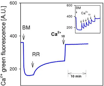
lead to a sequential and reversible increase of Ca Green fluorescence due to respective changes in extramitochondrial Ca2+
(Fig. 3, insertion). In line with previous reports, addition of RR to isolated brain mitochondria induced a significant increase in extramitochondrial Ca2+
which was caused by a net Ca2+
release from mitochondria (Fig. 3A) [21]. The subsequent addition of 10mM Ca2+ induced a sustained increase of Ca2+ Green fluorescence, confirming effective inhibition of the Ca2+
uniporter by RR, which is also in accordance with earlier reports [21].
Figure 2. Exclusive and reversible activation of glutamate-dependent respiration by extramitochondrial Ca2+at low levels of ADP.
(A) Isolated rat brain mitochondria (0.06 mg/ml) were incubated in EGTA-free medium (0.6mM Ca2+free) with 10 mM glutamate and 2 mM malate as substrates, but in the presence of 250 nM RR. Additions: M, 0.06 mg/ml rat brain mitochondria; A, 150mM ADP; EGTA, 100mM EGTA (0.15mM Ca2+
free); Ca2+4.9, 4.9mM Ca2+free. Horizontal arrows indicate the actual Ca2+freeconcentration. (B–D). Means of phosphorylating respiration6S.E. were calculated as stationary state 3 respiration rate minus state 4 respiration rate from measurements as shown for glutamate and malate in A at defined extramitochondrial Ca2+. Different substrates were used as indicated. *
In the next series of experiments, we determined the KM of mitochondrial Ca2+uptake via Ca2+uniporter under conditions
used in here. The estimated KMof 3.760.9mM Ca 2+
freeexceeds the S0.5of Ca
2+-activated respiration (360 nM Ca2+
free, Fig. 1F) about 10-fold. Such a big difference between KMand S0.5suggests that mitochondrial Ca2+
accumulation cannot take place as long as extramitochondrial Ca2+
remains within the nM concentration range. To verify this important conclusion, Ca2+
freewas monitored directly with Fura-2 under conditions otherwise equivalent to those in respirometric experiments with glutamate/malate and 100mM EGTA-medium (not shown). At Ca2+
free levels up to 1.2mM, mitochondrial Ca2+
accumulation was not detectable. Only after further Ca2+
additions did mitochondrial Ca2+
uptake become visible (not shown).
Discussion
It is widely believed that increased cytosolic Ca2+exerts a parallel
activation of extramitochondrial ATPases and OXPHOS, thereby balancing exactly ATP consumption and production without major changes in ADP concentration [2,3,6,7,8,10]. Ca2+transport into
the mitochondrial matrix and subsequent activation of distinct intramitochondrial dehydrogenases [2,3,6,7,22,23] and F0F1 AT-Pase [8,9] are assumed to constitute the regulatory mechanism of mitochondrial respiration and OXPHOS. However, an exclusive activation of OXPHOS by intramitochondrial Ca2+
is questionable in the light of following arguments. (i) Computer modeling of intramitochondrial Ca2+
activation of OXPHOS was unable to simulate the OXPHOS activation in response to physiological changes of work load in vivo [10]. (ii) The low affinity of the
mitochondrial Ca2+
uniporter to Ca2+
free (KM= 3.760.9mM) should not allow an effective increase in intramitochondrial Ca2+
effectively under conditions of only slightly elevated Ca2+
. Therefore, detectable mitochondrial Ca2+
uptake at nanomolar Ca2+
levels was explained by spatial heterogeneity of cytosolic Ca2+
concentration [24] and/or by a spermine-induced increase in the uniporter’s affinity for extramitochondrial Ca2+
[25,26]. (iii) Moreover, the relative insensitivities of intramitochondrial dehy-drogenases to Ca2+ (S
0.5= 0.4 - 13mM Ca 2+
free) [22,23] require significant higher Ca2+
freelevels for their activation compared with extramitochondrial Ca2+
activation of state 3glu/maland OXPHOS. Thus, the function of mitochondrial Ca2+
uptake and accumulation appears rather to serve as reversible Ca2+ buffer, ensuring
intracellular Ca2+
homeostasis, than to regulate state 3glu/maland OXPHOS [14].
This study reveals a novel mechanism of extramitochondrial Ca2+
activation of state 3glu/mal and OXPHOS mediated by aralar. This finding is supported by several earlier observations. (i) RR inhibits cardiac function only slightlyin vivo[27,28], suggesting
that mitochondrial Ca2+
uptake is not obligatory for stimulation of mitochondrial ATP productionin vivo. (ii) In contrast, AOA, an
inhibitor of MAS, attenuates the respiration of isolated synapto-somes [29] and suppresses the contractile function of the perfused, working heart [30], when glucose or lactate are oxidized. On the other hand, full contractile functionality can be observed if pyruvate is used in the presence of AOA [30].
Since pyruvate formation and aralar function are tightly interconnected in intact cells [11], extramitochondrial Ca2+,
beside its regulation of MAS [11-13], also regulates pyruvate formation from glucose or lactate. Since pyruvate is the main substrate of brain mitochondria [31], extramitochondrial Ca2+
is able to adjust the supply of OXPHOS with its main substrates precisely and reversibly, like a physiological ‘‘gas pedal’’, acting in response to distinct, Ca2+
-mediated cellular demands.
Taken together, our results imply a new and consistent feature of OXPHOS regulation in brain mitochondria in which the mitochondrial glutamate/aspartate carrier aralar controls mito-chondrial substrate supply and OXPHOS according to the extramitochondrial level of Ca2+
.
Materials and Methods
Mitochondria
Brain mitochondria (containing synaptosomal and nonsynapto-somal fractions) were isolated from 3–4-month-old Wistar WU rats (Charles River Laboratories, Germany) according to the protocol by Kudinet al., which includes permeabilization of synaptosomes
with digitonin [32]. Isolation and incubation media did not contain bovine serum albumin (BSA). Before final suspension, the mitochondrial Ca2+
content was routinely diminished by extrac-tion with nitriloacetic acid using the method of Brandtet al. [33].
For some experiments shown in Fig. 1G, mitochondria were isolated without digitonin. These mitochondria were also used to prepare mitoplasts by short term incubation with 1.2 mg digitonin/mg mitochondrial protein similarly as described previ-ously for heart mitoplasts [17]. All research and animal-care procedures were performed according to European guidelines.
Respirometry
Mitochondrial respiration was measured with a Clark-type oxygen electrode by means of high-resolution respirometry [34,35] using an OROBOROS oxygraph-2k (Oroboros, Innsbruck, Austria) at 30uC. Respiration of mitochondria (0.06 mg protein/ ml) was measured in a medium containing 120 mM mannitol, 40 mM MOPS, 5 mM KH2PO4, 60 mM KCl, 5 mM MgCl2, and either 0 or 100mM EGTA, pH 7.4. Ca2+
freeconcentrations in the various media were measured with Fura-2 as described below. EGTA-free medium contained 0.6mM Ca2+
free. 100mM EGTA medium contained 0.15mM Ca2+
free.
Figure 3. Brain mitochondria do not accumulate, but rather lose, Ca2+in the presence of ruthenium red.
Fluorimetric measurement of extramitochondrial Ca2+
with Ca2+
green. Brain mitochondria were incubated in EGTA-free medium with 10 mM glutamate and 2 mM malate. Additions: BM, 0.25 mg/ml brain mitochondria; RR, 250 nM ruthenium red (RR); Ca2+
10, 10mM Ca2+free, Insertion: Control experiment without RR demonstrating normal Ca2+
accumulation of brain mitochon-dria after repeated Ca2+additions.
Ca2+accumulation measurements
Ca2+
accumulation by isolated mitochondria (0.25 mg protein/ ml) was monitored fluorimetrically in the presence of 0.5mM Calcium Green-5N (Invitrogen) in a medium containing 120 mM
mannitol, 40 mM MOPS, 5 mM KH2PO4 and 60 mM KCl.
Measurements were performed in stirred and thermostatted (30uC) cells using a Carry Eclipse fluorimeter (Varian Deutschland GmbH) as described previously [36]. Excitation and emission wavelengths were set to 506 and 532 nm, respectively.
Measurement of Ca2+
freein EGTA medium
Ca2+
in EGTA medium was measured fluorimetrically with Fura-2 (10mM) as described previously [37]. The dissociation
constant (Kd) of the Ca2+-Fura-2 complex was determined experimentally under these conditions and was found to be 0.3mM, which was similar to that found in a previous study [37].
Protein determination
Mitochondrial protein concentrations were determined by the bicinchoninic acid assay [38], with BSA used as standard.
Author Contributions
Conceived and designed the experiments: FNG SV ES. Performed the experiments: ZG OA DJ. Analyzed the data: ZG ES. Wrote the paper: FNG FS. Conception: SV. Design and interpretation of new experiments: SV.
References
1. Chance B, Williams GR (1955) Respiratory enzymes in oxidative phosphory-lation. III. The steady state. J Biol Chem 217(1): 409–427.
2. Heineman FW, Balaban RS (1990) Phosphorus-31 nuclear magnetic resonance analysis of transient changes of canine myocardial metabolismin vivo. J Clin
Invest 85(3): 843–852.
3. Sharma N, Okere IC, Brunengraber DZ, McElfresh TA, King KL, et al. (2005) Regulation of pyruvate dehydrogenase activity and citric acid cycle intermedi-ates during high cardiac power generation. J Physiol 562(Pt 2): 593–603. 4. Gellerich FN, Schlame M, Bohnensack R, Kunz W (1987) Dynamic
compartmentation of adenine nucleotides in the mitochondrial intermembrane space of rat heart mitochondria. Biochim Biophys Acta 890(2): 117–126. 5. Seppet EK, Kaambre T, Sikk P, Tiivel T, Vija H, et al. (2001) Functional
complexes of mitochondria with Ca,MgATPases of myofibrils and sarcoplasmic reticulum in muscle cells. Biochim Biophys Acta 1504(2–3): 379–395. 6. McCormack JG, Halestrap AP, Denton RM (1990) Role of calcium ions in
regulation of mammalian intramitochondrial metabolism. Physiol Rev 70(2): 391–425.
7. Hansford RG, Zorov D (1998) Role of mitochondrial calcium transport in the control of substrate oxidation. Mol Cell Biochem 184(1–2): 359–369. 8. Territo PR, Mootha VK, French SA, Balaban RS (2000) Ca(2+) activation of
heart mitochondrial oxidative phosphorylation: role of the F(0)/F(1)-ATPase. Am J Physiol Cell Physiol 278(2): C423–35.
9. Das AM (2003) Regulation of the mitochondrial ATP-synthase in health and disease. Mol Genet Metab 79(2): 71–82.
10. Korzeniewski B (2007) Regulation of oxidative phosphorylation through parallel activation. Biophys Chem 129(2–3): 93–110.
11. Satru´stegui J, Pardo B, Del Arco A (2007) Mitochondrial transporters as novel targets for intracellular calcium signaling. Physiol Rev 87(1): 29–67. 12. Pardo B, Contreras L, Serrano A, Ramos M, Kobayashi K (2006) Essential role
of aralar in the transduction of small Ca2+
signals to neuronal mitochondria. J Biol Chem 281(2): 1039–1047.
13. Palmieri L, Pardo B, Lasorsa FM, del Arco A, Kobayashi K (2001) Citrin and aralar1 are Ca(2+)-stimulated aspartate/glutamate transporters in mitochondria. EMBO J 20(18): 5060–5069.
14. Rizzuto R, Pozzan T (2006) Microdomains of intracellular Ca2+
molecular determinants and functional consequences. Physiol Rev 86(1): 369–408. 15. Moore C (1971) Specific inhibition of mitochondrial Ca++ transport by
ruthenium red. Biochem Biophys Res Commun 42(2): 298–305.
16. Schnaitman C, Greenawalt JW (1968) Enzymatic properties of the inner and outer membranes of rat liver mitochondria. 38(1): 158–175.
17. Gellerich FN, Khuchua ZA, Kuznetsov AV (1993) Influence of the mitochondrial outer membrane and the binding of creatine kinase to the mitochondrial inner membrane on the compartmentation of adenine nucleo-tides in the intermembrane space of rat heart mitochondria. Biochim Biophys Acta 1140(3): 327–334.
18. Gellerich FN, Laterveer FD, Korzeniewski B, Zierz S, Nicolay K (1998) Dextran strongly increases the Michaelis constants of oxidative phosphorylation and of mitochondrial creatine kinase in heart mitochondria. Eur J Biochem 254(1): 172–180.
19. Nicolli A, Basso E, Petronilli V, Wenger RM, Bernardi P (1996) Interactions of cyclophilin with the mitochondrial inner membrane and regulation of the permeability transition pore, and cyclosporin A-sensitive channel. J Biol Chem 271(12): 2185–2192.
20. LaNoue KF, Tischler ME (1974) Electrogenic characteristics of the mitochon-drial glutamate-aspartate antiporter. J Biol Chem 249(23): 7522–75228. 21. Rossi CS, Vasington FD, Carafoli E (1973) The effect of ruthenium red on the
uptake and release of Ca2+by mitochondria. Biochem Biophys Res Commun 50(3): 846–852.
22. Rutter GA, Denton RM (1988) Regulation of NAD+-linked isocitrate dehydrogenase and 2-oxoglutarate dehydrogenase by Ca2+ ions within toluene-permeabilized rat heart mitochondria. Interactions with regulation by adenine nucleotides and NADH/NAD+ratios. Biochem J 252(1): 181–189. 23. Rutter GA, Midgley PJ, Denton RM (1989) Regulation of the pyruvate
dehydrogenase complex by Ca2+
within toluene-permeabilized heart mitochon-dria. Biochim Biophys Acta 1014(3): 263–270.
24. Rizzuto R, Pozzan T (2006) Microdomains of intracellular Ca2+
molecular determinants and functional consequences. Physiol Rev 86(1): 369–408. 25. McCormack JG (1989) Effects of spermine on mitochondrial Ca2+transport and
the ranges of extramitochondrial Ca2+to which the matrix Ca2+-sensitive dehydrogenases respond. Biochem J 264(1): 167–174.
26. Lenzen S, Mu¨nster W, Rustenbeck I (1992) Dual effect of spermine on mitochondrial Ca2+transport. Biochem J 286(Pt 2): 597–602.
27. Unitt JF, McCormack JG, Reid D, MacLachlan LK, England PJ (1989) Direct evidence for a role of intramitochondrial Ca2+
in the regulation of oxidative phosphorylation in the stimulated rat heart. Studies using 31P n.m.r. and ruthenium red. Biochem J 262(1): 293–301.
28. Garcı´a-Rivas Gde J, Carvajal K, Correa F, Zazueta C (2006) Ru360, a specific mitochondrial calcium uptake inhibitor, improves cardiac post-ischaemic functional recovery in ratsin vivo. Br J Pharmacol 149(7): 829–837.
29. Kauppinen RA, Sihra TS, Nicholls DG (1987) Aminooxyacetic acid inhibits the malate-aspartate shuttle in isolated nerve terminals and prevents the mitochon-dria from utilizing glycolytic substrates. Biochim Biophys Acta 930(2): 173–178. 30. Bu¨nger R, Glanert S, Sommer O, Gerlach E (1980) Inhibition by (aminooxy)acetate of the malate-aspartate cycle in the isolated working guinea pig heart. Hoppe Seylers Z Physiol Chem 361(6): 907–914.
31. Brown GK, Otero LJ, LeGris M, Brown RM (1994) Pyruvate dehydrogenase deficiency MJ Med Genet. 31(11): 875–879.
32. Kudin AP, Bimpong-Buta NY, Vielhaber S, Elger CE, Kunz WS (2004) Characterization of superoxide-producing sites in isolated brain mitochondria. J Biol Chem 279(6): 4127–4135.
33. Johnston JG, Brand MD (1986) Some properties of rat liver mitochondria with low Ca2+
content. Biochem Soc Trans 14(4): 1182–1185.
34. Kuznetsov AV, Veksler V, Gellerich FN, Saks V, Margreiter R, et al. (2008) Analysis of mitochondrial function in situ in permeabilized muscle fibers, tissues and cells. Nat Protoc 3(6): 965–976.
35. Gnaiger E (2001) Bioenergetics at low oxygen: dependence of respiration and phosphorylation on oxygen and adenosine diphosphate supply. Resp Phys 128(3): 277–297.
36. Gizatullina ZZ, Chen Y, Zierz S, Gellerich FN (2005) Effects of extramito-chondrial ADP on permeability transition of mouse liver mitochondria. Biochim Biophys Acta 1706(1–2): 98–104.
37. Groden DL, Guan Z, Stokes BT (1991) Determination of Fura-2 dissociation constants following adjustment of the apparent Ca-EGTA association constant for temperature and ionic strength. Cell Calcium 12(4): 279–287.