in Models of Cardiac Ischemic Injury
Diana S. Nascimento1., Mariana Valente1,2., Tiago Esteves1,3
, Maria de Fa´tima de Pina1,4, Joana G. Guedes1, Ana Freire1,3, Pedro Quelhas1,3, Perpe´tua Pinto-do-O´1*
1Instituto de Engenharia Biome´dica (INEB), Universidade do Porto, Porto, Portugal,2Instituto de Cieˆncias Biome´dicas Abel Salazar, Universidade do Porto, Porto, Portugal,3Faculdade de Engenharia, Universidade do Porto, Porto, Portugal,4Departamento de Epidemiologia Clı´nica, Medicina Preditiva e Sau´de Pu´blica, Faculdade de Medicina da Universidade do Porto, Porto, Portugal
Abstract
Background:The cardiac regenerative potential of newly developed therapies is traditionally evaluated in rodent models of surgically induced myocardial ischemia. A generally accepted key parameter for determining the success of the applied therapy is the infarct size. Although regarded as a gold standard method for infarct size estimation in heart ischemia, histological planimetry is time-consuming and highly variable amongst studies. The purpose of this work is to contribute towards the standardization and simplification of infarct size assessment by providing free access to a novel semi-automated software tool. The acronymMIQuantwas attributed to this application.
Methodology/Principal Findings:Mice were subject to permanent coronary artery ligation and the size of chronic infarcts was estimated by area and midline-length methods using manual planimetry and with MIQuant. Repeatability and reproducibility ofMIQuantscores were verified. The validation showed high correlation (rmidline length= 0.981;rarea= 0.970 ) and agreement (Bland-Altman analysis), free from bias for midline length and negligible bias of 1.21% to 3.72% for area quantification. Further analysis demonstrated that MIQuant reduced by 4.5-fold the time spent on the analysis and, importantly,MIQuanteffectiveness is independent of user proficiency. The results indicate thatMIQuantcan be regarded as a better alternative to manual measurement.
Conclusions: We conclude that MIQuant is a reliable and an easy-to-use software for infarct size quantification. The widespread use of MIQuant will contribute towards the standardization of infarct size assessment across studies and, therefore, to the systematization of the evaluation of cardiac regenerative potential of emerging therapies.
Citation:Nascimento DS, Valente M, Esteves T, de Pina MdF, Guedes JG, et al. (2011)MIQuant– Semi-Automation of Infarct Size Assessment in Models of Cardiac Ischemic Injury. PLoS ONE 6(9): e25045. doi:10.1371/journal.pone.0025045
Editor:Marcello Rota, Brigham & Women’s Hospital - Harvard Medical School, United States of America
ReceivedJune 30, 2011;AcceptedAugust 23, 2011;PublishedSeptember 30, 2011
Copyright:ß2011 Nascimento et al. This is an open-access article distributed under the terms of the Creative Commons Attribution License, which permits unrestricted use, distribution, and reproduction in any medium, provided the original author and source are credited.
Funding:This work was supported by Fundac¸a˜o para a Cieˆncia e a Tecnologia (FCT), Fundo Europeu de Desenvolvimento Regional (FEDER), Programa Operacional Factores de competitividade-COMPETE, Quadro de Refereˆncia Estrate´gico Nacional (QREN) [grant PTDC/SAU-OSM-68473/2006], and by the grants SFRH/BD/64715/2009 to Dr. Freire, SFRH/BPD/42254/2007 to Dr. Nascimento, and SFRH/BD/74218/2010 to Dr. Valente. Dr. Pinto-do-O´ and Dr. Quelhas are Cieˆncia2007 and Cieˆncia2008 awardees, respectively. Travel funds were awarded by Boehringer Ingelheim Fonds to Dr. Nascimento, and by APBRF-American Portuguese Biomedical Research Fund to Dr. Pinto-do-O´ and to Dr. Nascimento. The funders had no role in study design, data collection and analysis, decision to publish, or preparation of the manuscript.
Competing Interests:The authors have declared that no competing interests exist.
* E-mail: [email protected]
.These authors contributed equally to this work.
Introduction
Cardiovascular disease is a leading cause of morbidity and mortality worldwide. Heart failure due to ischemic coronary artery disease is currently the most common cardiac disorder and it correlates with a worse prognosis [1,2]. The physiological, histological and molecular changes associated with clinical ischemic heart disease have been clarified with the use of experimental models of myocardial infarction (MI) developed in both large animals, including dogs and swine, as well as in small rodents [3,4]. The latter are more applicable for high-throughput screening of novel therapeutic approaches, due to the easy maintenance, short reproductive cycle and to the latest advances in gene-targeting and transgenic technologies. In recent years, the evaluation of cardiac regenerative potential of newly developed
therapies, as is the case of gene-delivery and transplantation of stem/progenitor-cells, has been primarily explored in rat and mouse models of surgically-induced myocardial ischemia [2,5,6,7,8]. The so-called left anterior descending (LAD) coronary artery ligation is the prominent model in these studies, and the infarct size has been considered a key parameter for assessing the success of the novel therapy. A strong correlation between the infarction size and the functional and hemodynamic alterations following myocardial infarction is generally observed [9,10,11] and therefore considered a fundamental measure in the assessment of the morphological and functional consequences of infarction.
area [13] of infarcted versus non-infarcted left-ventricle (LV) regions. Despite the widespread use of the aforementioned approaches, the infarct size can vary depending on the used method [10,14] and therefore no direct comparison can be withdrawn across laboratories. Moreover, several aspects of MI size quantification that can also account for infarct size variation are inconsistent across studies and not always clearly defined, e.g. the number of sections used for the calculation, the histological staining and criteria used to identify the infarcted region. Thus, the purpose of the present work is to contribute towards standardi-zation and simplification of the infarct size assessment in experimental models of MI by making available, as freeware, an easy-to-use semi-automatic software application, which we devel-oped and validated at the ‘‘bench’’. This tool will contribute for the systematization of the evaluation of cardiac regenerative potential of newly developed therapies. The acronymMIQuantthat stands for MI quantification was attributed to the herein software application.
Methods
Animals
Male and female C57BL/6 mice aged 8 to 12 weeks were used for this study. All the procedures were subjected to approval by the IBMC-INEB (Instituto de Biologia Molecular e Celular – Instituto de Engenharia Biome´dica) Animal Ethics Committee and to the National Direc¸a˜o Geral de Veterina´ria (permit no: 022793), and are in conformity with the Directive 2010/63/EU of the European Parliament. Humane endpoints were followed in accordance to the Organisation for Economic Co-operation and Development (OECD) Guidance Document on the Recognition, Assessment, and Use of Clinical Signs as Humane Endpoints for Experimental Animals Used in Safety Evaluation (2000).
Surgical Induction of Myocardial Infarction
MI was experimentally induced by ligation of the LAD coronary artery as described elsewhere [13] with minor alter-ations. Following anesthesia by intraperitoneal injection (ip) of medetomidine (Sededorm, 1 mg/Kg) and ketamine (Clorketam, 75 mg/kg), animals were subjected to endotracheal intubation and were mechanically ventilated using a small-animal respirator (Minivent 845, Harvard Apparattus). Animals were maintained on warming pads during surgical procedure and until full recovery to prevent hypothermia. Under a stereomicroscope (Leica EZ4, Leica Microsystems) the heart was exposed (Ø 5–7 mm) via left thoracotomy on the third intercostal space and the pericardial sac was gently disrupted. After identification of the LAD coronary artery a non-absorbable 7-0 suture (SilkamH, B. Braun) was passed under the artery and the ligation was performed. The intercostal incision was closed by an absorbable 6-0 suture (SafilH, B. Braun) and surgical staples were used for skin closure. Anesthesia was reverted by atipamezole (ip, Revertor, 5 mg/Kg) and analgesia was achieved by butorphanol (ip, Butador, 1 mg/Kg). Analgesia and fluid therapy were performed by ip delivery of butorphanol (Butador, 1 mg/kg) and 5% glucose physiological saline, respec-tively. This procedure was repeated every 12 h up to 72 h post-surgery or until full animal recovery.
For organ collection animals were deeply anesthetized by ip injection of pentobarbital (Eutasil, 70 mg/kg). At 21days post-surgery, hearts were harvested, briefly washed in phosphate buffer saline and fixed in 10% Formalin neutral buffer (VWR BDH & Prolabo) up to 24 hours prior to paraffin-embedding. The sampling procedure herein described results on hearts arrested at variable stages of heart cycle, which may contribute to increased
variability of infarct size. Whenever normalization is a require-ment, hearts should be arrested in diastole following injection with potassium chloride.
Histological procedures
Representative sampling of the LV (approx. 12 sections) was obtained by transverse sectioning (3mm) from the apex to the base
(atrium region) of paraffin-embedded hearts with an interval of 300mm among each section (Figure 1A).
Paraffin sections were stained with modified Masson’s trichrome staining (MT). MT staining was performed according to the Trichrome (Masson) Stain kit (Sigma-Aldrich) with the following modifications: nuclei were pre-stained with Celestine Blue solution following staining with Gill’s Hematoxylin and incubation for 1 hour in aqueous Bouin’s solution to promote a uniform staining.
Myocardial infarct size calculation
For infarct size determination the collagen deposition, high-lighted (blue) in MT-stained sections collected at 21 days post-infarction, was used to define the LV scarred region. Images of histological sections were captured with an Olympus SZX10 stereomicroscope and Olympus DP21 camera. The percentage of affected LV wall was calculated by two different and previously validated methods: thearea measurement(calculated by dividing the infarct area by the total LV area) [13] and the midline length measurement (calculated by dividing the midline length of the infarcted LV wall by the midline length of total LV wall). Only regions with infarct in .50% of the whole thickness of the myocardium were considered for infarct midline [10]. The MI size determination was performed either manually, by drawing points to outline different anatomical/pathological regions using the Image J 1.42 software (Figure 1B), or by usingMIQuant(Figure 1C).
Software design
TheMIQuantsoftware was implemented in MATLABTMand a MS WindowsTM 32-bit compiled version is available online at http://paginas.fe.up.pt/,quelhas/MIQuant/MIQuant.zip. With
the objective of developing an approach for automatic infarct size estimation several image processing methodologies were tested [15] and, within all tested semi-supervised methods, region growing was found to work best and also faster, being selected for the final software implementation.
Data and statistical analysis
To validate MIQuant, four expert researchers analyzed five hearts (twelve sectionsperheart) using midline and area methods, manually and withMIQuant. Allexpertsrepeated measures at three distanced moments (one month between 1stand 2ndmeasure and one week between 2ndand 3rd). A one-way repeated measures analysis of variance (ANOVA) was conducted to evaluate repeatability. Seven non-trained volunteers measured the same samples using MIQuant. The association between manual and
Results
Software overview and availability
MIQuantis a user-friendly software application that assists on the infarct size quantification in an experimental MI-setting. The infarct size, defined as the percentage of the LV affected by coronary artery occlusion, is estimated with representative cross-sections of the LV stained with MT that enables the identification of collagen deposition, a hallmark of established infarction. The software allows the upload of single or multiple images and enables the computation of the MI size of each image, calculated by area
[13] and midline length [10] methods, and the total infarct size mean value that can be saved in excel file-format.
MIQuant was designed by applying the region growing image segmentation method, which exploits the spatial context of pixels with similar pixel-color properties. The main criterion for the algorithm of region growing is homogeneity, similar pixels (or regions) that are neighbors are joined together. For each image, region growing requires initial image points (or seeds) that define the region of interest. From these initial points the algorithm grows until no more neighbors can be joined to the region of interest, therefore regions/pixels are merged if they satisfy the chosen Figure 1. Manual andMIQuantsemi-automated calculation of MI size in chronic infarcts.(A) LV representative MT stained sections, numbered from the apex to the LV base, were obtained from an infarcted heart harvested at 21 days post-surgery. (B) Histological infarct size calculation by the area method requires manual tracing of the LV myocardium (light gray) and of the scarred LV tissue (black). The infarct size, expressed as a percentage, is the division of the infarct area by the LV area multiplied by 100. For the midline length approach (right) the midline, herein defined as the mid-region between the epicardial and endocardial surfaces, of the total LV (dashed line) and of scarred region (full line) are manually traced. The infarct size, expressed as a percentage, is the division of the infarct midline length by the LV midline length multiplied by 100. The total LV infarct extent is the average of infarct size obtained for the LV representative cross-sections (A). (C) Screen shot ofMIQuantlayout following infarct size calculation. Multiple images can be uploaded in TIFF or JPEG file-formats and the software calculates the intermediate values of infarct size for each image (bottom right). A total MI size is also generated assuming that the uploaded images were representative sections of the LV. For selection of the scarred myocardium (top right) the software requires the user to double-click in a normal tissue region and in the LV lumen, if applicable, over the uploaded image (top left).
criterion and no merging occurs when the criterion is not met [17,18]. In theMIQuantsoftware the user is asked to provide input seed points for the LV lumen (if present in the image) and the viable myocardium (if present in the image), prior to automated segmentation. The choice of not requiring the user to select the infarcted LV region has to do with the heterogeneity of the ischemic tissue. The user clicks with the mouse on the heart section image and gives as many input points as desired. Following selection of the viable myocardium and/or LV lumen the segmentation is generated and displayed on the screen. This will be the support for the infarct size computation. User adjustments to the segmentation are accessible by varying the merging criteria and the segmentation process can be repeated until the user is satisfied with the results. When the segmentation is complete the user can request computation of infarct size results by both midline length and area methods. For the midline length measurement, the MIQuant software automatically traces lines from the lumen centre outwards and identifies the middle distance between tissue boundaries. The midline of the infarcted region was considered when the LV wall was affected in more than 50% in radial direction. The midline generated by the software can be adjusted by the user prior to MI size calculation.
Commands for image edition are available on the ‘‘edit menu’’, which permits the removal of tissue regions/artifacts that may interfere with tissue automated segmentation, e.g. the right ventricle or blood within the LV lumen.
TheMIQuantsoftware was implemented in MATLABTMand a MS WindowsTM 32 bit compiled version is available online at http://paginas.fe.up.pt/,quelhas/MIQuant/MIQuant.zip. The
archive should be downloaded and unzipped into a specific folder.
TheMIQuantmanual reading is recommended prior to beginning with the software, available at http://www.fe.up.pt/,quelhas/
MIQuant/MIQuant_manual.pdf. MIQuant requires the installa-tion of MATLABTMor of the MATLABTMComponent Runtime (MCR) installer (http://paginas.fe.up.pt/,quelhas/MIQuant/
MCRInstaller.zip). The application can be initiated by double-click on the executable ‘‘MIQuant’’ file. More information about the software usage and installation is available at the MIQuant
website http://www.fe.up.pt/,quelhas/MIQuant/.
MIQuant repeatability and reproducibility
Manual andMIQuantinfarct size quantification was assessed by two well-validated methods, i.e. the area and the midline length measurement (Figure 2A). Visual inspection of the infarct size scores across methods demonstrate that MIQuant results are consistent with the manual assessment, and thus infarct values obtained with the area measurement were significantly smaller than the midline length infarct scores. The similarity between the manual andMIQuantapproaches demonstrate that the latter might constitute an alternative for the histological quantification of infarct size. Further validation ofMIQuantis detailed bellow.
Intra- (repeatability) and inter-observer (reproducibility) vari-ability was considered in the experimental design, thus three independent measures were conducted by four different users. Repeated measures one-way ANOVA was applied to compare the repeatability of the area and midline length-based methods, both calculated manually and by usingMIQuant. The LV infarct size means with standard deviations and ANOVA results are detailed in table 1. No significant effect of the repetition was found on the infarct size obtainedpersection andperheart, i.e. mean value of 12
Figure 2. Consistency and reproducibility ofMIQuant infarct size calculation.(A) Consistency of manual andMIQuantinfarct size results obtained using the area and midline length measurements. Hearts were harvested at 21 days post-surgery and infarct size determinations are the mean value of 12 cross-sections representative of the LV. Mann-Whitney statistical analysis demonstrated significant differences between the area and midline length methods, as already described by Takagawa [10]. (B) Reproducibility ofMIQuantmeasurements. Although ANOVA demonstrated no significant influence of the observer on the LV infarct size scores obtained, neither manually nor usingMIQuant, the latter displays a tendency for lower discrepancy between operators.eindicates the mean value of each group. *p,0.05.
sections representative of the LV, demonstrating the consistency of
MIQuantmeasurements obtained at different instances.
Inter-observer variability for each analyzed sample is displayed on Fig. 2B. A two-way ANOVA was conducted to investigate whether the observer influences (inter-observer variability) infarct size measurements manually or usingMIQuant. Post-hoc compar-ison using the Tukey HSD test indicated that the mean score of infarct size, for each heart, did not differ significantly (p.0.05) among observers in any of the tested infarct size quantification methods. However, a tendency for increased variability of the manual results when compared toMIQuantwas observed and was particularly evident on heart C, which is the sample that retrieved more deviation amongst users (Figure 2B).
Validation of MIQuant infarct size quantification
A scatter diagram of the infarct size values measured manually and by MIQuant is shown in Fig. 3A. The Pearson Product-moment correlation for the individual data points wasr= 0.981 for the midline length and r= 0.970 for the area methods, with a significance level ofp,0.01, hence the infarct size values obtained byMIQuant are strongly associated to the manual quantification. The strong correlation between manual and MIQuant results prompted further analysis to evaluate the magnitude and direction of the differences between methods.
The gold-standard statistical analysis applied to method-comparison studies is the Bland-Altman plot, which determines the agreement of two methods that measure the same variable [16,19]. Manual andMIQuantresults were subjected to the Bland-Altman agreement statistical method that predicts the bias, i.e. difference in values obtained by the two methods, and the limits of agreement between methods (Figure 3B and C). Bias and concordance limits of 62% and 67%, respectively, were establisheda priorias the maximum parameters for acceptance of
MIQuant regarding per heart infarct size quantifications. These values were selected on the basis of acceptance limits addressed for infarct size methods on published studies [20,21,22,23]. The a prioriestablishment of acceptable agreement limits for infarct size
persection was conditioned by the fact that, to our best knowledge, no previous comparison was performed for single sections. Hence, since it is expected higher degree of discordance across sections, when compared to the mean value, a low-stringency predeter-mined bias and concordance limits of 62% and 615%, respectively, were established.
Bland-Altman analysis was conducted with manual and
MIQuantresults obtainedperLV section (Figure 3B). The estimated bias is 0.36% with concordance limits of 210.72% and 11.45% for the midline length method, whereas for the area approach the bias is 2.68% with limits of agreement of 27.59% and 12.94%
(Figure 3B). Hence, for both methodological approaches, the predicted confidence interval is within acceptance limits and so
MIQuant is considered equivalent to the established manual quantification method.
The visual inspection of Bland-Altman plot denoted that differences between MIQuant and manual measurements are scattered around the bias with no obvious pattern for the midline length results whereas, the area differences appear to increase for higher infarction values (Figure 3B). To determine whether an association exists between the methods discrepancies and the size of infarction, the Pearson coefficient was calculated and a small, non-statistically significant correlation between the two variables was observed (r= 0.063;p= 0.337).
Measurements of the infarct sizeperheart, i.e. mean value of 12 sections representative of the LV, obtained by the manual and
MIQuantcalculation were also compared accordingly to the Bland-Altman concordance analysis. For the midline length the predicted bias is 0.25% and the limits of agreement are23.60% and 4.09%, resulting on 7.74% amplitude of concordance (Figure 3C). The analysis of the area measurements retrieves a mean difference of 2.47% (95% confidence interval (CI) from 1.21% to 3.72%), suggesting thatMIQuanttends to give a higher reading from 1.21% to 3.72% (Figure 3C). The area method concordance interval ranges from22.79% to 7.72%. Thus, forMIQuant perheart infarct size results the confidence interval of the predicted bias and concordance limits are within acceptance limits (bias 62%, concordance limits 67%) for both midline length- and area-measurements, which show that the performance ofMIQuant is equivalent to the manual infarct size calculation.
Although the differences between MIQuant and manual measurements are scattered around the bias with no obvious pattern, the association between the two variables was investigated using the Pearson Product-moment-correlation coefficient. Small and non-statistically significant correlations were found for both midline length (r= 0.149; p= 0.531) and area (r=20.315;
p= 0.176) approaches, consequently discrepancies between the manual and semi-automated quantification are independent of the sample infarction size.
Validation of MIQuant by non-trained volunteer-users To address whether previous experience with the MIQuant
application and knowledge on infarct size calculation are strict requirements for the correct software usage, a comparison was established betweenMIQuantresults obtained by users with distinct proficiency. Five hearts were independently analyzed by four competent users (experts), i.e. investigators with extensive training on MI size quantification either manually or usingMIQuant, and by volunteer-users with no previous experience on MI size Table 1.Repeatability analysis of the manual andMIQuantresults by repeated measures one-way ANOVA.
MI size (%) Midline length measurement1 Area measurement2
Measurement 1 Measurement 2 Measurement 3 Measurement 1 Measurement 2 Measurement 3
Manuala 44.24
613.01 44.84612.62 45.03612.82 31.2369.60 32.5169.29 31,9269.25
MIQuantb 44.66
613.26 44.58613.00 45.61613.01 34.0268.71 34.0068.16 35,0468.72
Values area mean6STDEV;n= 20;1, aPerLV: Wilks’Lambda = 0.818, F(2, 18) = 2.0001, p = 0.164, multivariate partial eta squared = 0.18;Persection:
Wilks’Lambda = 0.990, F(2, 234) = 2.0001, p = 0.140, multivariate partial eta squared = 0.322;1, bPerLV: Wilks’Lambda = 0.757, F(2, 18) = 2.892, p = 0.081, multivariate partial eta squared = 0.24;Persection: Wilks’Lambda = 0.977, F(2, 234) = 2.734, p = 0.067, multivariate partial eta squared = 0.023;2, aPerLV: Wilks’Lambda = 0.848, F(2, 18) = 1.617, p = 0.226, multivariate partial eta squared = 0.15;Persection: Wilks’Lambda = 0.969, F(2, 234) = 3.737, p = 0.025, multivariate partial eta squared = 0.031;2, b PerLV: Wilks’Lambda = 0.827, F(2, 18) = 1.886, p = 0.180, multivariate partial eta squared = 0.17;Persection: Wilks’Lambda = 0.981, F(2, 234) = 2.286, p = 0.104, multivariate partial eta squared = 0.019.
calculation, but to whom free-access to theMIQuantmanual was provided. An independent-samples t-test was conducted and no significant differences were observed on the midline length and area measurements obtained by eitherexpertsorvolunteers(Figure 4).
In addition, a two–way ANOVA analysis of variance was conducted to explore the impact of the observer type (expert or volunteer) and the heart sample on MIQuant infarct size measurement, obtained by either midline length (ML) or area Figure 3. Validation ofMIQuantfor infarct size assessment.(A) Infarct size scatter diagram and the Pearson coefficient demonstrated strong association between manual andMIQuantresults for area (right) and midline length (left) approaches. (B) Bland-Altman concordance analysis of the manual and MIQuantinfarct size measurements demonstrated acceptable limits of agreement between methods. Average values of the three independent measures of infarct sizepersection (B) andperheart (C) were subject to the analysis. Differences between the infarct sizes retrieved by each method (MIQuant-manual) are displayed in they-axis and the mean infarct size values are plotted in thex-axis. The limits of agreement (- -) and bias (&&&) and respective 95% confidence intervals ([ ]) are shown.
(A) approaches. There was no statistically significant effect for the observer type (MLp= 0.267; Ap= 0.77), whereas the effect of the heart sample was found to be statistically significant (p,0.05).
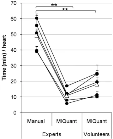
Time-efficiency of MIQuant infarct size quantification The manual quantification of MI size is a time-consuming and laborious endeavor, thus the simplification of this task is highly desired and was a major drive for the development ofMIQuant. The time required for manual and MIQuant-assisted infarct size calculation was compared (Figure 5). The latter was additionally compared forexpertsand volunteer operators. Despite the required definition of initial parameters by the user prior to MIQuant
segmentation, this method resulted on a significant overall 4.5-and 3-fold decrease in the time period spent on the analysis when performed by competent and volunteer users, respectively.
Discussion
In this study, the development and validation ofMIQuant, a simple and user-friendly software application that calculates the infarct size on cardiac models of induced-ischemia, is reported. To our best knowledge,MIQuantconstitutes the first computer-assisted tool to ease the arduous and time-consuming endeavor of manual infarct size calculation by classical planimetry.
The view of the heart as a post-mitotic organ has been challenged in recent years by reports of cardiomyocyte renewal in humans [24], cardiomyocytic-cell replacement after injury in mouse [25] and of myocardium-resident Sca-1+
/c-Kit+
/MDR1+
progenitor/stem-like
cells [26,27,28]. These findings, together with the fact that cardiovascular diseases are a major cause of morbidity/mortality, have encouraged the publication of studies on the evaluation of cardio-regenerative potential of novel therapies. The latter are commonly tested on rodent models of MI and the infarct size has been regarded as a decisive parameter for the determination of the success of the therapy under test. Although histological planimetry is the gold standard for infarct size quantification, methodological discrepancies are frequent across publications due to a general lack of standardized protocols/methods. The most common methods used to quantify infarct extension are either based on the infarcted area or on the length of the infarction circumference. Both methodologies show limitations related to the infarct size estimation accuracy using parameters that are affected and distorted by cardiac remodeling subsequent to MI [29]. RegardingMIQuant, we decided to make available two methods for infarct quantification: the area-based quantification first described by Michael [13] and the midline length measurement that was extensively validated recently [10]. In accordance with Takagawa’s [10] observations on manual infarct size quantification, with MIQuant we obtained a statistically significant compression of the area results when compared to the midline-length method. Overall, obvious consistency was achieved between manual andMIQuantinfarct size quantification, which was further illustrated by the excellent correlation between both and by Bland-Altman analysis. Bland-Altman analysis indicated good agreement free from systematic bias for midline-length MIQuant
infarct scores (0.2563.84). Regarding the area measurements, although MIQuant overestimates infarct size by 1.21–3.27% as Figure 4.MIQuantefficacy is not affected by user proficiency.
MIQuantinfarct size values obtained by competent (experts) and non-trained (volunteer) users were compared and the mean values are displayed as graph bars. Independent-samples t-test showed no significant differences between infarct scores calculated by theexperts vs. volunteers; furthermore, a two–way ANOVA demonstrated no significant influence of the user on the obtained infarct size value. doi:10.1371/journal.pone.0025045.g004
Figure 5.MIQuantimproves the time-efficiency of infarct size quantification.The time consumption of the infarct size determina-tionperheart (mean value of 12 representative sections of the LV) was compared between the manual and MIQuant approaches. The D
compared to the manual quantification, the biological relevance of this overestimation is negligible. Moreover, a random dispersion of results around the predicted bias was observed, demonstrating that
MIQuantresults are reliable independently of the size of infarction. The repeatability and reproducibility ofMIQuantresults were also confirmed by the use of three independent measures obtained by four independent observers. Overall these results indicate that
MIQuant is a reliable alternative to the manual quantification of infarct size.
Despite being a determinant factor for an accurate estimation of the infarct size[10], the number of transverse sections used for such analysis is extremely variable across studies. One of the advantages ofMIQuantover the classical manual quantification is the 4.5 fold reduction on the time spent on the analysis, thus improving time-efficiency and allowing the investigator to increase the number of sectionsperanalysis and consequently the accuracy of results.
MIQuant is available as freeware for research use. The widespread use of MIQuant will constitute by itself a major improvement towards normalization of infarct size assessment by restricting the methods to the area and midline length, by standardizing the histological stain used and by restricting the criteria for the identification of the infarcted region. Our results also indicated a tendency, although not statistically significant, for reduced inter-observer variability in MIQuant infarct size scores when compared to manual analysis. This may well be underes-timated given that the observers in this study were investigators that received similar training on infarct size calculation. It is therefore expected that the diversity of criteria on infarct identification/calculation of observers with different backgrounds will result in increased variability for the manual outcome. In contrast, we demonstrated thatMIQuantefficacy is independent of previous training with the software and experience on MI size calculation. An interesting experiment would be a comparative analysis betweenMIQuantand manual quantification with experts from different laboratories to therefore undoubtedly clarify whether MIQuant contributes to the homogenization of infarct size results. Our attempts to engage in this task experts with
previous published work on infarct size histological quantification, met with little success and the intent was therefore aborted.
For the interpretation of this study several limitations should be considered: firstly a single species (mouse) was used for the validation of MIQuant, and secondly the only model of cardiac induced-ischemia performed was the permanent LAD coronary artery ligation. However, the pathophysiological and morpholog-ical alterations following MI are similar in the rat and the mouse [9,30,31], supporting the applicability of MIQuant for the quantification of rat infarcts. The extension ofMIQuant to other infarction models, e.g. ischemia-reperfusion or the cryoinjury, is of major interest. Hence, because the software recognizes the infarction region by the collagen deposition, a hallmark of established infarction, we are confident on the software applica-bility to other models. Indeed, in hearts with non-transmural infarction that very much resembles the reperfusion scenario,
MIQuantinfarct scores were similar to manual quantification (data not shown).
We conclude thatMIQuant is a valid and easy-to-use software application that assists on infarct size calculation. The widespread use ofMIQuantwill contribute to the reduction of time spent on the analysis and for the standardization of infarct size quantification across studies and, therefore, to a more systematic evaluation of the cardiac regenerative potential of newly developed therapies.
Acknowledgments
The authors are indebted to Dr. Dirk J. Duncker for providing mouse cardiac surgical training to DSN at his Laboratory. Dr. Pedro Oliveira is acknowledged by statistical coaching. This work would not have been possible without the collaboration of all those who volunteered as non-trained operators for the statistical validation of theMIQuantsoftware.
Author Contributions
Conceived and designed the experiments: DSN PQ PPO´ . Performed the experiments: DSN MV TE JGG AF. Analyzed the data: DSN MV TE MFP PQ PPO´ . Contributed reagents/materials/analysis tools: PQ PPO´ . Wrote the paper: DSN MV PQ PPO´ .
References
1. Lloyd-Jones D, Adams RJ, Brown TM, Carnethon M, Dai S, et al. (2010) Executive summary: heart disease and stroke statistics–2010 update: a report from the American Heart Association. Circulation 121: 948–954.
2. Gonzales C, Pedrazzini T (2009) Progenitor cell therapy for heart disease. Experimental Cell Research 315: 3077–3085.
3. Zaragoza C, Gomez-Guerrero C, Martin-Ventura JL, Blanco-Colio L, Lavin B, et al. (2011) Animal models of cardiovascular diseases. J Biomed Biotechnol. 497841 p.
4. Sun Y (2009) Myocardial repair/remodelling following infarction: roles of local factors. Cardiovasc Res 81: 482–490.
5. Ramani R, Nilles K, Gibson G, Burkhead B, Mathier M, et al. (2011) Tissue Inhibitor of Metalloproteinase-2 Gene Delivery Ameliorates Postinfarction Cardiac Remodeling. Clinical and Translational Science 4: 24–31.
6. Ahmed RP, Haider KH, Shujia J, Afzal MR, Ashraf M (2010) Sonic Hedgehog gene delivery to the rodent heart promotes angiogenesis via iNOS/netrin-1/ PKC pathway. PLoS One 5: e8576.
7. Smits AM, van Laake LW, den Ouden K, Schreurs C, Szuhai K, et al. (2009) Human cardiomyocyte progenitor cell transplantation preserves long-term function of the infarcted mouse myocardium. Cardiovasc Res 83: 527–535. 8. Yoon YS, Wecker A, Heyd L, Park JS, Tkebuchava T, et al. (2005) Clonally
expanded novel multipotent stem cells from human bone marrow regenerate myocardium after myocardial infarction. J Clin Invest 115: 326–338. 9. Pfeffer MA, Pfeffer JM, Fishbein MC, Fletcher PJ, Spadaro J, et al. (1979)
Myocardial infarct size and ventricular function in rats. Circ Res 44: 503–512. 10. Takagawa J, Zhang Y, Wong ML, Sievers RE, Kapasi NK, et al. (2007) Myocardial infarct size measurement in the mouse chronic infarction model: comparison of area- and length-based approaches. J Appl Physiol 102: 2104–2111.
11. Gao X-M, Dart AM, Dewar E, Jennings G, Du X-J (2000) Serial echocardiographic assessment of left ventricular dimensions and function after myocardial infarction in mice. Cardiovascular Research 45: 330–338.
12. Patten RD, Aronovitz MJ, Deras-Mejia L, Pandian NG, Hanak GG, et al. (1998) Ventricular remodeling in a mouse model of myocardial infarction. American Journal of Physiology - Heart and Circulatory Physiology 274: H1812–H1820.
13. Michael LH, Entman ML, Hartley CJ, Youker KA, Zhu J, et al. (1995) Myocardial ischemia and reperfusion: a murine model. Am J Physiol 269: H2147–2154.
14. Minicucci MF, Azevedo PS, Duarte DR, Matsubara BB, Matsubara LS, et al. (2007) Comparison of different methods to measure experimental chronic infarction size in the rat model. Arq Bras Cardiol 89: 83–87, 93–88. 15. Esteves T, Valente M, Nascimento DS, Perpe´tua PO, Quelhas P (2011)
Automatic and semi-automatic analysis of the extension of myocardial infarction in an experimental murine model. Proceedings of Iberian Conference on Pattern Recognition and Image Analysis (IbPria).
16. Bland JM, Altman DG (1986) Statistical methods for assessing agreement between two methods of clinical measurement. Lancet 1: 307–310.
17. Wu Q, Merchant F, Castleman K (1996) Microscope Image Processing; 7 C, ed. USA: Elsevier.
18. Gonzalez R, Woods R, Eddins S (2004) Digital Image Processing Using MAT-LAB. Pearson Education.
19. Hanneman SK (2008) Design, analysis, and interpretation of method-comparison studies. AAN Adv Crit Care 19: 223–234.
20. Bohl S, Lygate CA, Barnes H, Medway D, Stork L-A, et al. (2009) Advanced methods for quantification of infarct size in mice using three-dimensional highfield late gadolinium enhancement MRI. American Journal of Physiology -Heart and Circulatory Physiology 296: H1200–H1208.
22. Dawson D, Lygate CA, Saunders J, Schneider JE, Ye X, et al. (2004) Quantitative 3-Dimensional Echocardiography for Accurate and Rapid Cardiac Phenotype Characterization in Mice. Circulation 110: 1632–1637.
23. Protti A, Sirker A, Shah AM, Botnar R (2010) Late gadolinium enhancement of acute myocardial infarction in mice at 7T: Cine-FLASH versus inversion recovery. Journal of Magnetic Resonance Imaging 32: 878–886.
24. Bergmann O, Bhardwaj RD, Bernard S, Zdunek S, Barnabe-Heider F, et al. (2009) Evidence for cardiomyocyte renewal in humans. Science 324: 98–102. 25. Hsieh PC, Segers VF, Davis ME, MacGillivray C, Gannon J, et al. (2007)
Evidence from a genetic fate-mapping study that stem cells refresh adult mammalian cardiomyocytes after injury. Nat Med 13: 970–974.
26. Barile L, Messina E, Giacomello A, Marban E (2007) Endogenous cardiac stem cells. Prog Cardiovasc Dis 50: 31–48.
27. Beltrami AP, Barlucchi L, Torella D, Baker M, Limana F, et al. (2003) Adult cardiac stem cells are multipotent and support myocardial regeneration. Cell 114: 763–776.
28. Leri A, Kajstura J, Anversa P (2005) Cardiac Stem Cells and Mechanisms of Myocardial Regeneration. Physiol Rev 85: 1373–1416.
29. Zornoff LA, Paiva SA, Minicucci MF, Spadaro J (2009) Experimental myocardium infarction in rats: analysis of the model. Arq Bras Cardiol 93: 434–440, 426–432.
30. Fishbein MC, Maclean D, Maroko PR (1978) Experimental myocardial infarction in the rat: qualitative and quantitative changes during pathologic evolution. Am J Pathol 90: 57–70.