Advanced Application of Porcine
Intramuscular Adipocytes for Evaluating
Anti-Adipogenic and Anti-Inflammatory Activities
of Immunobiotics
Masahiko Suzuki1☯, Asuka Tada1☯, Paulraj Kanmani1☯, Hitoshi Watanabe2, Hisashi Aso2,
Yoshihito Suda4, Tomonori Nochi2, Kenji Miyazawa5, Kazutoyo Yoda5, Fang He5, Masataka Hosoda5, Tadao Saito1, Julio Villena3*, Haruki Kitazawa1*
1Food and Feed Immunology Group, Graduate School of Agricultural Science, Tohoku University, Sendai 981-8555, Japan,2Cell Biology Laboratory, Graduate School of Agricultural Science, Tohoku University, Sendai 981-8555, Japan,3Laboratory of Immunobiotechnology, Reference Centre for Lactobacilli (CERELA-CONICET), Tucuman, Argentina,4Department of Food, Agriculture and Environment, Miyagi University, Sendai 982-0215, Japan,5Technical Research Laboratory, Takanashi Milk Products Co., Ltd, Yokohama Kanagawa, 241-0023, Japan
☯These authors contributed equally to this work.
*[email protected](HK);[email protected](JV)
Abstract
We previously established a clonal porcine intramuscular preadipocyte (PIP) line and we were able to establish a protocol to obtain functional mature adipocytes from PIP cells. We hy-pothesized that both PIP cells and mature adipocytes are likely to be usefulin vitrotools for in-creasing our understanding of immunobiology of adipose tissue, and for the selection and study of immunoregulatory probiotics (immunobiotics) able to modulate adipocytes immune responses. In this study, we investigated the immunobiology of PIP cells and mature adipo-cytes in relation to their response to TNF-αstimulation. In addition, we evaluated the possibili-ty that immunobiotic microorganisms modify adipogenesis and immune functions of porcine adipose tissue through Peyer’s patches (PPs) immune-competent cells. We treated the por-cine PPs immune cells with different probiotic strains; and we evaluated the effect of condi-tioned media from probiotic-stimulated immune cells in PIP cells and mature adipocytes. The
LactobacillusGG andL.gasseriTMC0356 showed remarkable effects, and were able to sig-nificantly reduce the expression of pro-inflammatory factors and negative regulators (A20, Bcl-3, and MKP-1) in adipocytes challenged with TNF-α. The results of this study demonstrat-ed that the evaluation of IL-6, and MCP-1 production, and A20 and Bcl-3 down-regulation in TNF-α-challenged adipocytes could function as biomarkers to screen and select potential immunobiotic strains. Taking into consideration that severalin vivoandin vitrostudies clearly demonstrated the beneficial effects ofLactobacillusGG andL.gasseriTMC0356 in adipose inflammation, the results presented in this work indicate that the PIP cells and porcine adipo-cytes could be used for the screening and the selection of new immunobiotic strains with the potential to functionally modulate adipose inflammation when orally administered.
OPEN ACCESS
Citation:Suzuki M, Tada A, Kanmani P, Watanabe H, Aso H, Suda Y, et al. (2015) Advanced Application of Porcine Intramuscular Adipocytes for Evaluating Anti-Adipogenic and Anti-Inflammatory Activities of Immunobiotics. PLoS ONE 10(3): e0119644. doi:10.1371/journal.pone.0119644
Academic Editor:Markus M. Heimesaat, Charité, Campus Benjamin Franklin, GERMANY
Received:December 14, 2014
Accepted:February 2, 2015
Published:March 19, 2015
Copyright:© 2015 Suzuki et al. This is an open access article distributed under the terms of the Creative Commons Attribution License, which permits unrestricted use, distribution, and reproduction in any medium, provided the original author and source are credited.
Data Availability Statement:All relevant data are within the paper.
Introduction
The incidence of obesity has risen continuously over the last decades, and the associated medi-cal and economic costs to society are substantial. Obesity is often accompanied with metabolic syndromes and increased risk for development of various life threatening health complications such as inflammation, type 2 diabetes, cardiovascular diseases, hypercholesterolaemia, cancer, hypertension, and respiratory problems [1–3]. Adipose tissue inflammation is proposed as a central factor connecting obesity with its metabolic and vascular complications. In fact, obesi-ty-induced inflammation exerts profound effects on metabolic pathways, playing one of the central roles in the development of insulin resistance [4,5].
Adipose tissue is considered as a major storage compartment for lipid accumulation in mammals. This tissue is not homogenous, it contains various cellular components such as pre-adipocytes, mature pre-adipocytes, fibroblasts, macrophages and endothelial cells; capable of differ-entiate into other cell types; being mature adipocytes the dominant cell type [6,7].
Preadipocytes are able to proliferate and differentiate into lipid-laden or insulin responsive ma-ture adipocyte, determining the number of fat cells that will exist throughout the entire lifespan [7]. Adipose tissue is constituted by remarkable active endocrine cells that secrets a number of adipokines: adiponectins, leptin, visfatin, resistin, serum amyloid A3, omentin and RBP4, and inflammatory cytokines: tumor necrosis factor (TNF)-α, interleukin (IL)-6, IL-1, IL-10, mono-cyte chemoattractant protein (MCP)-1 and interferon (IFN)-γ. Those factors play pivotal roles in the regulation of various physiological and pathological processes in which adipose tissue is involved [6,8].
TNF-αis a multifactorial regulatory cytokine, which has been implicated as mediator in in-duction of insulin resistance and adipose tissue inflammation [9–11]. This cytokine is elevated in the adipose tissues of obese mice and humans [10]. TNF-αis believed to regulate adipocyte metabolism and immune activities by modulating glucose and fatty acid metabolism, inflam-matory genes expression, transcriptional regulation and hormone receptor signaling [8,9]. Studies reported that administration of TNF-αincreased the glucose homeostasis and insulin resistance in animals and humans [12,13]. Moreover, some reports described that deletion or lacking of TNF-αgene allowed the protection against the development of insulin resistance in obese mice [14]. Some human studies demonstrated that treatment of obese subjects with TNF-αantagonists is able to beneficially modulate glucose metabolism and inflammation [15, 16]. Then, regulation of TNF-αsignaling pathway in adipocytes could be one strategy to con-trol undesirable metabolic and immune effects of obesity.
Healthy food and life style habits have been recommended to avoid obesity-associated dis-eases. Thus, finding natural and safe dietary supplements able to modulate adipocytes function in general, and TNF-αsignaling pathway in particular, would be of value to prevent obesity as-sociated diseases. Probiotics are one of the functionally proved effective and safe dietary sup-plements to restrain body obesity and insulin resistance. Some scientificin vivostudies reported that probiotics supplementation reduced high fat diet induced obesity, decreased in-sulin resistance, and beneficially modulated inflammatory response in rodent models [17,18]. High-fat diet induced obese mice treated withLactobacillus rhamnosusGG improved insulin sensitivity and reduced lipid accumulation. Those effects were associated to reductions of glu-cose transporter (GLUT4) expression and secretion of adiponectin [17]. Recently, it was re-ported that the administration ofL.coryniformisCECT5711 to obese mice induced marked changes in microbiota composition, reduced the metabolic endotoxaemia as it decreased lipo-polysaccharide (LPS) and TNF-αplasma levels, and improved endothelial dysfunction and vascular oxidative stress [18].
Adipogenic and Anti-Inflammatory Activities of Immunobiotics
Products Co., Ltd., provided support in the form of salaries for authors KM, KY, FH and MH but did not have any additional role in the study design, data collection and analysis, decision to publish, or preparation of the manuscript. The specific roles of these authors are articulated in the "author contributions" section.
In a previous work, we demonstrated that the murine macrophage-like cell line J774.1 treat-ed withL.rhamnosusGG orL.gasseriTMC0356 improved the production of IL-6 and IL-12 [19]. The conditioned medium from lactobacilli-cultured J774.1 cells transferred to the preadi-pocyte cell line 3T3-L1 significantly suppressed lipid accumulation and decreased peroxisome proliferator activated receptorγ(PPAR-γ) [19]. Similarly, Park et al. [20] showed that exposure of 3T3-L1 adipocytes toL.brevisOPK-3 inhibited intracellular lipid accumulation by decreas-ing PPAR-γand CCAAT/enhancer binding proteinα. Development of established preadipo-cyte cell lines, such as 3T3-L1, greatly facilitated the study of molecular mechanisms of adipocyte differentiation and inflammatory response under defined conditions. These cell lines are derived from mouse, and preadipocyte cell lines of other species have not yet been main-tained in culture long enough to study differentiation or immune responses. Some porcine pre-adipocytes cell lines have been developed which maintain a normal phenotype without transforming spontaneously even after long-term maintenance in culture [21,22]. In this re-gard, we have established a clonal porcine intramuscular preadipocyte (PIP) line from the Mus-culus longissimus thoractisof a Duroc pig. Moreover, we used this cell line for the investigation of adipogenic differentiation and we were able to establish a protocol to obtain functional ma-ture adipocytes from PIP cells [21]. Both PIP cells and mama-ture adipocytes are likely to be useful
in vitrotools for increasing our understanding of adipogenesis and immunobiology of adipose tissue.
In this study, we investigated the immunobiology of PIP cells and mature adipocytes in rela-tion to their response to TNF-αstimulation. In addition, we investigated the possibility of immunoregulatory probiotics that modify adipogenesis and immune functions of porcine adi-pose tissue through Peyer´s patches (PPs) immune-competent cells. We treated the porcine PPs immune cells with different immunobiotic strains [23]; and we evaluated the effect of con-ditioned media from immunobiotic-stimulated immune cells in porcine preadipocytes (PIP cells) and mature adipocytes.
Materials and Methods
Cell culture and induction of adipogenesis
Porcine intramuscular preadipocyte (PIP) cells, which are derived from marbling muscle tissue of themusculus longissimus thoracisfrom female Duroc pig [21], were maintained in Dulbec-co’s modified Eagle medium (DMEM, Gibco, Paisley, Scotland, UK) supplemented with 10% fetal calf serum (FCS), 100 mg/ml penicillin, and 100 U/ml streptomycin as a growth medium. PIP cells were plated at density of 2.5×10⁴/cm2in 6-well cell culture plates (BD Falcon, Tokyo, Japan) and incubated at 37°C in a humidified atmosphere of 5% CO2. The 4-day
post-conflu-ent PIP cells were fed with differpost-conflu-entiation medium for another 4 more days to induce differen-tiated adipocyte. The differentiation medium was DMEM containing 10% FBS, 50 ng/ml insulin (swine, Sigma), 0.25μM dexamethasone (Sigma), 2 mM octanoate (Wako), 200μM
ole-ate (Ardorich, Milwaukee, WI, USA), 100 U/ml penicillin, and 100μg/ml streptomycin. The
medium was changed every day. All experiments were performed between the 26thand 35th passages of PIP cells.
Oil red O and hematoxylin stain
with Mayer's Hematoxylin Solution (MERCK, Darmstadt, Germany). The stained cells were then washed with DW and immersed in aqueous ammonia. Then, the cells were dried to ob-serve and photograph lipid droplet accumulation under the biological microscope (OLYMPUS IX70). Photographs of cells were also imaged using Image J (National Institutes of Health, Be-thesda, MD) and measured Oil red O stained area and evaluated area per cell.
Quantitative Real-time Polymerase Chain Reaction (RT-PCR)
The expression of pattern recognition receptors (PRRs), cytokines and chemokines (IL-1α, IL-1β, IL-6, IL-8 and MCP-1) and negative regulators (MKP-1, A20 and Bcl-3) in both PIP and dif-ferentiated adipocyte were evaluated by RT-PCR under non-inflammatory conditions and after the challenge with different concentration of TNF-α(2.5, 25, 250 ng/ml) for 24 h. Total RNA was isolated from each cell sample using TRIzol reagent (Invitrogen) and cDNAs were synthe-sized using a Quantitect reverse transcription (RT) kit (Qiagen, Tokyo, Japan) according to the manufacturer’s recommendations. Real-time quantitative PCR was carried out using a 7300 real-time PCR system (Applied Biosystems, Warrington, UK) and the Platinum SYBR green qPCR SuperMix uracil-DNA glycosylase with 6-carboxyl-X-rhodamine (Invitrogen). The primer se-quences for TNF receptors are as follows: TNFR1 (sense 5’-GGTCCATTTGCTGCACGAA-3’, antisense 5’-GGGCCCAGACAGTCATTGTG-3’), TNFR2, (sense 5’- GCTCCACGAGGGACA CAGA-3’, antisense 5’- AAGGGACCTGCTCATCTTTGG-3’), Nod1 (Sense 5’-CTGTCGTCAA CACCGATCCA-3’), antisense 5’-CCAGTTGGTGACGCAGCTT-3, and Nod2 (sense 5’-GA GCGCATCCTCTTAACTTTCG-3’, antisense 5’- ACGCTCGTGATCCGTGAAC-3’). The primers used for the analysis of PRRs, cytokines, chemokines and TLR negative regulators were described previously [24,25]. The PCR cycling conditions were 5 minutes at 50°C, followed by 5 minutes at 95°C, and then 40 cycles of 15 seconds at 95°C, 30 seconds at 60°C, and 30 seconds at 72°C. The volume of reaction mixtures was 10μl, which contains 2.5μl of cDNA and 7.5μl of
master mix included the sense and antisense primers. Expression ofβ-actin in each sample was assessed, and theβ-actin data was used as an internal control to normalize differences between samples and to calculate relative expression levels.
Preparation of lactic acid bacterial (LAB) strains
Lactobacillus gasseriTMC0356,L.rhamnosusGG,L.rhamnosusLA-2,L.paracaseiTMC0409,
Streptococcus thermophilusTMC1543,Bifidobacterium bifidumTMC3108, andB.bifidum
TMC3115 were used in this study, which were kindly gifted by Takanashi milk product compa-ny, Japan. The lactobacilli and bifidobacterial strains were grown in deMan-Rogosa-Sharp (MRS, Difco, Detroit, MI, USA) medium for 16 hours at 37°C.S.thermophiluswere cultured in Elliker medium (Difco) at 37°C for 16 hours. All these bacterial strains were washed with PBS and sterilized by heating at 60°C for 30 minutes. After washing twice with PBS, the cells resus-pended in DMEM (10% FCS, 1% streptomycin/penicillin) and adjusted cell concentration with 2.5×10⁹cells/ml.
Immunomodulatory activity of LAB in PIP cells and differentiated
adipocytes
For the evaluation of anti-inflammatory and anti-adipogenic activity of LAB strains, we performed severalin vitrosteps to simulate LAB-immune cells-adipocytes interaction. For this purpose, we first stimulated mononuclear cells isolated from porcine Peyer’s patches with the different LAB strains. The immunocompetent cells isolated from porcine Peyer’s patches according to our previous method [26] were placed in a 6-well cell culture plate (BD Falcon, 1 x 10⁶cells/well). Then the cells were stimulated with LAB (5 x 10⁷cells /well) at 5% CO2, 37°C, for 24 hours, in a
complete RPMI 1640 medium (Sigma) supplemented with 2% fetal bovine serum (FCS). After stimulation, cell free supernatant (CFS) was collected for further experiments. Both PIP and dif-ferentiated adipocyte were pre-stimulated for 48 hours with CFS. Then, cells were post-stimulat-ed with TNF-αfor 3, 6, and 12 hours at 37°C, 5% CO2. Accumulation of intracellular lipid
droplets were quantified after staining with Oil red O. In addition, the expression of immune re-ceptors, cytokines and negative regulators was quantified by RT-PCR.
Statistical Analysis
Analysis of variance (One-way ANOVA) was performed by GLM procedure of SAS programs (Version 9.1). The mean comparisons between the PIP cells and adipocyte copy numbers, and mean comparisons among the values of relative indices, and the normalized fold expressions were carried out using tukey-kramer’s method. Relative indices were calculated as the ratio of fat deposition in adipocyte cells; and the values are normalized by common logarithmic trans-formation and confirmed as approximate values included significantly into normal distribu-tion. Then, they were adjusted similarly to the mean of control groups. The mRNA expression in the stimulated time showed as the ratio of cytokine expressions induced by TNF-α, which was normalized by common logarithmic transformation.
Results
Immune characterization of PIP cells and differentiated adipocytes
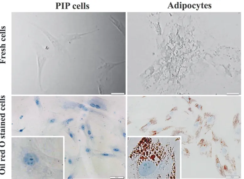
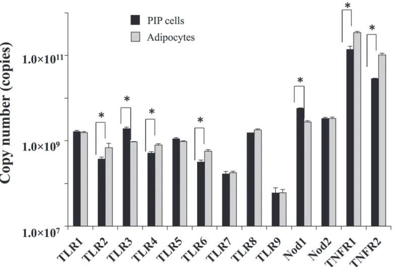
We first aimed to morphologically characterize PIP cells and differentiated porcine adipocytes. Fresh cells as well as cells stained with Oil red O were observed. As shown inFig. 1, remarkable differences were observed between PIP cells and adipocytes. PIP cells were found to be small cells with a clear nucleus at the center surrounded by abundant cytoplasm. On the contrary, differentiated adipocytes were observed as bigger cells than PIP cells and showed lipid droplets that pushed nucleus to the periphery.In addition, expression of various PRRs (TLR1–9, NOD-1 and 2) and TNFR-1 and 2 were analyzed in PIP cells and differentiated adipocytes (Fig. 2). Both PIP cells and adipocytes ex-pressed TLR1–9, NOD-1, NOD-2, TNFR-1 and TNFR-2. However, TNFR-1 and TNFR-2 were highly expressed when compared with other receptors in both PIP cells and differentiated adi-pocytes. We also observed that PIP cells showed a higher expression of NOD-1 and TLR3 when compared with adipocytes (Fig. 2). No significant differences were observed in the ex-pression of TLR1, TLR5, TLR7–9 and NOD-2; while the exex-pression of TLR2, TLR4, TLR6, TNFR-1 and TNFR-2 was higher in adipocytes than in PIP cells (Fig. 2).
Response of PIP cells and adipocytes to TNF-
α
stimulation
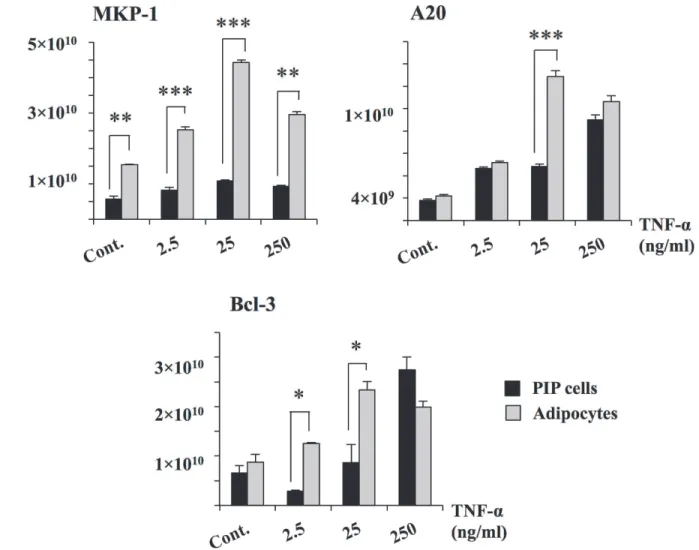
significantly increased the expression of A20 and Bcl-3 while no effect was observed in the ex-pression of MKP-1. Similarly, stimulation of differentiated adipocytes with TNF-αincreased the expression of A20 and Bcl-3 reaching levels that were significantly higher than those ob-served in PIP cells (Fig. 4). Moreover, TNF-αincreased mRNA levels of MKP-1 in differentiat-ed adipocytes. As observdifferentiat-ed with cytokines, for negative regulators, higher levels of expression were also observed with 25 ng/ml of TNF-α. Therefore, this concentration of TNF-αwas select-ed for further experiments.
Evaluation of anti-inflammatory and anti-adipogenic activity of LAB
strains
For the evaluation of the anti-adipogenic and immunoregulatory activity of LAB, we per-formed severalin vitrosteps to simulate LAB-immune cells-adipocytes interaction as described in materials and methods. Then, PIP cells and adipocytes were stimulated with CFS from por-cine Peyer’s patche immunocompetent cell cultures stimulated with different LAB strains. PIP
Fig 1. Photographic pictures of PIP and adipocyte cells.The PIP cells were grown at 37°C for 4 days, and subsequently fed with differentiation medium for another 4 days to differentiate into adipocyte cells. Before and after adipocyte differentiation, the cells were stained with Oil red O stain. Presence of red and blue color indicates accumulation of lipid droplets and nucleus in the adipocyte cells at 8 day.
doi:10.1371/journal.pone.0119644.g001
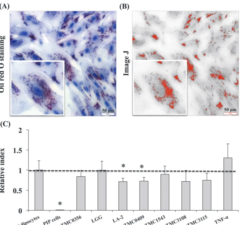
cells and adipocytes were pre-stimulated with the different CFS for 48 hours and then chal-lenged with TNF-αfor 3, 6, and 12 hours. The effect of LAB on PIP cells differentiation and adipogenesis was investigated by using Oil red O staining (Fig. 5). The deposition of fats in the mature adipocytes was quantified by image J. Deposition of lipids increased with differentia-tion of PIP cells into mature adipocytes, and fat deposit were significantly increased after stim-ulation with TNF-α(Fig. 5). In mature adipocytes coming from PIP cells treated with culture free supernatants from LA-2 and TMC0409 strains, significantly lower deposition of lipids was observed when compared to control cells (Fig. 5).
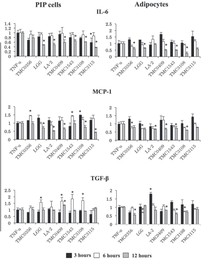
In addition, the mRNA levels of TLR2, TLR4, IL-6, MCP-1, and TGF-βwere determined in PIP cells and adipocytes as shown inFig. 6. As mentioned earlier, TNF-αwas capable to induce mRNA expressions of IL-6, MCP-1, TLR2 and TLR4 in both PIP cells and differentiated adipo-cytes (Fig. 3), while no modification was observed in the expression of TGF-β(data not shown). A strain dependent effect was observed in the levels of IL-6, MCP-1, and TGF-βwhen PIP cells were pretreated with cell free supernatants. PIP cells pretreated with cell free superna-tant fromL.rhamnosusGG,L.paracaseiTMC0409 orB.bifidumTMC3108 showed increased expression of TGF-βearlier at hour 6 post-TNF-αchallenge, while those cells showed reduced levels of IL-6 and MCP-1 at hour 12 (Fig. 6).L.rhamnosusLA-2 was able to significantly
Fig 2. Expression of pattern of PPRs (TLR1–9, NOD 1, 2) and TNFR1, 2 in PIP, and differentiated adipocyte cells.Copy number of mRNA in 25 ng of
cDNA prepared from PIP and adipocyte cells were calculated. Three independent experiments were conducted for each case and the average values (mean±S.D) were represented. The presence of stars (*) indicates statistical differences with significant levels of p<0.05.
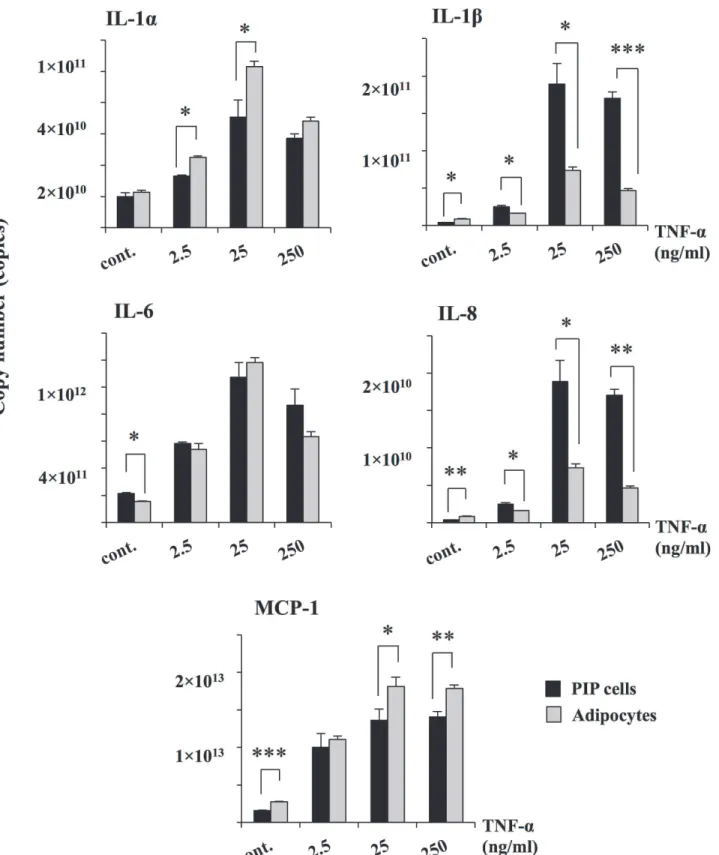
Fig 3. TNF-αinduced inflammatory cytokines in PIP and differentiated adipocyte cells.Both PIP cells and adipocytes were incubated with different
concentrations of TNF-α(2.5, 25, 250 ng/ml) for 24 hours and the level of pro-inflammatory cytokines (IL-1α, IL-1β, IL-6, IL-8, and MCP-1) were determined. Cells without addition of TNF-αwere used as controls. Three independent experiments were conducted for each case and the average values (mean±S.D) were represented as inflammatory effect of TNF-α. The presence of various stars (*,**and***) indicates statistical differences with significant levels of p<0.05, p<0.01 and p<0.001, respectively.
doi:10.1371/journal.pone.0119644.g003
reduce levels of IL-6 and MCP-1 but did not induce changes in TGF-βexpression (Fig. 6).L.
gasseriTMC0356 significantly increased MCP-1. In addition,S.thermophilusTMC1543 re-duced IL-6 levels but increased the expression of MCP-1; whileB.bifidumTMC3115 reduced IL-6 and MCP-1 levels (Fig. 6). Interestingly, almost all the studied LAB strains induced similar effects in differentiated adipocytes after TNF-αchallenge.L.gasseriTMC0356,L.rhamnosus
GG,L.paracaseiTMC0409,S.thermophilusTMC1543 andB.bifidumTMC3108 significantly reduced levels of IL-6, MCP-1, and TGF-βin TNF-α-challenged porcine adipocytes (Fig. 6). On the contrary,L.rhamnosusLA-2 significantly increased levels of TGF-βin adipocytes.
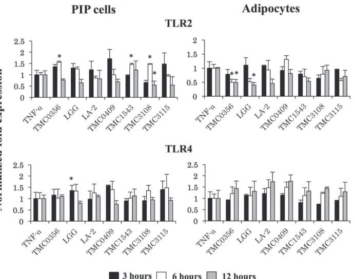
Strain dependent effects were also observed when analyzing TLR2 and TLR4 expressions in PIP cells and adipocytes pretreated with cell free supernatants (Fig. 7).L.gasseriTMC0356,S.
thermophilusTMC1543 andB.bifidumTMC3108 significantly increased TLR2 expression in PIP cells, whileL.rhamnosusGG augmented mRNA levels of TLR4. In addition,L.gasseri
TMC0356 andL.rhamnosusGG reduced TLR2 levels in differentiated adipocytes while no
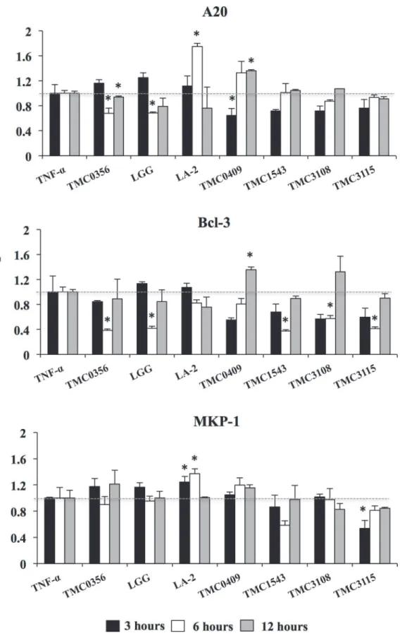
Fig 4. Expression of negative regulators in PIP and differentiated adipocyte cells.Both PIP cells and adipocytes were challenged with different concentrations of TNF-α(2.5, 25, and 250 ng/ml) for 24 hours and the level of negative regulators (MKP-1, A20, and Bcl-3) were determined. Cells without TNF-αstimulation were used as controls. Three independent experiments were conducted for each case and the average values (mean±S.D) were represented as inflammatory effect of TNF-α. The presence of various stars (*,**and***) indicates statistical differences with significant levels of p<0.05, p<0.01 and p<0.001, respectively.
significant changes were observed in the expression of TLR4 in these cells with CFS treatments (Fig. 7).
Finally we aimed to evaluate whether CFS was able to modulate the expressions of negative regulators after TNF-αchallenge in porcine adipocytes (Fig. 8). Again, strain dependent effects were observed.L.gasseriTMC0356 andL.rhamnosusGG significantly reduced the expression
Fig 5. Anti-adipogenic activity of various LABs.The PIP and differentiated adipocytes were incubated with CFS of various LABs treated with PPs at 37°C for 48 hours. Mature adipocyte cells were Oil red O stained and deposition of lipid droplets in the mature adipocytes was quantified by image J. (A) Oil red O stained mature adipocytes. (B) Photograph of image J with lipid accumulation. (C) Anti-adipogenic activity of LABs. Three independent experiments were conducted for each case and the average values (mean±S.D) were represented as anti-adipogenic effect of LABs. The presence of stars (*) indicates statistical differences with significant levels of p<0.05.
doi:10.1371/journal.pone.0119644.g005
Fig 6. Anti-inflammatory activity of various LABs in PIP cells and differentiated adipocytes.Both PIP cells and differentiated adipocytes were pre-stimulated with CFS of various LABs for 48 h and then post-pre-stimulated with TNF-αfor 3, 6, and 12 hours. Cells without any TNF-αstimulation were used as cell controls. The expressions of IL-6, MCP-1, and TGF-βmRNAs were evaluated. Cells treated only with TNF-αwere used as TNF-αcontrols. Three or four independent experiments were conducted for each case and the average values (mean±S.D) were represented as immunomodulatory effect of LABs. The presence of stars (*) indicates statistical differences with significant levels of P<0.05.
of A20 and Bcl-3;S.thermophilusTMC1543 andB.bifidumTMC3108 reduced levels of Bcl-3 and;B.bifidumTMC3115 reduced Bcl-3 and MKP-1 (Fig. 8). On the contrary,L.rhamnosus
LA-2 significantly increased levels of A20 and MKP-1 in adipocytes whileL.paracasei
TMC0409 increased A20 and Bcl-3.
Discussion
From the histological point of view, adipose tissue is composed of adipocytes (mature fat cells) and the interadipocytar stromal-vascular fraction formed by extracellular matrix with dis-persed fibroblasts, preadipocytes, endothelial, and immune cells [27]. Excessive growth of adi-pose tissue in obesity is the result from enlargement of existing adipocytes (hypertrophy) and formation of new adipocytes (hyperplasia) through differentiation of stromal preadipocytes (adipogenesis) [28]. Mature adipocytes represent 50–85% of the total cellular components of adipose tissue. Obese subjects are characterized by a higher total adipocyte number than lean individuals [29]. Moreover, the hypertrophic adipocytes in obese individuals shift their im-mune balance towards the production of pro-inflammatory molecules [30,31]. Accordingly,
Fig 7. Induction of TLRs expression in PIP cells and differentiated adipocytes.Both PIP cells and differentiated adipocytes were pre-stimulated with CFS of various LABs for 48 h and then post-stimulated with TNF-αfor 3, 6, and 12 hours. The expressions of TLR2 and TLR4 were evaluated. Cells treated only with TNF-αwere used as TNF-αcontrols. Three or four independent experiments were conducted for each case and the average values (mean±S.D) were represented as immunomodulatory effect of LABs. The presence of stars (*) indicates statistical differences with significant levels of P<0.05.
doi:10.1371/journal.pone.0119644.g007
microarray profiling of isolated adipocytes from obeseversusnon-obese Pima Indians revealed an increased expression of inflammation-related genes in obese adipocytes [32].
In this work, the expression profile of immune receptors and pro-inflammatory cytokines were examined in porcine preadipocytes (PIP cells) and differentiated adipocytes. TLRs that are usually expressed immune cells, but their expression has been also documented in non-im-mune cells like intestinal epithelial cells [23,24,26]. The expression of TLRs was also observed in adipose tissue [33], although this expression was mainly attributed to infiltrated macro-phages [34]. However, murine derived preadipocyte and differentiated adipocyte cell lines (3T3-L1) have been shown to express TLRs in response to TLR ligands [33,35]. Khazen et al. [36] reported an augmented expression of TLRs when 3T3-F442A cells were differentiated into adipocytes. TLRs are also expressed and are functional in human adipose tissue [37]. That work demonstrated that TLR2 and TLR4 were expressed at relatively high levels (compared to a monocyte cell line) on the surface of human adipose cells. Moreover, stimulation of human adipocytes with LPS, or with lipoteichoic acid, two specific ligands of TLR4 and TLR2, respec-tively, induced a strong increase in TNF-αproduction. Our results are in line with these previ-ous observations since mRNAs from TLR1 to TLR9 were detected in both porcine adipose cells. Some preliminary experiments with TLRs agonists showed that these receptors are func-tional in porcine adipocytes, however further experiments are needed to fully evaluate TLRs signaling pathways in PIP cells and mature adipocytes.
Our work also demonstrated the expression of TNF-αreceptors (TNFR-1 and TNFR-2) in both PIP cells and mature porcine adipocytes. Since the first description of enhanced expres-sion and secretion of TNF-αby adipose tissue of obese rodents that linked inflammation to obesity and insulin resistance [38], several works have clearly demonstrated that TNF-αhas a crucial role in the pathogenesis of inflammation and insulin resistance by promoting immune and metabolic complications. It is known that most effects of TNF-αon adipose tissue are me-diated by the TNFR-1 and subsequent activation of various transduction pathways [11]. Two transcription factor-signaling pathways have been linked to the pro-inflammatory effects of obesity and insulin resistance: the NF-κB (nuclear factor-κB) pathway and the c-Jun NH2-ter-minal kinase (JNK) pathway. It is believed that overproduction of TNF-αin obesity is an ulti-mate attempt to control adiposity, since increased TNF-αmay help at limiting further weight gain through lipolysis and insulin resistance, impaired preadipocyte differentiation and in-creased adipocyte apoptosis [39]. However, these effects of TNF-αoccur at the expense of worsening insulin resistance and inflammation. TNF-αproduced by adipocytes further up-reg-ulate pro-inflammatory adipokines and cytokines [40,41]. Culturing human adipocytes in media previously conditioned by TNF-αresulted in pro-inflammatory adipokine overproduc-tion. This was abrogated by immunoneutralization of TNF-αin these media, indicating that TNF-αis a crucial and proximal contributor to adipokine dysregulation in adipocytes [40]. Ac-tivation of TNFR-1 pathway in adipocytes increases the production of other inflammatory fac-tors such as IL-6 and MCP-1. Thein vivorelease of IL-6 by whole-body adipose tissue could contribute to 15–35% of the systemic IL-6 in human and circulating levels and adipocytes pro-duction of IL-6 are increased in obesity [42]. Infiltration of adipose tissue by macrophages is an important event in the increased inflammatory process in obesity. MCP-1 and its receptor (CCR2) are required for macrophage infiltration into adipose tissue. MCP-1-or CCR2-defi-cient mice fed a high-fat diet exhibited fewer macrophages and a lower inflammatory gene pro-file in adipose tissue together with reduced insulin resistance [43]. Treatment of 3T3-L1 differentiated adipocytes with MCP-1 decreased insulin-stimulated glucose uptake and the ex-pression of several adipogenic genes [44]. Also, they report that the capacity of TNF-αto in-crease pro-inflammatory factors in adipose cells. Similarly, both PIP cells and mature
adipocytes showed significant up-regulation in the expressions of MCP-1, IL-1α, IL-1β, IL-6, and IL-8.
Additionally, stimulation of porcine mature adipocytes with TNF-αsignificantly changed the expression of negative regulators of the TNFR-1 pathway. The zinc-finger protein A20 is a key player in the negative feedback regulation of the NF-κB pathway in response to multiple stimuli. It is known that TNF-αdramatically increases A20 expression in all tissues, including adipose tissue [45]. Interestingly, it was reported that the NADPH oxidase Nox4 acts as a switch from insulin-induced proliferation to differentiation by controlling MKP-1 expression, which limits ERK1/2 signaling in human and mouse preadipocytes [46]. The work clearly showed that Nox4 controls the expression of MKP-1 and thereby limits the contribution of the proliferative Ras–Raf-ERK1/2 pathway to insulin signaling. ERK1/2 phosphorylates IRS-1 on serine-residues and thereby prevents IRS-1 tyrosine phosphorylation. The Nox4-dependent in-duction of MKP-1 prevents this effect and therefore promotes insulin-induced differentiation but attenuated insulin-induced proliferation. Those works demonstrated that negative regula-tors have important roles in the biology of adipocytes, with impacts not only in immune re-sponses but in proliferation and differentiation as well.
The data presented in this work showed that our porcinein vitrosystems (PIP cells and ma-ture adipocytes) share all the immunological characteristics that have been attributed to these cells in other species, especially human. Then PIP cells and the porcine mature adipocytes ob-tained from them, could be useful laboratory tools to gain insight into the immunobiology of adipose tissue, as well as for the screening and evaluation of potential therapies aimed to bene-ficially modulate adipose immune response. In relation to this last assumption, we demonstrat-ed here that our porcinein vitrosystems are of value for the evaluation of immunobiotic effects.
Recently, gut microbiota has been identified as an important modifier of systemic inflam-matory reactions influencing remote tissues [47]. Interestingly, different gut microbiota-de-rived products can exert both pro- and anti-inflammatory effects. It was described that translocation of LPS and peptidoglycans from microbiota into systemic circulation leads to metabolic endotoxemia, suggested as one of the main triggers of adipose tissue and systemic low-grade inflammation [47,48]. On the contrary, products of gut bacterial fermentation such as short-chain fatty acids (SCFA) were shown to have anti-inflammatory effects and influence energy homeostasis [49]. In addition, several works demonstrated that orally administered pro-biotics are able to modulate tissues distant from the gut including the respiratory tract [50], blood [51], bone marrow [52], and adipose tissue [17,18]. These studies showed that in addi-tion to translocated microbial products, immunobiotics are able to modulate distant tissues through their ability to modify cytokine’s profiles [50–52].
In this work we investigated the possibility that LAB modify the response of porcine adipo-cytes to TNF-αstimulation through host’intestinal immune-competent cells. We treated the porcine immune cells from Peyer’s patches with different LAB strains and, tested conditioned media from LAB-stimulated immune cells to determine the regulatory effects on porcine prea-dipocytes (PIP cells) and differentiated aprea-dipocytes. As it is described for other probiotics’ ef-fects, we found strain specific effects of LAB on PIP cells and differentiated adipocytes.
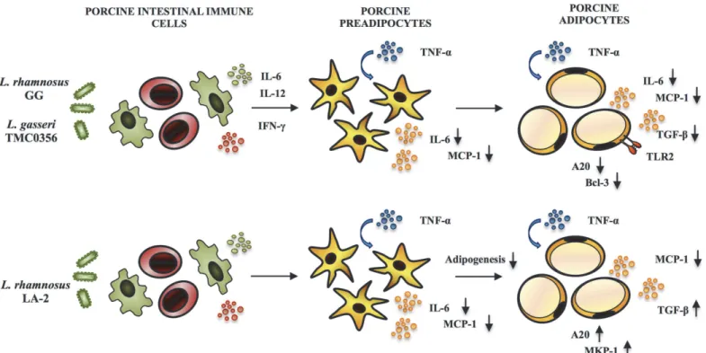
TheLactobacillusGG,L.gasseriTMC0356, andL.rhmanosusLA-2 showed remarkable ef-fects with significant reduction in the expression of pro-inflammatory cytokines and chemo-kines in adipocytes challenged with TNF-α(Fig. 9). The strainsLactobacillusGG andL.gasseri
mechanism behind theL.rhmanosusLA-2 mediated up-regulation was unknown. In addition, we previously used the conditioned medium of murine macrophage-like cell line J774.1 cul-tured with LGG or TMC0356 strains to stimulate mouse preadipocyte cell line 3T3-L1 and found a suppressed lipid accumulation and reduced PPAR-γmRNA expression [19]. More-over, the J774 cells treated withLactobacillusGG orL.gasseriTMC0356 increased production of cytokines IL-6 and IL-1, suggesting that lactobacilli may suppress differentiation of preadi-pocytes through macrophage activation and production of Th1 cytokines. Severalin vivo stud-ies have comparatively evaluated the immunoregulatory effects ofL.gasseriTMC0356 and
LactobacillusGG. Kawase et al. [53] demonstrated that oral administration ofLactobacillusGG orL.gasseriTMC0356 alleviate nasal allergic symptoms by suppressing the increase in nasal vascular permeability caused by local inflammation associated with allergic rhinitis in rodents. Moreover, in an allergic rhinitis guinea pig model, both LAB strains were able to decrease the total numbers of leukocytes, particularly eosinophils and neutrophils from the nasal cavity la-vage fluid, and the OVA-specific IgE concentration in the serum [54].In vitrostudies of the immune responses of murine Peyer's patches stimulated withLactobacillusGG orL.gasseri
TMC0356 showed the capacity of both strains to increase the production of IL-6, IL-12 and IFN-γby intestinal immune cells [55]. Those studies clearly indicate that bothLactobacillus
GG andL.gasseriTMC0356 are equally effective in improving Th1 response not only in the gut by in the systemic compartment as well. Similarly, in this work, CFS from cultures of por-cine Peyer's patches with GG or TMC0356 strains were able to functionally modulate the re-sponse of differentiated porcine adipocytes to TNF-αchallenge. Then, our data suggest that Th1 cytokines produced by intestinal immune cells will be also capable of downregulating ex-pression of pro-inflammatory genes in mature adipocytes (Fig. 9). In line with this assumption,
L.gasseriTMC0356 was found to be able to stimulate the respiratory immune responses in a
Fig 9. Possible immunomodulatory activity ofL.rhamnosusGG,L.gasseriTMC0356, andL.rhamnosusLA-2 in both PIP cells and differentiated adipocytes after stimulation with TNF-α.
doi:10.1371/journal.pone.0119644.g009
diet-induced obese mouse model, indicating that this immunobiotic strain may protect host animals from the lung immune dysfunction caused by obesity [56].
Concluding Remarks
Taking into consideration that severalin vivoandin vitrostudies clearly demonstrated the ben-eficial effects ofLactobacillusGG andL.gasseriTMC0356 in adipose inflammation, the results presented in this work indicate that the PIP cells and porcine adipocytes could be used for the screening and the selection of new immunobiotic strains with the potential to functionally modulate adipose inflammation when orally administered. Moreover, the results of this work demonstrated that the regulation of IL-6, MCP-1, A20 and Bcl-3 induction in TNF-α -chal-lenged porcine adipocytes could be certain biomarkers to screen and select potential immunobiotic strains.
Acknowledgments
We would like to thanks Grant-in-Aid for Scientific Research, Japan Society for the Promotion of Science (JSPS), Development of Agricultural Products and Foods with Health-promoting benefits (NARO), and all our colleagues who cooperate this study.
Author Contributions
Conceived and designed the experiments: MS AT PK FH HA TN TS JV HK. Performed the ex-periments: MS AT PK HW. Analyzed the data: MS AT PK YS JV HK. Contributed reagents/ materials/analysis tools: HW AT KM KY MH. Wrote the paper: MS AT PK JV HK.
References
1. DiBaise JK, Zhang H, Crowell M, Krajmalnik-Brown R, Decker GA, Ritmann BE. Gut microbiota and its possible relationship with obesity. Mayo Clinic Proceed. 2008; 83: 460–469.
2. Huebner L, Engeli S, Wrann CD, Goudeva L, Laue T, Kielstein H. Human NK cell subset functions are differentially affected by adipokines. PLoS One. 2013; 8(9): e75703. doi:10.1371/journal.pone. 0075703PMID:24098717
3. Chi W, Dao D, Lau TC, Henriksbo BD, Cavallari JF, Foley KP, et al. Bacterial peptidoglycan stimulates adipocyte lipolysis via NOD1. PLoS One. 2014; 9(5): e97675. doi:10.1371/journal.pone.0097675
PMID:24828250
4. Heilbronn LK, Campbell LV. Adipose tissue macrophages, low grade inflammation and insulin resis-tance in human obesity. Cur Pharma Design. 2008; 14: 1225–1230.
5. Oliver E, McGillicuddy F, Phillips C, Toomey S, Roche HM. The role of inflammation and macrophage accumulation in the development of obesity-induced type 2 diabetes mellitus and the possible thera-peutic effects of long-chain n-3 PUFA. Proceed Nut Soc. 2010; 69: 232–243.
6. Desruisseaux MS, Nagajyothi, Trujillo ME, Tanowitz HB, Scherer PE. Adipocyte, adipose tissue, and infectious disease. Infect. Immunit. 2007; 75: 1066–1078. PMID:17118983
7. Zhu HJ, Ding HJ, Deng JY, Pan H, Wang LJ, Li NS, et al. Inhibition of preadipocyte differentiation and adipogenesis by zinc-a2-glycoprotein treatment in 3T3-L1 cells. J. Diabet Invest. 2013; 4: 252–260. 8. Tsai YT, Cheng PC, Pan TM. Anti-obesity effects of gut microbiota are associated with lactic acid
bacte-ria. Appl Microbiol Biotechnol. 2014; 98: 1–10. PMID:24232731
9. Sethi JK, Hotamisligil GS. The role of TNF-αin adipocyte metabolism. Sem Cell. Develop Biol. 1999; 10: 19–29.
10. Moller DE. Potential Role of TNF-αin the pathogenesis of insulin resistance and Type 2 diabetes. Trend Endocrinol. Metab. 2000; 11: 212–217. PMID:10878750
11. Cawthorn WP, Sethi JK. TNF-a and adipocyte biology. FEBS Lett. 2008; 582: 117–131. PMID:
18037376
13. Van der Poll T, Romijn OA, Endert E, Borm JJJ, Buller HR, Buller HR. Tumor necro- sis factor mimics the metabolic response to acute infection in healthy humans. Am. J. Physiol. 19991; 261: E457–E465 PMID:1928337
14. Uysal KT, Wiesbrock SM, Marino MW, Hotamisligil GS. Protection from obesity-induced insulin resis-tance in mice lacking TNF-αfunction. Nature. 1997; 389: 610–614. PMID:9335502
15. Stanley TL, Zanni MV, Johnsen S, Rasheed S, Makimura H, Lee H, et al. TNF-alpha antagonism with etanercept decreases glucose and increases the proportion of high molecular weight adiponectin in obese subjects with features of the metabolic syndrome. J Clin Endocrinol Metab. 2011; 96: E146– E150. doi:10.1210/jc.2010-1170PMID:21047923
16. Lo J, Bernstein LE, Canavan B, Torriani M, Jackson MB, Ahima RS, et al. Effects of TNF-alpha neutrali-zation on adipocytokines and skeletal muscle adiposity in the metabolic syndrome. Am J Physiol Endo-crinol Metab. 2007; 293: E102–E109. PMID:17374698
17. Kim SW, Park KY, Kim B, Kim E, Hyun CK.Lactobacillus rhamnosusGG improves insulin sensitivity and reduces adiposity in high-fat diet-fed mice through enhancement of adiponectin production. Bio-chem. Biophy Res Comm. 2013; 431: 258–263. doi:10.1016/j.bbrc.2012.12.121PMID:23313485
18. Toral M, Gómez-Guzmán M, Jiménez R, Romero M, Sánchez M, Utrilla MP, et al. The probiotic Lacto-bacillus coryniformisCECT5711 reduces the vascular pro-oxidant and pro-inflammatory status in obese mice. Clin Sci (Lond). 2014; 127(1): 33–45. doi:10.1042/CS20130339PMID:24410749
19. Miyazawa K, He F, Yoda K, Hiramatsu M. Potent effects of, and mechanisms for, modification of cross-talk between macrophages and adipocytes by lactobacilli. Microbiol Immunol. 2012; 56: 847–854. doi:
10.1111/j.1348-0421.2012.00512.xPMID:23017059
20. Park JE, Oh SH, Cha YS.Lactobacillus brevisOPK-3 isolated from kimchi inhibits adipogenesis and exerts anti-inflammation in 3T3-L1 adipocyte. J. Sci Food Agric. 2014; 94(12): 2514–2520. doi:10. 1002/jsfa.6588PMID:24453065
21. Sanosaka M, Minashima T, Suzuki K, Watanabe K, Ohwada S, Hagio A, et al. A combination of octano-ate and oleoctano-ate promotes in vitro differentiation of porcine intramuscular adipocytes. Comp. Biochem. Physiol A. 2008; 149: 285–292. PMID:17977041
22. Nobusue H, Kano K. Establishment and characteristics of porcine preadipocyte cell lines derived from mature adipocytes. J Cell Biochem. 2010; 109(3): 542–552. doi:10.1002/jcb.22431PMID:20013788
23. Villena J, Kitazawa H. Role of Toll-like Receptors in the Modulation of Intestinal Inflammation by Immu-nobiotics, Probiotics. In: Kitazawa H, Villena J, Alvarez S, editors. Immunobiotics and Immunogenics. CRC Press; 2013. pp. 89–127.
24. Moue M, Tohno M, Shimazu T, Kido T, Aso H, Suda Y, et al. Toll-like receptor 4 and cytokine expres-sion involved in functional immune response in an originally established porcine intestinal epitheliocyte cell line. Biochim Biophys Acta. 2008; 1780: 134–144. PMID:18082146
25. Shimazu T, Villena J, Tohno M, Fujie H, Hosoya S, Shimosato T, Aso H, et al. Immunobiotic Lactobacil-lus jenseniielicit anti-inflammatory activity in porcine intestinal epithelial cells by modulating negative regulators of the toll-like receptor signaling pathway. Infect Immun. 2012; 80: 276–288. doi:10.1128/ IAI.05729-11PMID:22083706
26. Tomosada Y, Villena J, Murata K, Chiba E, Shimazu T, Aso H, et al. Immunoregulatory effect of bifido-bacteria strains in porcine intestinal epithelial cells through modulation of ubiquitin-editing enzyme A20 expression. PLoS One. 2013; 8(3): e59259. doi:10.1371/journal.pone.0059259PMID:23555642
27. Curat CA, Miranville A, Sengenes C, Diehl M, Tonus C, Busse R, et al. From blood monocytes to adi-pose tissue-resident macrophages: induction of diapedesis by human mature adipocytes. Diabetes. 2004; 53: 1285–1292. PMID:15111498
28. Constant VA, Gagnon A, Landry A, Sorisky A. Macrophage-conditioned medium inhibits the differentia-tion of 3T3-L1 and human abdominal preadipocytes. Diabetologia. 2006; 49: 1402–1411. PMID:
16609875
29. Spalding KL, Arner E, Westermark PO, Bernard S, Buchholz BA, Bergmann O, et al. Dynamics of fat cell turnover in humans. Nature. 2008; 453: 783–787. doi:10.1038/nature06902PMID:18454136
30. Jernas M, Palming J, Sjoholm K, Jennische E, Svensson PA, Gabrielsson BG, et al. Separation of human adipocytes by size: hypertrophic fat cells display distinct gene expression. FASEB J. 2006; 20: 1540–1542. PMID:16754744
31. Skurk T, Alberti-Huber C, Herder C, Hauner H. Relationship between adipocyte size and adipokine ex-pression and secretion. J. Clin. Endocrinol. Metab. 2007; 92: 1023–1033. PMID:17164304
32. Lee YH, Nair S, Rousseau E, Allison DB, Page GP, Tataranni PA, et al. Microarray profiling of isolated abdominal subcutaneous adipocytes from obese vs non-obese Pima Indians: increased expression of inflammation-related genes. Diabetologia. 2005; 48: 1776–1783. PMID:16059715
33. Poulain-Godefroy O, Bacquer OL, Plancq P, Lecœur C, Pattou F, Fruhbeck G, et al. Inflammatory role
of Toll-like receptors in human and murine adipose tissue. Medi. Inflam. 2010;1–10. doi:10.1155/2010/ 823486
34. Zhang HM, Chen LL, Wang L, Xu S, Wang X, Yi LL, et al. Macrophage infiltrates with high levels of Toll-like receptor 4 expression in white adipose tissues of male Chinese. Nut. Metab. Card Dis. 2009; 19: 736–743.
35. Batra A, Pietsch J, Fedke I, Glauben R, Okur B, Stroh T, et al. Leptin-dependent toll-like receptor ex-pression and responsiveness in preadipocytes and adipocytes. Am J. Pathol. 2007; 170: 1931–1941. PMID:17525261
36. Khazen W, M’Bika JP, Collinet M, Tramoni M, Chany C, Achour A, et al. Differentiation-dependent ex-pression of interferon gamma and toll-like receptor 9 in 3T3-F442A adipocytes. Biochimie. 2007; 89: 669–675. PMID:17331636
37. Bes-Houtmann S, Roche R, Hoareau L, Gonthier MP, Festy F, Caillens H, et al. Presence of functional TLR2 and TLR4 on human adipocytes. Histochem Cell Biol. 2007; 127: 131–137. PMID:16988837
38. Hotamisligil GS, Shargill NS, Spiegelman BM. Adipose expression of tumor necrosis factor-α: direct role in obesity-linked insulin resistance. Science. 1993; 259: 87–91. PMID:7678183
39. Prins JB, Niesler CU, Winterford CM, Bright NA, Siddle K, O’Rahilly S, et al. Tumor necrosis factor-alpha induces apoptosis of human adipose cells. Diabetes. 1997; 46: 1939–1944. PMID:9392477
40. Maury E, Noel L, Detry R, Brichard SM. In vitro hyper-responsiveness to TNF-{alpha}contributes to adi-pokine dysregulation in omental adipocytes of obese subjects. J. Clin. Endocrinol. Metab. 2009; 94: 1393–1400. doi:10.1210/jc.2008-2196PMID:19174496
41. Bruun JM, Lihn AS, Verdich C, Pedersen SB, Toubro S, Astrup A, et al. Regulation of adiponectin by adipose tissue-derived cytokines: in vivo and in vitro investigations in humans. Am. J. Physiol. Endocri-nol. Metab. 2003; 285: E527–E533. PMID:12736161
42. Bastard JP, Lagathu C, Caron M, Capeau J. Point-counterpoint: interleukin-6 does/does not have a beneficial role in insulin sensitivity and glucose homeostasis. J. Appl. Physiol. 2007; 102: 821–822. PMID:17323465
43. Kanda H, Tateya S, Tamori Y, Kotani K, Hiasa K, Kitazawa R, et al. MCP-1 contributes to macrophage infiltration into adipose tissue, insulin resistance, and hepatic steatosis in obesity. J. Clin. Invest. 2006; 116: 1494–1505. PMID:16691291
44. Sartipy P, Loskutoff DJ. Monocyte chemoattractant protein 1 in obesity and insulin resistance. Proc. Natl. Acad. Sci. U.S.A. 2003; 100: 7265–7270. PMID:12756299
45. Dorronsoro A, Lang V, Jakobsson E, Ferrin I, Salcedo JM, Fernandez-Rueda J, et al. Identification of the NF-κB inhibitor A20 as a key regulator for human adipogenesis. Cell Death Dis. 2013; 19: 972. doi:
10.1038/cddis.2013.494
46. Schroder K, Wandzioch K, Helmcke I, Brandes RP. Nox4 acts as a switch between differentiation and proliferation in preadipocytes. Arterioscler Thromb Vasc Biol. 2009; 29(2): 239–245. doi:10.1161/ ATVBAHA.108.174219PMID:19057021
47. Burcelin R, Serino M, Chabo C, Garidou L, Pomie C, Courtney M, et al. Metagenome and metabolism: the tissue microbiota hypothesis. Diab. Obes. Metab. 2013; 15: 61–70.
48. Carvalho BM, Saad MJ. Influence of gut microbiota on subclinical inflammation and insulin resistance. Med Inflam. 2013; 1–13.
49. Kim S, Kim JH, Park BO, Kwak YS. Perspectives on the therapeutic potential of short-chain fatty acid receptors. BMB Reports. 2014; 47: 173–178. PMID:24499669
50. Villena J, Chiba E, Tomosada Y, Salva S, Marranzino G, Kitazawa H, et al. Orally administered Lacto-bacillus rhamnosusmodulates the respiratory immune response triggered by the viral pathogen-associ-ated molecular pattern poly(I:C). BMC Immunol. 2012; 13: 53. PMID:22989047
51. Salva S, Villena J, Alvarez S. Differential immunomodulatory activity ofLactobacillus rhamnosus
strains isolated from goat milk: impact on intestinal and respiratory infections. Int. J. Food Microbiol. 2010; 141: 82–89. doi:10.1016/j.ijfoodmicro.2010.03.013PMID:20395002
52. Salva S, Marranzino G, Villena J, Agüero G, Alvarez S. Probiotic Lactobacillus strains protect against myelosuppression and immunosuppression in cyclophosphamide-treated mice. Int. Immunopharma-col. 2014; 22(1): 209–221. doi:10.1016/j.intimp.2014.06.017PMID:24975836
53. Kawase M, He F, Kubota A, Hata JY, Kobayakawa S, Hiramatsu M. Inhibitory effect ofLactobacillus gasseriTMC0356 andLactobacillusGG on enhanced vascular permeability of nasal mucosa in experi-mental allergic rhinitis of rats. Biosci Biotechnol Biochem. 2006; 70(12): 3025–3030. PMID:17151443
54. Kawase M, He F, Kubota A, Harata G, Hiramatsu M. Orally administratedLactobacillus gasseri
55. Harata G, He F, Kawase M, Hosono A, Takahashi K, Kaminogawa S. Differentiated implication of Lac-tobacillusGG andL.gasseriTMC0356 to immune responses of murine Peyer's patch. Microbiol Immu-nol. 2009; 53(8): 475–480. doi:10.1111/j.1348-0421.2009.00146.xPMID:19659932
56. Yoda K, He F, Miyazawa K, Kawase M, Kubota A, Hiramatsu, M. Orally administered heat-killed Lacto-bacillus gasseriTMC0356 alters respiratory immune responses and intestinal microbiota of diet-in-duced obese mice. J Appl Microbiol. 2012; 113(1): 155–162. doi:10.1111/j.1365-2672.2012.05316.x
PMID:22519947