Thrombocytes
Craig E. Hughes1*, Uvaraj P. Radhakrishnan2, Marie Lordkipanidze´1, Stuart Egginton1,
Johannes M. Dijkstra3, Pudur Jagadeeswaran2, Stephen P. Watson1
1Centre for Cardiovascular Sciences, Institute for Biomedical Research, The College of Medical and Dental Sciences, University of Birmingham, Edgbaston, Birmingham, United Kingdom,2Department of Biological Sciences, University of North Texas, Denton, Texas, United States of America,3Institute for Comprehensive Medical Science, Fujita Health University, Toyoake, Aichi, Japan
Abstract
Collagen activates mammalian platelets through a complex of the immunoglobulin (Ig) receptor GPVI and the Fc receptor c-chain, which has an immunoreceptor tyrosine-based activation motif (ITAM). Cross-linking of GPVI mediates activation through the sequential activation of Src and Syk family kinases and activation of PLCc2. Nucleated thrombocytes in fish are activated by collagen but lack an ortholog of GPVI. In this study we show that collagen activates trout thrombocytes in whole blood and under flow conditions through a Src kinase driven pathway. We identify the Ig receptor G6f-like as a collagen receptor and demonstrate in a cell line assay that it signals through its cytoplasmic ITAM. Using a morpholino for
in vivoknock-down of G6f-like levels in zebrafish, we observed a marked delay or absence of occlusion of the venous and
arterial systems in response to laser injury. Thus, G6f-like is a physiologically relevant collagen receptor in fish thrombocytes which signals through the same ITAM-based signalling pathway as mammalian GPVI, providing a novel example of convergent evolution.
Citation:Hughes CE, Radhakrishnan UP, Lordkipanidze´ M, Egginton S, Dijkstra JM, et al. (2012) G6f-Like Is an ITAM-Containing Collagen Receptor in Thrombocytes. PLoS ONE 7(12): e52622. doi:10.1371/journal.pone.0052622
Editor:Elad Katz, University of Edinburgh, United Kingdom
ReceivedAugust 17, 2012;AcceptedNovember 20, 2012;PublishedDecember 21, 2012
Copyright:ß2012 Hughes et al. This is an open-access article distributed under the terms of the Creative Commons Attribution License, which permits unrestricted use, distribution, and reproduction in any medium, provided the original author and source are credited.
Funding:This work was supported by the Wellcome Trust (073107 and 088410), the British Heart Foundation (PG/07/116 and PG/05/134), and the National Institutes of Health (HL077910). SPW holds a BHF Chair (CH/03/003). The funders had no role in study design, data collection and analysis, decision to publish, or preparation of the manuscript.
Competing Interests:The authors have declared that no competing interests exist.
* E-mail: [email protected]
Introduction
Mechanisms for the cessation of bleeding are an important evolutionary pressure, and complex organisms have all evolved haemostatic pathways. Studying human haemostatic and throm-botic disorders are possible due to the similarities between the pathways in mammals (i.e. mice); however less is known about the mechanisms in other species.
The mechanism of activation of mammalian platelets by the extracellular matrix protein collagen has been studied in great detail. In the high shear environment of the mammalian circulatory system, platelets are captured on von Willebrand factor (VWF)-coated collagen fibres through the GPIb-IX-V complex. Once arrested, the low affinity immunoglobulin (Ig) receptor, GPVI, binds to collagen and induces robust platelet activation though an immunoreceptor tyrosine-based activation motif (ITAM) in its associated Fc receptor c-chain [1]. This is followed by the release of the powerful secondary agonists, ADP and thromboxane A2, allowing the recruitment of further platelets
and the subsequent growth of a thrombus.
The use of zebrafish as a model organism for haemostasis and thrombosis has increased in recent years, even though we know much less about haemostasis in fish and there are significant differences to haemostasis in mammals. For example, fish thrombocytes are nucleated cells and are generally larger than the small, anucleate platelets of mammalian circulatory systems. Furthermore, orthologs of several key platelet receptors are absent
in the fish genome, including the collagen receptor, GPVI, and the C-type lectin receptor, CLEC-2. Despite these differences, however, fish thrombocytes readily form thrombi in response to vessel wall injury and are activated by collagen, suggesting the presence of a novel collagen receptor [2,3,4,5,6,7].
An ITAM has two YxxLs separated by 6–12 amino acids.
Clustering of ITAM receptors such as the collagen receptor GPVI leads to Src family kinase-dependent phosphorylation of the two conserved tyrosines and subsequent binding and activation of Syk tyrosine kinase [1]. This initiates a signalling cascade that culminates in the activation of PLCc2, which in turn mediates cellular activation through production of the secondary messen-gers inositol-1,4,5-trisphosphate (IP3) and 1,2-diacylglycerol
(DAG). Syk is highly conserved throughout evolution and orthologs have been found in the first vertebrate, the jawless fish, which date back to the Cambrian era, over 500 million years ago [8].
tail containing a conserved Grb2 binding domain YxN, and an
ITAM [9]. The ITAM is split over two exons, a notable feature of classical ITAMs.
In the present study we demonstrate that G6fL is a functional receptor for collagen in thrombocytes in zebrafish, signalling through a similar pathway to the mammalian collagen receptor GPVI. This is a novel example of convergent evolution of the signalling pathway.
Methods
Reagents
Horm collagen was purchased from Nycomed (Munich, Germany). Human fibrinogen was purchased from Enzyme Research Laboratories (Swansea, UK). Horseradish-peroxidase-conjugateda-rabbit secondary antibody, and enhanced chemilu-minescence reagents were purchased from Amersham Biosciences (GE Healthcare, Bucks, UK). DT40 cells [10] were provided by Dr M. Tomlinson (The University of Birmingham, UK). A polyclonal antibody for G6fL was produced by BioGenes (Berlin, Germany). Two rabbits were immunised with a peptide corresponding to residues 203–217 in the extracellular domain of zebrafish G6fL. Serum was tested by ELISA against the peptide and subsequently against zebrafish thrombocytes. All other reagents were purchased from Sigma-Aldrich (Poole, UK) or from previously described sources [11].
Constructs
Zebrafish RNA was isolated with TRIzol from the anterior body part and then reverse transcribed into cDNA with random hexamers using the Superscript First-Strand Synthesis System for RT-PCR (Invitrogen, Paisley, UK). PCR was performed using PrimeSTAR HS DNA Polymerase (Takara Bio, Otsu, Japan) with the primers G6FL-59UTR-FWD (59 -CTA-ACT-ATT-AAG-GAT-ATA-TTG-CTT-CCT-AGA-AGC-ATC-39) and G6FL-39UTR-REV (59 -TGT-CCC-ATT-ATT-TAT-GGA-CCT-AAC-TGT-AAC-39). PCR fragments were blunt-end cloned into the HincII site of vector pUC118 and the sequence of individual clones were determined using CEQ Dye terminator Cycle Sequencing Kit (Beckman Coulter, High Wycombe, UK) and an automated sequencer CEQTM2000 DNA Analysis System (Beck-man Coulter). The clone that was used for further experiment was a common splicoform identical with GenBank accession GU393012, except for an extra nucleotide triplet adding an alanine to the ectodomain stalk region [9].
G6fL was sub-cloned into the pEF6 expression vector using the following primers G6fL-FWD(59 -TAG-TAG-GGT-ACC-ACC-ATG-AGG-AAA-AAT-GAT-CAA-GCT-TTT-39), G6fL-REV (59 -TAG-TAG-GCG-GCC-GCT-CAA-CCA-CAG-CAC-TGG-TTG-TAT-CTC-TCC-39). Subsequent mutations were made using the Quikchange II Site Directed Mutagenesis kit (Agilent Technologies, Stockport, UK) with the following primers G6fL-Y499F-FWD (59 -CGG-AGA-GGT-GGA-GAA-CAT-CTT-TGA-AAA-TCC-TGA-TG-39), G6fL-Y499F-REV (59 -CAT- CAG-GAT-TTT-CAA-AGA-TGT-TCT-CCA-CCT-CTC-CG-39), G6fL-Y515F-FWD (59 -CCC-AAG-GCG-CAG-TCT-TCA-TGG-ATC-TCA-AGC-C-39), G6fL-Y515F-REV (59 -GGC-TTG-AGA-TCC-ATG-AAG-ACT-GCG-CCT-TGG-G-39), G6fL-Y527F-FWD (59 -CCA-ACG-GGT-GAA-ATG-GAT-GTA-TTT-AAA-GAA-TTG-GAG-AGA-TAC-AAC-39), G6fL-Y527F-REV (59 -GTT-GTA-TCT-CTC-CAA-TTC-TTT-AAA-TAC-ATC-CAT-TTC-ACC-CGT-TGG-39).
Preparation of Thrombocytes from Trout Blood and Whole Blood Aggregometry
Blood was drawn from farmed rainbow trout (Hathersage, UK) in accordance with ‘The Humane Killing of Animals under Schedule 1 to the Animals (Scientific Procedures) Act 1986’ by concussion of the brain, into heparin (100 U/ml). Trout blood was kept on ice until required for study and all experiments were performed at room temperature. Anticoagulated blood from trout was diluted 1:1 using normal saline (0.9%) in a mini-cuvette (Verum Diagnostica, Munich, Germany) and stirred for 3 min at room temperature. Dasatinib or DMSO vehicle control was pre-incubated at room temperature for 1 min followed by addition of collagen. Following agonist addition, aggregation was monitored at room temperature for 12 min, calculated by impedance across two electrode pairs (Multiplate analyser, Instrumentation Labora-tory UK Ltd, Warrington, UK), and reported in arbitrary impedance units (AU).
Static Adhesion Assay
Coverslips were coated with matrix proteins as previously reported [12] followed by blocking with 5 mg/ml of heat-inactivated bovine serum albumin (BSA) in PBS for 1 hr at room temperature. Trout thrombocytes were allowed to spread for 90 min at room temperature, before washing with PBS followed by fixation with paraformaldehyde (3.7%). Thrombocytes were imaged by DIC microscopy on a Zeiss Axiovert 200 M microscope.
Flow Adhesion Assay
Trout blood was drawn into sodium heparin (100 U/ml). Glass capillary tubes (Camlab, Cambridge, UK) were coated with matrix proteins for 1 hr at room temperature followed by blocking with 5 mg/ml of heat-inactivated BSA in PBS for 1 hr at room temperature, and then mounted on the stage of an inverted microscope (DM IRB; Leica). Anticoagulated whole blood was pre-incubated with the fluorescent dye DiOC6 (3,39 -dihexylox-acarbocyanine iodide –2mM) for 10 min before perfusion through the chamber at room temperature for 10 min. The thrombocytes were then washed with modified-Tyrodes buffer followed by imaging using DIC microscopy on a Zeiss Axiovert 200 M microscope.
Western Blotting
Zebrafish larvae were lyophilised 48 hrs following injection and subsequently lysed in RIPA buffer with mechanical crushing to fully dissolve proteins. The equivalent of 4 larvae per well were separated by SDS-PAGE, electro-transferred, and western blotted. Transfected DT40 cells were lysed in RIPA buffer. 106cells per well were separated by SDS-PAGE, electro-transferred, and western blotted.
Cell Culture and Transfection
assays were carried out as described previously [15]. Luciferase activity was measured with a Centro LB 960 microplate luminometer (Berthold Technologies, Germany).
Zebrafish Knockdowns
Zebrafish (Ekkwell, Gibsonton, FL, USA) were maintained under standard conditions. An antisense G6fL Vivo-MO (59 -GCT-TGA-TCA-TTT-TTC-CTC-ATG-ATG-C-39) against the zebrafish G6fL gene and a control Vivo-MO (59 -CCT-CTT-ACC-TCA-GTT-ACA-ATT-TAT-A-39) were produced by Gene-Tools LLC (Philomath, OR, USA). Adult zebrafish (.3 months) were anesthetized and approximately 5ml Vivo-MOs (either the original solution supplied by the vendor, 0.5 or 0.05 mM diluted with PBS, pH 7.4) were taken into a 27KG needle such that the only Vivo-MO solution remained in the needle. For injection, the needle was placed into the region located between the second and third body stripes closer to the anal pore and at right angles to the location of the inferior vena cava. It was then gently inserted into the vessel and the contents immediately injected [16]. Zebrafish larvae (3 days post fertilisation (dpf)) were injected with 5–10 nl Vivo-MO with glass pulled needles and microinjected intrave-nously using a Nanoject II (Drummond, Broomall, PA, USA).
Zebrafish Blood Sample Preparation and Aggregation 48 hrs post injection blood collection was performed by gently poking the lateral surface of the fish body where the caudal artery and the caudal vein anastomose with a needle. A micropipette set was used for collecting 1mL blood welling out from the vessel. This 1ml blood was immediately dispensed into a tube containing 1ml of 3.8% sodium citrate in PBS. Thrombocyte aggregation was measured by adding 1ml of citrated blood to 8ml of 0.63% sodium citrate in PBS and either 1ml ADP (200mM) or collagen (1 mg/ ml) in a conical well of a micro-titre plate. The plate was tilted manually every 5 min for 1–1.5 hrs at 25uC to determine the time taken to stop the flow of blood down the walls of the well, i.e. time taken for thrombocyte aggregation [7].
Laser-induced Injury and Time to Occlusion Analysis 48 hrs following injection zebrafish larvae that showed normal blood circulation (late phenotype) were placed in agarose blocks and a pulsed nitrogen laser beam pumped through coumarin 440 dye (Micro Point Laser system, Photonic Instrument, St Charles, IL, USA) was used to injure vessels around 5–7 somites from the anal pore to measure time to occlusion as reported previously [17]. Time to occlusion was recorded up to 2 min after injury.
Statistical Analysis
Where applicable, data is expressed as mean6standard error. Statistical analysis was carried out using unpaired Student’s t-tests. Significance was taken P,0.05.
Results
Collagen Activates Trout Thrombocytes through a Src Kinase-dependent Pathway
We used blood from rainbow trout to provide a sufficient sample of blood for investigation of thrombocyte activation by collagen using whole blood impedance aggregometry (Multiplate), a method that is routinely used in the investigation of human platelets. Trout were one of the species of fish used in the initial identification of G6fL on thrombocytes and have previously been shown to be responsive to collagen, whereas leukocytes and erythrocytes are unresponsive to collagen [2,9]. We were unable to
investigate activation of thrombocytes using light transmission aggregometry as there is no straightforward way to separate thrombocytes from other blood cells due to their similar size.
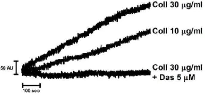
Collagen stimulates aggregation of trout thrombocytes at room temperature (Figure 1), which is approaching the upper temper-ature for trout. The response was similar to that in human platelets [18], although the time course was noticeably slower. The response to 30mg/ml collagen was faster than that to 10mg/ml indicating a similar concentration-response relationship to that seen in human platelets. Aggregation was inhibited by the Src kinase inhibitor dasatinib (5mM) which is effective in whole blood in contrast to many other Src family kinase inhibitors [19]. This demonstrates that collagen stimulates aggregation rather than agglutination.
Trout Thrombocytes Adhere to Collagen
In trout blood, only thrombocytes bind to collagen [2] and thus they can be readily identified from other blood cells. Under static conditions, trout thrombocytes underwent adhesion and lamelli-podia formation on collagen and also on fibrinogen-coated surfaces (Figure 2A). In both cases, spreading, but not adhesion, was completely inhibited by dasatinib (5mM) consistent with a Src kinase mediated activation event. Furthermore, single thrombo-cytes were able to adhere and spread on collagen at shear rates ranging from 100–1000 s21 (Figure 2B). In some instances, clusters of cells could be seen, although the number of cells in each cluster was small and they were present as a monolayer in contrast to the three dimensional aggregates observed in mammalian platelets under flow. The majority of the adherent cells in the clusters had formed lamellipodia which were inhibited in the presence of dasatinib. These results show that thrombocytes are able to adhere and spread on collagen under static and flow conditions, and that activation is blocked by Src family kinase inhibition. This result is similar to that seen in mammalian platelets where adhesion to collagen is not dependent on intracellular signalling as it is mediated by GPIb anda2b1 [20], but signalling through GPVI is required for spreading and aggregation.
G6fL Responds to Collagen through an ITAM in a Src Dependent Manner
The observation that collagen activates thrombocytes through a Src dependent pathway led us to search for a functional equivalent
Figure 1. Trout thrombocytes aggregate to collagen in a Src dependent manner.Anticoagulated whole blood from rainbow trout was diluted 1:1 with normal saline (0.9%) and stirred for 3 min at room temperature in a Multiplate mini-cuvette. Dasatinib (5mM) or DMSO vehicle control was pre-incubated at room temperature for 1 min followed by addition of collagen, 30 or 10mg/ml, and aggregation was monitored for 12 min at room temperature. Aggregation was measured in arbitrary impedance units (AU).
to the mammalian collagen receptor GPVI which is expressed in thrombocytes. The key signalling motif in the GPVI/FcRc-chain complex is an ITAM, which has a conserved sequence of Yxx(L/
I)x6–12Yxx(L/I), and initiates signalling through Src and Syk family
kinases. A novel ITAM-containing receptor, G6fL, has recently been described on thrombocytes in teleost fish, and additionally on Figure 2. Trout thrombocytes adhere to and spread on collagen in a Src dependent manner.A) Trout blood was diluted by a factor of 100 to reduce the number of cells, and then allowed to adhere to either collagen or fibrinogen coated coverslips for 90 min at room temperature, either in the presence of 20mM dasatinib or DMSO vehicle control. B) Anticoagulated whole blood was flowed through collagen coated glass capillary tubes at a range of shear rates (i) or at 100 s21for inhibitor studies (ii). Blood was pre-treated with either 20
mM dasatinib or DMSO vehicle control.
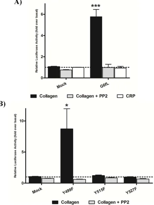
the erythrocytes of crucian carp, although its function is not known [9]. To investigate whether G6fL is a receptor for collagen, we used an NFAT-luciferase reporter assay that has been previously used to monitor activation of GPVI by collagen [21]. Zebrafish G6fL was cloned into an expression vector, and subsequently co-transfected into chicken DT40 B-cells along with an NFAT-luciferase reporter construct. Cells transfected with GPVI and FcR c-chain expression vectors were used as a positive control. Six hours post stimulation luciferase activity was measured as a readout of receptor activation. Collagen (10mg/ml) stimulated a robust signal in the GPVI/FcR c-chain transfected cells, which was blocked by the Src kinase inhibitor PP2 (20mM) (Figure S1), as previously reported [13]. Strikingly, collagen also stimulated a marked increase in luciferase activity in G6fL transfected cells compared to mock transfected cells (Figure 3A) and this response was again blocked by PP2, as expected for an ITAM signalling mechanism.
We used site-directed mutagenesis to determine the signalling motif that underlies activation. G6fL has two distinct signalling motifs, namely a Grb2 binding site, YxN (Y499) and an ITAM
with tyrosines at Y515 and Y527. Mutation of either of the conserved ITAM tyrosines to phenylalanine abrogated signalling to collagen (Figure 3B) thereby demonstrating that both tyrosines are required for signalling, consistent with an ITAM signalling pathway [11]. In contrast, mutation of the conserved tyrosine in the Grb2 binding site (Y499F) had no significant effect on the NFAT-reporter assay demonstrating that this tyrosine is not required for activation. These data demonstrate that G6fL requires an ITAM for signalling in response to collagen.
Activation of GPVI by collagen is mediated through a repeat glycine-proline-hydroxyproline (GPO) motif and synthetic triple helical peptides which contain this motif, known as collagen related peptides (CRP), are powerful activators of the Ig receptor (Figure S1) [22]. However, CRP (3mg/ml) had no effect on G6fL-transfected cells demonstrating that collagen induces activation through a distinct collagen recognition sequence (Figure 3A). Consistent with this, CRP was unable to cause aggregation of trout thrombocytes (not shown) providing further evidence that G6fL recognising a distinct collagen motif. Thus, the motif in collagen for activation of G6fL is distinct to that for GPVI. The snake venom toxin convulxin, which also induces powerful activation of GPVI, also had no effect on G6fL-transfected cells (not shown) as anticipated given that they are encoded by distinct genes.
G6fL is Required for Haemostasis in Zebrafish
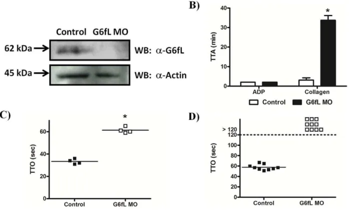
We used a Vivo-morpholino (MO) to knock-down G6fL levels
in vivoin zebrafish to investigate a possible role for the receptor in
the activation of thrombocytes and in haemostasis. A specific antibody to G6fL was raised and shown to detect a band at the predicted molecular weight of 59 kDa and this band was absent in the MO-treated larvae revealing a high degree of knock-down (Figure 4A). Equal loading was confirmed by reprobing the samples for actin. The ability of collagen to stimulate aggregation of thrombocytes in whole blood from zebrafish was measured using a plate-tilt assay, which measures the time taken for a drop of blood to aggregate following the addition of an agonist. The time to aggregation (TTA) by collagen in this assay is increased from less than 2 min in control MO-treated larvae to over 30 min in the G6fL MO-treated larvae, consistent with a critical role of G6fL in mediating thrombocyte activation (Figure 4B). In comparison, the time to aggregation in response to ADP was not altered. It was noted that the blood cell counts were not affected by the knock-down (not shown). These results confirm that G6fL is a collagen
receptor on thrombocytes and that it plays a critical role in mediating thrombocyte activation by the matrix protein.
We used a laser injury assay to investigate the role of G6fL in thrombus formationin vivoin both arteries and veins using
MO-treated larvae. We found that the G6fL MO-MO-treated larvae took longer to occlude their veins compared to controls (Figure 4C) and that they were unable to occlude their arteries within the 120 sec experimental window (Figure 4D). This result confirms a critical role for G6fL in haemostasis in zebrafish.
Discussion
In this study we have sought to understand the molecular mechanism of activation of fish thrombocytes by collagen. Zebrafish, a member of the teleost family, is an increasingly used model organism for mammalian haemostasis due to the speed and inexpensive nature of their use and genetic manipulation via morpholino technology. There are broad similarities which make this comparison possible to mammalian haemostasis such as the observation that thrombocytes will occlude vessels in response to an injury and that they have many orthologs of mammalian receptors. However, there are gaps in our understanding of fish thrombocytes, including the lack of a GPVI ortholog, although they do express an ortholog of the second mammalian collagen receptor, the integrina2b1 complex [9]. The question therefore is whether this is sufficient to mediate platelet adhesion and activation by collagen or whether fish thrombocytes express a second receptor which mediates powerful activation, as is the case in mammalian platelets.
Here, we have shown that trout thrombocytes will respond to collagen in analogous ways to mammalian platelets i.e. via spreading and aggregation, and that this response is mediated by a Src family kinase. This is a new finding as the majority of the current literature on fish thrombocytes focuses on zebrafish, from which only tiny amount of blood can be removed for analysis. The collagen-binding integrina2b1 has been reported at the gene level in teleost fish, and the integrin is known to signal weakly through Src in mammalian platelets although on its own this is insufficient to mediate platelet activation [20]. This raises the question as to whether activation of thrombocytes in fish is mediated solely through integrina2b1 or whether they express a second activation receptor for collagen bearing in mind the absence of an ortholog of GPVI.
In the present study, we show that the novel ITAM-containing receptor G6fL, which has previously been shown to be expressed at the mRNA level in a number of teleost fish [9], functions as a collagen receptor in a cell line reporter assay and in zebrafish thrombocytes. G6fL has a number of similarities to the mamma-lian collagen receptor, the GPVI/FcRc-chain complex, including the presence of extracellular Ig domains (four in the case of G6fL compared with 2 for GPVI) and a cytosolic ITAM which is similar to that on FcRc-chain. In addition, it has sequence similarities to the Ig membrane protein G6f which is expressed on mammalian platelets, including the presence of a cytosolic Grb2-binding sequence, YxN [9,23].
homologs, consistent with their structural differences and genomic locations [9]. This is an example of convergent evolution of the use of an ITAM to rapidly activate thrombocytes/platelets upon binding to collagen.
Definitive evidence that G6fL is a physiologically relevant collagen receptor mediating thrombocyte activation was achieved using anin vivo morpholino knock down approach in zebrafish.
The ex vivo activation of platelets by collagen was markedly
reduced following the knock-down of G6fL, leaving the response to ADP unaffected, and larvae were unable to occlude their arteries in a laser induced injury, while the response in the venous
system was significantly delayed. While there is no direct evidence that the laser injury model exposes collagen, these results give strong indirect evidence that collagen is exposed as the result is reminiscent of that with GPVI or FcR c-chain-deficient mice, which have abrogated platelet activation and marked reduction in thrombus formation, the extent of which is dependent on the
in vivoassay injury [24,25,26]. However, we cannot rule out the
possibility that although collagen is demonstrated here to be an agonist for G6fL, there may be other agonists in vivo which
mediate thrombus formation.
Figure 3. G6fL responds to collagen in a cell line assay through its ITAM.DT40 cells were transfected with wild type zebrafish G6fL (A) or with point mutants of G6fL (Y499F, Y515F and Y527F) (B) and stimulated with either collagen (10mg/ml) or CRP (3mg/ml). Where stated, cells were pre-incubated with 20mM PP2. 6 hrs following stimulation, cells were lysed and luciferase activity was measured as a readout of signalling. Data is expressed as fold over basal (dotted line). Statistical significance was calculated with a Student’s t-test (*P.0.05, ***P.0.005). Similar levels of expression of all G6fL constructs was confirmed by western blotting (not shown).
G6fL is named after its mammalian ortholog G6f, although there are a significant number of differences between the two proteins including the reduced number of Ig domains and the absence of an ITAM in the mammalian protein. Furthermore, G6f does not appear to be a functional collagen receptor despite undergoing tyrosine phosphorylation following collagen or CRP treatment, as it is unable to support adhesion of transfected cells to a collagen surface or allow signalling to collagen of transfected cells [23]. The most striking similarities between the two proteins is the presence of the cytoplasmic Grb2-binding motif which suggests a novel pathway of regulation of Grb2-effector proteins including p42/44 MAP kinases, which in mammals play a critical role in platelet formation in and are implicated in the regulation of several platelet responses, including phosphorylation of cytosolic phos-pholipase A2under certain conditions [27]. We hypothesise that if
the mammalian receptor plays a signalling role in platelets it is either downstream of either a novel agonist or downstream of the collagen receptor GPVI.
In summary, in the present study, we have demonstrated the presence of a novel signalling receptor for collagen in teleost fish, G6fL, which plays an analogous role to mammalian GPVI. The structural features and gene location of G6fL and GPVI suggests that they are unlikely to be orthologs and it is therefore possible that these disparate receptors independently evolved to respond to collagen and signal through an ITAM allowing the rapid signalling required for thrombocyte/platelet activation and rapid
haemosta-sis, an example of convergent evolution. Furthermore, as the mammalian ortholog of G6fL appears to not be a collagen receptor, this demonstrates divergent evolution.
Supporting Information
Figure S1 GPVI responds to collagen and CRP in a cell line assay.DT40 cells were transfected with GPVI/FcRcand stimulated with either collagen (10mg/ml) or CRP (3mg/ml). Where stated, cells were pre-incubated with 20mM PP2. 6 hrs following stimulation, cells were lysed and luciferase activity was measured as a readout of signalling. Data is expressed as fold over basal (dotted line). Statistical significance was calculated with a Student’s t-test (*P.0.05, **P.0.01).
(TIF)
Acknowledgments
The authors would like to thank Drs. Sonia Severin and Steve Thomas for assistance with flow-adhesion studies.
Author Contributions
Conceived and designed the experiments: CEH PJ SPW. Performed the experiments: CEH UPR ML SE JMD. Analyzed the data: CEH UPR JMD PJ SPW. Wrote the paper: CEH SPW.
Figure 4. Zebrafish G6fL is required for haemostasis.A) A novela-zebrafish G6fL antibody was used to determine knock-down of G6fL in morpholino (MO) treated larvae. 48 hrs post injection, 3 dpf larvae were lyophilised and lysed. Proteins were separated by SDS-PAGE and Western blotted with the antibody. The membrane was subsequently stripped and reprobed with ana-actin antibody to confirm equal loading. B) Control and G6fL MO treated adult fish were bled, and the blood used for a plate tilt aggregation assay. Time to aggregation (TTA) was measured following addition of either ADP (20mM) or collagen (100mg/ml). C) Control and G6fL MO treated larvae were immobilised on a microscope stage and then injured with a laser in the vessel wall of either veins or arteries (D). Time to occlusion (TTO) of vessels was measured for up to 2 min at which time the experiment was ceased. Statistical significance was calculated with a Student’s t-test (*P.0.05).
References
1. Gibbins J, Asselin J, Farndale R, Barnes M, Law CL, et al. (1996) Tyrosine phosphorylation of the Fc receptor gamma-chain in collagen-stimulated platelets. J Biol Chem 271: 18095–18099.
2. Kollner B, Fischer U, Rombout JH, Taverne-Thiele JJ, Hansen JD (2004) Potential involvement of rainbow trout thrombocytes in immune functions: a study using a panel of monoclonal antibodies and RT-PCR. Dev Comp Immunol 28: 1049–1062.
3. Kim S, Carrillo M, Kulkarni V, Jagadeeswaran P (2009) Evolution of primary hemostasis in early vertebrates. PLoS One 4: e8403.
4. Jagadeeswaran P, Carrillo M, Radhakrishnan UP, Rajpurohit SK, Kim S (2011) Laser-induced thrombosis in zebrafish. Methods Cell Biol 101: 197–203. 5. Jagadeeswaran P, Gregory M, Day K, Cykowski M, Thattaliyath B (2005)
Zebrafish: a genetic model for hemostasis and thrombosis. J Thromb Haemost 3: 46–53.
6. Jagadeeswaran P, Cykowski M, Thattaliyath B (2004) Vascular occlusion and thrombosis in zebrafish. Methods Cell Biol 76: 489–500.
7. Jagadeeswaran P, Sheehan JP, Craig FE, Troyer D (1999) Identification and characterization of zebrafish thrombocytes. Br J Haematol 107: 731–738. 8. Guo P, Hirano M, Herrin BR, Li J, Yu C, et al. (2009) Dual nature of the
adaptive immune system in lampreys. Nature 459: 796–801.
9. Ohashi K, Takizawa F, Tokumaru N, Nakayasu C, Toda H, et al. (2010) A molecule in teleost fish, related with human MHC-encoded G6F, has a cytoplasmic tail with ITAM and marks the surface of thrombocytes and in some fishes also of erythrocytes. Immunogenetics 62: 543–559.
10. Baba TW, Giroir BP, Humphries EH (1985) Cell lines derived from avian lymphomas exhibit two distinct phenotypes. Virology 144: 139–151. 11. Hughes CE, Pollitt AY, Mori J, Eble JA, Tomlinson MG, et al. (2010) CLEC-2
activates Syk through dimerization. Blood 115: 2947–2955.
12. Hughes CE, Auger JM, McGlade J, Eble JA, Pearce AC, et al. (2008) Differential roles for the adapters Gads and LAT in platelet activation by GPVI and CLEC-2. J Thromb Haemost 6: 2152–2159.
13. Mori J, Pearce AC, Spalton JC, Grygielska B, Eble JA, et al. (2008) G6b-B inhibits constitutive and agonist-induced signaling by glycoprotein VI and CLEC-2. J Biol Chem 283: 35419–35427.
14. Fuller GL, Williams JA, Tomlinson MG, Eble JA, Hanna SL, et al. (2007) The C-type lectin receptors CLEC-2 and Dectin-1, but not DC-SIGN, signal via a novel YXXL-dependent signaling cascade. J Biol Chem 282: 12397–12409. 15. Tomlinson MG, Kane LP, Su J, Kadlecek TA, Mollenauer MN, et al. (2004)
Expression and function of Tec, Itk, and Btk in lymphocytes: evidence for a unique role for Tec. Mol Cell Biol 24: 2455–2466.
16. Kim S, Radhakrishnan UP, Rajpurohit SK, Kulkarni V, Jagadeeswaran P (2010) Vivo-Morpholino knockdown of alphaIIb: A novel approach to inhibit thrombocyte function in adult zebrafish. Blood Cells Mol Dis 44: 169–174. 17. Gregory M, Hanumanthaiah R, Jagadeeswaran P (2002) Genetic analysis of
hemostasis and thrombosis using vascular occlusion. Blood Cells Mol Dis 29: 286–295.
18. Dawood BB, Wilde J, Watson SP (2007) Reference curves for aggregation and ATP secretion to aid diagnose of platelet-based bleeding disorders: effect of inhibition of ADP and thromboxane A(2) pathways. Platelets 18: 329–345. 19. Nash CA, Severin S, Dawood BB, Makris M, Mumford A, et al. (2010) Src
family kinases are essential for primary aggregation by G(i) -coupled receptors. J Thromb Haemost 8: 2273–2282.
20. Inoue O, Suzuki-Inoue K, Dean WL, Frampton J, Watson SP (2003) Integrin alpha2beta1 mediates outside-in regulation of platelet spreading on collagen through activation of Src kinases and PLCgamma2. J Cell Biol 160: 769–780. 21. Tomlinson MG, Calaminus SD, Berlanga O, Auger JM, Bori-Sanz T, et al.
(2007) Collagen promotes sustained glycoprotein VI signaling in platelets and cell lines. J Thromb Haemost 5: 2274–2283.
22. Smethurst PA, Onley DJ, Jarvis GE, O’Connor MN, Knight CG, et al. (2007) Structural basis for the platelet-collagen interaction: the smallest motif within collagen that recognizes and activates platelet Glycoprotein VI contains two glycine-proline-hydroxyproline triplets. J Biol Chem 282: 1296–1304. 23. Garcia A, Senis YA, Antrobus R, Hughes CE, Dwek RA, et al. (2006) A global
proteomics approach identifies novel phosphorylated signaling proteins in GPVI-activated platelets: involvement of G6f, a novel platelet Grb2-binding membrane adapter. Proteomics 6: 5332–5343.
24. Munnix IC, Strehl A, Kuijpers MJ, Auger JM, van der Meijden PE, et al. (2005) The glycoprotein VI-phospholipase Cgamma2 signaling pathway controls thrombus formation induced by collagen and tissue factor in vitro and in vivo. Arterioscler Thromb Vasc Biol 25: 2673–2678.
25. Kalia N, Auger JM, Atkinson B, Watson SP (2008) Critical Role of FcR gamma-Chain, LAT, PLCgamma2 and Thrombin in Arteriolar Thrombus Formation upon Mild, Laser-Induced Endothelial Injury In Vivo. Microcirculation 15: 325–335.