the N-Terminal Region
Anand Srivastava1,2,3,4,5, Ste´phane Gangnard1,2,3,4,5, Se´bastien Dechavanne1,2,3,4,5, Farroudja Amirat1,2, Anita Lewit Bentley1,2, Graham A. Bentley1,2, Benoıˆt Gamain3,4,5*
1Institut Pasteur, Unite´ d’Immunologie Structurale, F-75015 Paris, France,2CNRS, URA2185, F-75015 Paris, France,3INSERM, U665, F-75739 Paris, France,4Institut National de la Transfusion Sanguine, F-75739 Paris, France,5Univ Paris Diderot, Sorbonne Paris Cite´, Prote´ines de la membrane e´rythrocytaire, UMR-S665, F-75739 Paris, France
Abstract
Var2CSA, a key molecule linked with pregnancy-associated malaria (PAM), causes sequestration ofPlasmodium falciparum infected erythrocytes (PEs) in the placenta by adhesion to chondroitin sulfate A (CSA). Var2CSA possesses a 300 kDa extracellular region composed of six Duffy-binding like (DBL) domains and a cysteine-rich interdomain region (CIDRpam) module. Although initial studies implicated several individual var2CSA DBL domains as important for adhesion of PEs to CSA, new studies revealed that these individual domains lack both the affinity and specificity displayed by the full-length extracellular region. Indeed, recent evidence suggests the presence of a single CSA-binding site formed by a higher-order domain organization rather than several independent binding sites located on the different domains. Here, we search for the minimal binding region within var2CSA that maintains high affinity and specificity for CSA binding, a characteristic feature of the full-length extracellular region. Accordingly, truncated recombinant var2CSA proteins comprising different domain combinations were expressed and their binding characteristics assessed against different sulfated glycosamino-glycans (GAGs). Our results indicate that the smallest region within var2CSA with similar binding properties to those of the full-length var2CSA is DBL1X-3X. We also demonstrate that inhibitory antibodies raised in rabbit against the full-length DBL1X-6etarget principally DBL3X and, to a lesser extent, DBL5e. Taken together, our results indicate that efforts should
focus on the DBL1X-3X region for developing vaccine and therapeutic strategies aimed at combating PAM.
Citation:Srivastava A, Gangnard S, Dechavanne S, Amirat F, Lewit Bentley A, et al. (2011) Var2CSA Minimal CSA Binding Region Is Located within the N-Terminal Region. PLoS ONE 6(5): e20270. doi:10.1371/journal.pone.0020270
Editor:Silvia N. Moreno, Univ. Georgia, United States of America
ReceivedFebruary 24, 2011;AcceptedApril 17, 2011;PublishedMay 19, 2011
Copyright:ß2011 Srivastava et al. This is an open-access article distributed under the terms of the Creative Commons Attribution License, which permits unrestricted use, distribution, and reproduction in any medium, provided the original author and source are credited.
Funding:This work was supported by an ATIP-AVENIR grant from the Institut National de la Sante´ et de la Recherche Me´dicale and Sanofi-Aventis (BG). The research leading to these results has received funding from the European Community’s Seventh Framework Programme Grant ([FP7/2007–2013]) (to GAB and BG) under Grant agreement 201222. AS was supported by a grant from the Fondation pour la Recherche Me´dicale (FRM). The funders had no role in study design, data collection and analysis, decision to publish, or preparation of the manuscript.
Competing Interests:Although Sanofi-Aventis is supporting the INSERM program ATIP-Avenir, the authors have declared that this did not alter their adherence to all the PLoS ONE policies on sharing data and materials.
* E-mail: [email protected]
Introduction
Pregnancy-associated malaria (PAM) causes adverse pregnancy outcomes, including anemia and hypertension in first-time pregnant women, and low birth weight due to premature delivery and fetal growth restriction, which are associated with a higher risk of fetal and neonate morbidity and mortality [1,2]. Complications arising from PAM have been attributed to massive accumulation ofPlasmodium falciparum-infected erythrocytes (PEs) in the placenta mediated by adhesion to chondroitin sulfate A (CSA) [3]. Significantly, after one or two pregnancies, women acquire transcendent antibodies recognizing placental PEs from different geographic regions that inhibit placental adhesion, thus correlating with protection against malaria [4,5].
Several lines of evidence support var2CSA, a member of the Plasmodium falciparumErythrocyte Membrane Protein 1 (PfEMP1) adhesins encoded by the var gene family [6,7,8], as the leading PAM vaccine candidate. Indeed, var2CSA is the only var gene transcribed in CSA-binding laboratory isolates and placental PEs [9,10,11,12,13,14]. Importantly, gene disruption studies have clearly demonstrated that var2CSA is the primary var gene
placental sequestration by avidity effects. However, more recent studies revealed that these individual domains have low affinity to CSA and that they can also bind to other sulfated glycosamino-glycans (GAGs), in some cases with higher affinity than to CSA [28]. Moreover, while most of these individual domains elicited antibodies that reacted with CSA-binding parasite isolates, only few induced an adhesion-blocking response [29,30,31,32,33], suggesting that individual domains are not sufficient to exhibit the full binding phenotype.
Recently, we and others have shown that the complete extracellular region of PfEMP1 possesses a higher-order organi-sation that is determined by well defined inter-domain contacts and is a prerequisite for creating the native high affinity
CSA-specific binding site [34,35]. Indeed, unlike the individual DBL domains, the full-length var2CSA extracellular region binds with high affinity and specificity to CSA. In addition, small angle X-ray scattering and analytical ultracentrifugation experiments showed that var2CSA forms a more compact structure than expected from an extended ‘‘beads-on-a-string’’ model [34], favoring the view that a single high affinity CSA-binding site is formed by the association of several var2CSA domains rather than an ensemble of independent sites residing on individual domains.
Although the full-length protein possesses binding properties characteristic of placental PEs, its size and polymorphism pose a challenge for vaccine development. There is therefore a need to identify which domain or combination of domains is required to form the high affinity CSA-binding site. Such knowledge would provide a rational basis for accelerating vaccine and therapeutic developments aimed at blocking the adhesion of CSA-binding parasites to the placenta. In order to address this question, we expressed various var2CSA DBL domains and multi-domain proteins in HEK293 andE. coliheterologous expression systems and tested their binding properties to CSA and other sulfated GAGs. In addition, we used these recombinant proteins to map the domains recognized by the inhibitory IgG raised in rabbits against the full-length extracellular region of var2CSA using antibody depletion and elution experiments.
Our results suggest that the high affinity CSA-binding site lies within the DBL1X-3X segment of var2CSA and that DBL3X and, to some extent, DBL5eare the principal targets of the inhibitory antibodies. Taken together, our results indicate that DBL3X is an important target for inhibitory antibodies and that strategies aimed at blocking PE adhesion to CSA should focus on the N-terminal region of var2CSA. These results present an important new step towards the design of vaccine and therapeutic strategies to combat PAM.
Methods
Ethics statement
All animal work was conducted according to relevant national and international guidelines. Immunizations were performed by a custom vendor (Proteogenix, France), and all animal experiments were approved and conducted in accordance with the Institut Pasteur and Proteogenix Biosafety Committees. Animals were housed under controlled laboratory conditions by qualified personnel who were licensed by the French Agricultural Ministry (agreement B 75 15-08 dated May 22, 2008). All researchers performing animal experiments in this study were directly responsible for the experimental protocols and had obtained individual licenses from the French Ministry of Agriculture.
Expression and Purification of Recombinant Protein
(i) HEK 293-F cell Recombinant Protein Expression and Purification. Synthetic var2csa genes for 3D7-DBL1X-3X (residues 59–1577) and 3D7-DBL4e-6e (residues 1578–2630) (accession PFL0030c) were designed with optimized codons for human cell expression, as previously described [34], and were cloned into the pTT3 vector [36]. pTT3-3D7-DBL1X-6e (residues 59–2630) were prepared by incorporation of 3D7-DBL4e-6e into pTT3-3D7-DBL1X-3X using compatible restriction sites. Genes encoding 3D7-DBL1X (residues 59–430) and 3D7-DBL1X-CIDR (residues 59–1209) were PCR amplified from pTT3-3D7-DBL1X-3X and cloned into the pTT3 vector using EcoRI/HindIII restriction sites for expression in FreeStyle 293-F cells, as previously described (Invitrogen) [34]. The oligonucleotide primers used for amplification were as follows
Figure 1. Various var2CSA recombinant proteins expressed in HEK293 cells andE. coli.(A). Schematic view of the var2CSA domain organization and sequence limits of the recombinant domains studied (3D7-DBL1X, 3D7-DBL2X, FCR3-DBL3X, 3D7-DBL5e, 3D7-DBL1X-2X,
FCR3-DBL3X-4e, 3D7-DBL1X-CIDR, 3D7-DBL1X-3X, 3D7-DBL1X-4e,
3D7-DBL1X-5e and 3D7-DBL1X-6e). Var2CSA comprises six DBL domains
(DBL1X to DBL6e), a CIDRpam domain and three inter-domain regions
(INT1-3) in the extracellular region, together with a trans-membrane segment and acidic C terminus sequence (ATS). DBL1X, DBL2X, DBL3X, DBL4e, DBL5e and DBL6e are shown in green; CIDR in orange; N-terminal sequence (NTS) and inter-domain regions (INT) in grey; the trans-membrane and ATS regions in blue. The length of each bar corresponds to the domain size. (B). Purification of var2CSA derived proteins expressed in HEK293 andE. coli. Nu-SDS-PAGE Precast 4–12% Bis-Tris gel under nonreducing (B) and reducing (C) conditions was loaded with purified recombinant proteins. Lane 1: Marker, lane 2: 3D7-DBL1X, lane 3: 3D7-DBL2X, lane 4: FCR3-DBL3X, lane 5: 3D7-DBL5e, lane 6: 3D7-DBL1X-2X, lane 7: FCR3-DBL3X-4e, lane 8: 3D7-DBL1X-CIDR, lane
9: 3D7-DBL1X-3X, lane 10: 3D7-DBL1X-4e, lane 11: 3D7-DBL1X-5eand lane 12: 3D7-DBL1X-6e. Proteins were visualized with Coomassie blue.
(restriction sites are represented in lower case): 3D7-DBL1X: DBL1XF (59-AGC gaattc ATG GAG ACC GAC ACC C-39) and 3D7-DBL1XR (59-CAC aagctt CCC GCT TTC ATT GCA GTT GCA CAC-39); 3D7-DBL1X-CIDR: 3D7-DBL1X-CIDRF (59-AGC gaattc ATG GAG ACC GAC ACC C-39) and 3D7-DBL1X-CIDRR (59-CTC aagctt GCT GGT CTC AGA GCT TTT CAT C -39). Genes encoding 3D7-DBL1X-4e(residues 59– 1905) and 3D7-DBL1X-5e(residues 59–2286) were obtained by PCR amplification of 3D7-DBL4e and 3D7-DBL4e-5e from pTT3-3D7-DBL1X-6e prior to cloning into pTT3-DBL1X-3X, using HindIII/XbaI restrictions sites. The oligonucleotide primers used to amplify were as follows (restriction sites are represented in lower case): DBL4eF (59- AAC aagctt CTG TGC CAC GAG AAA GG -39) and DBL4eR (59-GTT tctaga GTT CTT GCA GCT ACA GCA GAT G-39), and DBL5eR (59- CTT tctaga GTC ATT GAA GCC GCA GGG GCA C-39) primers, respectively.
The pTT3 vector contains an N-terminal murine Igk-chain leader sequence and a C-terminal hexa-His tag. FreeStyle 293-F cells (Invitrogen) were grown in Freestyle 293 serum-free expression medium and transfected with the pTT3 vector containing the appropriate gene, as previously described [30]. Cells were centrifuged 96 h post-transfection and the culture medium was harvested, filtered by a 0.22mm filter; the
supernatants were concentrated twenty times using a 10 kDa cut-off Vivaflow 200 System (Vivasciences). All proteins were purified on a HisTrap Fast Flow Ni-affinity column (GE Healthcare). Proteins were further purified by ion exchange chromatography on an SP Sepharose column (GE Healthcare) followed by gel filtration chromatography on a Superdex 200 16/ 60 column (GE Healthcare) in 50 mM sodium phosphate buffer pH 6.8, 200 mM NaCl. Proteins were then concentrated using Macro- and Microsep concentrators (Millipore).
(ii) Escherichia coli Recombinant Protein Expression and Purification. The gene encoding FCR3-DBL3X (residues 1218–1577), cloned into a modified pET15b vector, was a kind gift from Dr. Matthew K. Higgins; expression was carried out as previously described [37]. The gene encoding 3D7-DBL5e was PCR amplified and cloned into the pET15b vector between the NdeI/XhoI restriction sites, in frame with the N-terminal hexa-His tag, using the following oligonucleotide primers DBL5eF (59 -ACTGGCAG catatg TCC AAG ATG AAG GTG TGC GAC C-39) and DBL5eR (59-GCAG ctcgag TTA CAT GTC ATT GAA GCC GCA GGG G -39). The gene encoding FCR3-DBL3X-4e was PCR amplified and cloned into a modified pET21b vector using the following oligonucleotide primers FCR3-DBL3XF (59 -ACTGGCAG gctagc ATG AGC GAG ACC AGC TGC GAC C-39) and FCR3-DBL4eR (59-ACTGGCAG gcggccgc TCA ATG ATG ATG ATG ATG ATG CAT CTT GTT CAG CAC CTC GTT C -39). Similarly, genes encoding DBL2X and 3D7-DBL1X-2X were PCR amplified and cloned into a modified pET21b vector in frame with a C-terminal hexa-His tag using the following oligonucleotide primers DBL2XF (59- GCAG gaattc CAG GAT TTC CTG CGC ATT CTG-39), DBL2XR (59-GGCAG gtcgac TTA ATG ATG ATG ATG ATG ATG ATT GGT AGG GAT TTT GCA CTG G -39) and DBL1X-2XF (59 -GCAG gaattc TGT AAG ATC ACA GTG AAC CAC AGC GAT TCC GGC ACA AAT GAT CCT TG -39) and DBL1X-2XR (59-GGCAG gtcgac TTA ATG ATG ATG ATG ATG ATG ATT GGT AGG GAT TTT GCA CTG G -39).
All proteins were expressed either in the Rosetta-Gami, Origami B or SHuffle strains of E. coli (Novagen) as soluble proteins at 20uC for 20 h after IPTG induction. Post-induction cells were centrifuged, resuspended in 20 mM Tris-HCl, 150 mM NaCl, pH 7.5 and lysed with an Emulsiflex homogeniser (Avestin).
The proteins were purified using a metal affinity column (TALON, Clontech). All proteins were further purified using a heparin affinity column (GE Healthcare) and were eluted in 20 mM Tris-HCl pH 7.5, 1 M NaCl, followed by gel filtration (Sephedex 75 16/60, GE Healthcare) in 20 mM Tris-HCl 150 mM NaCl, pH 7.5.
ELISA binding assays of recombinant proteins to various sulfated glycosaminoglycans
ELISA binding assays were performed as previously described [34]. Briefly, ELISA plates were coated overnight at 4uC with different sulfated glycosaminoglycans (GAG): 5mg/mL for decorin (Sigma, D8428); 50mg/mL for chondroitin sulfate A (CSA)
(Sigma, C8529), chondroitin sulfate C (CSC) (Seikagaku, 400670) and heparan sulfate (HS) (Sigma, H7640) in PBS (Gibco, NaCl 155 mM pH 7.2), using 100mL per well. BSA at 1% in PBS was
used as background measurement. After coating, the wells were blocked with 150mL of dilution buffer per well (PBS 1% BSA, 0.05% Tween20) for 1 h at 37uC. After removal of the blocking solution, each recombinant protein (3D7-DBL1X, 3D7-DBL2X, FCR3-DBL3X, 3D7-DBL5e, 3D7-DBL1X-2X, FCR3-DBL3X-4e, 3D7-DBL1X-CIDR, 3D7-DBL1X-3X, 3D7-DBL1X-4e, 3D7-DBL1X-5e and 3D7-DBL1X-6e) at serial dilutions of 0.3125– 20mg/mL in the dilution buffer, was added per well and
incubated for 1 h at 37uC with gentle shaking. After washing three times with PBST (PBS containing 0.05% Tween 20), 100mL
anti-His HRP conjugated antibody (diluted 1/2000 in dilution buffer) was added to each well and incubated for 1 h at 37uC. After washing three times with PBST, the interaction was quantified with TMB (3,39,5,59-tetramethylbenzidine) substrate (Biorad) using 100mL per well for 20 min or until saturation was reached. Absorbance was measured at 655 nm.
Adhesion inhibition assays of recombinant proteins to CSPG
Inhibition assays were performed using a protocol similar to that described above for ELISA, with decorin coated on the plate. Recombinant proteins (3D7-DBL1X-2X, 3D7-DBL1X-CIDR, 3D7-DBL1X-3X, 3D7-DBL1X-4e, 3D7-DBL1X-5e and 3D7-DBL1X-6e) at a concentration of 1mg/mL were premixed with
increasing amounts of BSA, CSA, CSC, or HS (0.156–100mg/
mL) and incubated for 30 min at room temperature with gentle shaking before addition to the coated ELISA plate.
Surface Plasmon Resonance
Interaction between the recombinant proteins and human placental CSPG was studied by surface plasmon resonance (SPR) using a BiacoreH 2000 system (GE Healthcare) as previously described [34]. Human placental CSPG (MR4 Reagents Re-source) was covalently coupled via primary amino groups of the protein moiety to the sensor chip (CM5 chip, GE Healthcare) surface using amine coupling kit (GE Healthcare) as described previously [38]. The amount of immobilized CSPG corresponded to 240 response units (RU). A separate flow channel on the same sensor chip without CSPG was used for control runs. For all SPR measurements, the recombinant domains were dialyzed against PBS buffer (Gibco), 0.005% P20 (GE Healthcare), and centrifuged immediately before the runs to minimize possible effects from nonspecific aggregation. The association was monitored by injecting different concentrations of the DBL analytes at 25uC with the flow rate of 20mL/min for 300 s to achieve steady-state
were corrected for nonspecific binding by subtraction of control curves obtained from injection of the corresponding protein through the blank flow channel. KD was determined from the concentration dependence of steady-state SPR response (after corrections for nonspecific binding) using the Biacore BIAevalua-tion 3.1 software (Biacore AB).
Animal immunization
Immunizations with 3D7-DBL1X-6eVAR2CSA recombinant protein were performed at Proteogenix, France, according to animal immunization guidelines. Briefly, a New Zealand white rabbit was immunized with recombinant protein in TiterMax Gold Adjuvant (Sigma) intradermally with 50mg of immunogen
for first immunization and subcutaneously with 25mg of
immunogen in subsequent injections. IgG were purified from rabbit plasma using Hitrap protein G (GE Healthcare) according to manufacturer’s instructions.
IgG purification and depletion
The recombinant proteins (3D7-DBL1X, 3D7-DBL2X, FCR3-DBL3X, 3D7-DBL5e, 3D7-DBL6e, DBL1X-2X, 3D7-DBL1X-CIDR, 3D7-DBL1X-3X, FCR3-DBL3X-4e, 3D7-DBL1X-4e, 3D7-DBL1X-5e and 3D7-DBL1X-6e) were coated on M-280 tosyl-activated Dynabeads (Invitrogen) according to manufacturer’s instructions. Briefly, 66mL of M-280
tosyl-activated Dynabeads were washed three times in 900mL PBS,
pH 7.4, and resuspended in the same buffer. Beads were then mixed with 20mg of each recombinant protein containing 1.2 M
ammonium sulfate and incubated for 12 h at 37uC with slow rocking. Following the incubation, the beads were washed five times with 1 mL PBS, incubated 1 h at room temperature with PBS, 0.5% BSA and washed 2 times in PBS and stored at 4uC in the same buffer.
For antibody depletion, 50mL of purified rabbit anti
3D7-DBL1X-6eIgG at 160mg/mL diluted in PBS were incubated for 1 h at 37uC with the different protein-coated beads. Unbound antibodies were collected and beads were washed three times with 1 mL PBS prior to elution of bound antibodies using 50mL 0.1 M glycine pH 2.5. Eluted fractions were subsequently neutralized with Tris-HCl 1 M pH 8.0.
Inhibition assays were performed using a protocol similar to that described above for ELISA using decorin-coated plates. Recom-binant proteins at a concentration of 0.25mg/mL were premixed
with either the unbound antibody fractions or the eluted antibody fractions, and incubated for 30 min at room temperature with gentle shaking before addition to the coated ELISA plate.
Results
Recombinant DBL domains were successfully purified to high purity
Var2CSA is a 350 kDa protein possessing a large extracellular region, a transmembrane region and a short, conserved intracel-lular domain (Fig. 1A). While individual domains of var2CSA have been expressed and purified in various heterologous expression systems [29,30,33,39,40], it has been difficult to produce multiple PfEMP1 domains due to their large size and the presence of many cystine bridges. Recently, we reported the expression of the complete extracellular region of var2CSA using the human embryonic kidney cell (HEK293) heterologous expression system [34]. In the present study, we attempted to express in the HEK293 heterologous expression system various single and multiple domain recombinant proteins from the 3D7 var2CSA ortholog (3D7-DBL1X, 3D7-DBL1X-CIDR, 3D7-DBL1X-3X, 3D7-DBL1X-4e,
3D7-DBL1X-5e and 3D7-DBL1X-6e). In addition, single (3D7-DBL2X, FCR3-DBL3X, 3D7-DBL5e) and double (3D7-DBL1X-2X, FCR3-DBL3X-4e) DBL domains were expressed as soluble proteins in E. coli. All these proteins were successfully expressed and purified to better than 95% homogeneity (Fig. 1B). Some domains, such as DBL4e, could not be obtained due to poor expression yields. In SDS-PAGE under reducing conditions, all proteins migrated according to their expected molecular weights: 3D7-DBL1X (45 kDa), 3D7-DBL2X (53 kDa), FCR3-DBL3X (43 kDa), 3D7-DBL5e (37 kDa), 3D7-DBL1X-2X (105 kDa), FCR3-DBL3X-4e (86 kDa), 3D7-DBL1X-CIDR (134 kDa), 3D7-DBL1X-3X (176 kDa), 3D7-DBL1X-4e (215 kDa), 3D7-DBL1X-5e(259 kDa), and 3D7-DBL1X-6e(300 kDa) (Fig. 1C), while under non-reducing conditions a shift in the migration of all proteins confirmed the presence of disulfide bridges (Fig. 1B). N-terminal sequencing and Western blots using anti-His antibodies confirmed that the proteins were not degraded (data not shown). The yields after purification varied from 0.1 to 20 mg per litre of culture media: 3D7-DBL1X (1 mg/L), 3D7-DBL2X (5 mg/L), FCR3-DBL3X (20 mg/L), 3D7-DBL5e(1 mg/L), 3D7-DBL1X-2X (1 mg/L), FCR3-DBL3X-4e (3 mg/L), 3D7-DBL1X-CIDR (0.25 mg/L), 3D7-DBL1X-3X (0.25 mg/L), 3D7-DBL1X-4e (0.15 mg/L), 3D7-DBL1X-5e(0.15 mg/L), and 3D7-DBL1X-6e (0.5 mg/L).
Minimal CSA-binding region lies within 3D7-DBL1X-3X
The adhesion properties of the different recombinant proteins were tested by direct ELISA using plates coated with different sulfated glycosaminoglycans (GAG), including chondroitin sulfate A (CSA), chondroitin sulfate C (CSC), decorin (a proteoglycan containing CSA), and heparan sulfate (HS) (Fig. 2A–J). Among the single DBL domains tested 3D7-DBL1X did not bind to any of the sulfated GAGs (data not shown) and 3D7-DBL3X bound weakly to HS and CSA at higher concentrations (Fig. 2B), like DBL6eas previously reported [34]. Interestingly, DBL2X and 3D7-DBL5ebound to decorin and CSA in a dose-dependent manner but these proteins also bound to BSA and heparan sulfate at higher concentrations (Fig. 2A and C), indicating some non-specific interactions. For 3D7-DBL2X, a much stronger signal was observed in binding to decorin in comparison to CSA, reaching saturation at very low concentrations. Binding properties of the double domain recombinant proteins were also tested. While DBL1X-2X bound with high affinity to decorin, the apparent affinity to CSA was lower (Fig. 2E). DBL3X-4e bound to the different GAGs in a weak, non-specific manner (Fig. 2D). Similarly, recombinant proteins possessing more than two domains (CIDR, 3X, 3D7-DBL1X-4e and 3D7-DBL1X-5e) were tested for their binding behavior with CSA and were compared with the full-length 3D7-DBL1X-6e protein. All multiple domain proteins bound to decorin and CSA in a dose-dependent manner with various degrees of affinity, except for DBL1X-4e which did not bind to CSA (Fig. 2F–J). DBL1X-3X and DBL1X-5eexhibited an affinity and specificity quite similar to the full-length 3D7-DBL1X-6eprotein while 3D7-DBL1X-CIDR had lower affinity to CSA and decorin.
Our results indicate that the high affinity CSA-binding site lies within the DBL1X-3X recombinant protein.
3D7-DBL1X-4e3D7-DBL1X-5eand 3D7-DBL1X-6e) to decorin was inhibited by CSA in a dose-dependent manner. Significant inhibition with the other sulfated GAGs, was also observed at higher concentrations for DBL1X-CIDR, and 3D7-DBL1X-4e (Fig. 3B and D). However, the binding of 3D7-DBL1X-2X and 3D7-DBL1X-3X (Fig. 3A and C) to decorin was specifically and efficiently inhibited by CSA and most closely
mimicked the binding inhibition of DBL1X-5e and DBL1X-6e (Fig. 3E and F).
Taken together, our results indicate that the minimal region within var2CSA for specific and high affinity binding to CSA lies within DBL1X-3X and that the binding site is mainly centred on the DBL2X domain. Although 3D7-DBL2X binds with high affinity to decorin and CSA, it shows poor specificity as it also
Figure 3. Competitive inhibition of recombinant DBL domains binding to decorin using various glycosaminoglycans.Recombinant proteins (A) 3D7-DBL1X-2X, (B) 3D7-DBL1X-CIDR, (C) 3D7-DBL1X-3X, (D) 3D7-DBL1X-4e, (E) 3D7-DBL1X-5eand (E) 3D7-DBL1X-6eat 1mg/mL were premixed with increasing amounts of BSA (%) or glycosaminoglycans, 0.156–100mg/mL of CSA (6), CSC (e) or HS (n) and incubated in plates previously coated with decorin.
doi:10.1371/journal.pone.0020270.g003
Figure 2. Binding of recombinant DBL domains from 3D7 and FCR3-DBL1X-6eto different glycosaminoglycans.ELISA-based direct binding assay was performed to identify the specificity of (A) 3D7-DBL2X, (B) FCR3-DBL3X, (C) 3D7-DBL5e, (D) FCR3-DBL3X-4e, (E) 3D7-DBL1X-2X, (F) 3D7-DBL1X-CIDR, (G) 3D7-DBL1X-3X, (H) 3D7-DBL1X-4e, (I) 3D7-DBL1X-5eand (J) 3D7-DBL1X-6eto different sulfated glycosaminoglycans. Increasing
concentrations of recombinant proteins at serial dilutions of 0.31–20mg/mL were added to wells previously coated with BSA (n) or different glycosaminoglycans: decorin (#), CSA (%), CSC (e), HS (6).
binds to other GAGs at higher concentrations (Fig. 2A). Thus, with our observations that DBL1X does not bind to any GAG (data not shown) and that DBL3X binds weakly to HS and CSA (Fig. 2B), we propose that the main CSA-binding residues are likely to be present on DBL2X and that the surrounding domains, DBL1X, CIDR and DBL3X, contribute to the affinity and specificity of this interaction by a higher-order organisation of the multidomain protein.
N-terminal multidomain constructs bind placental CSPG with high affinity
The binding of the recombinant proteins DBL2X,
3D7-DBL1X-2X, 3D7-DBL1X-CIDR, 3D7-DBL1X-3X,
3D7-DBL1X-5eand 3D7-DBL1X-6eto placental CSPG was examined more quantitatively by real-time SPR. Data from the kinetic association and dissociation curves did not fit a simple 1:1 binding model, thus precluding the use of kinetic constants konand koffto estimate the binding constant KD. We therefore performed concentration-dependent, steady-state SPR experiments to deter-mine the constants KD, which were calculated from the variation of the steady-state binding response with protein concentration, as shown for 3D7-DBL1X-2X (Fig. 4).
Results are summarized in Table 1, which also includes previously published constants for other var2CSA-derived recom-binant proteins for comparison. Not all domains could be tested since the expression yields of some were not sufficient for SPR experiments.
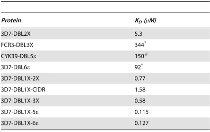
The calculated affinity for 3D7-DBL1X-5ewas similar to that of 3D7-DBL1X-6eindicating that DBL6ehas little or no role in the adhesion process. The affinities of DBL1X-2X and 3D7-DBL1X-3X for human placental CSPG are,5 times lower than that of 3D7-DBL1X-6e, but,10 times higher than that of 3D7-DBL2X and ,150 times higher than that of the single DBL domains FCR3-DBL3X and FCR3-DBL6e. KDfor the binding of DBL5e, derived from the placental strain CYK39, for placental CSPG was reported as 140mM [41], similar to that of the single
3D7-DBL3X and 3D7-DBL6edomains. Thus, 3D7-DBL1X-2X is the shortest var2CSA recombinant protein with an affinity for human placental CSPG that is the most comparable with that of 3D7-DBL1X-6e.
DBL3X and DBL5epossess epitopes for inhibitory
antibodies
Recently, we showed that immunization of rabbits with full-length var2CSA generated inhibitory antibodies capable of blocking var2CSA and PEs adhesion to CSA [42]. In order to determine which domains of var2CSA are preferentially targeted by these inhibitory antibodies, we used our var2CSA recombinant proteins immobilized on beads to deplete rabbit IgG; the depleted and protein-purified antibodies were then tested for their capacity to inhibit the interaction of the full-length protein with decorin. As expected, immobilized DBL1X-6e was able to deplete the inhibitory activity of the var2CSA-purified IgG fraction very efficiently, as inhibition dropped from 85% to less than 20% (Fig. 5A). Furthermore, antibodies eluted from the beads were able to inhibit the interaction between DBL1X-6eand decorin by 72%, almost completely recapitulating the initial level of inhibition. Among the single and multidomain var2CSA proteins, only DBL1X-CIDR, DBL1X-3X, DBL1X-4e and DBL1X-5e were able to significantly deplete an important fraction of inhibitory antibodies (Fig. 5A).
No significant inhibitory activity was observed in the fractions eluted from beads carrying DBL1X, DBL2X, DBL6e, DBL1X-2X or DBL1X-CIDR (Fig. 5B). However, antibody fractions eluted from the single domains DBL3X and DBL5e, as well as those eluted from the multidomains DBL3X-4e, 3X, DBL1X-4eand DBL1X-5e, had significant inhibitory activity ranging from 35% for DBL1X-4eup to 59% for DBL1X-3X (Fig. 5B). Of note, although we were not able to express DBL4e domain alone to assess its contribution, DBL3X-4ewas more effective than DBL3X and almost as effective as DBL1X-3X in retaining inhibitory antibodies, suggesting that DBL4e could also be a target of inhibitory antibodies, as previously reported [32]. Taken together, these results indicate that although the DBL1X-DBL2X region is central in forming the high affinity CSA-binding site, no inhibitory antibodies were generated against these domains, while most of the inhibitory antibodies targeted DBL3X and, to some extent, DBL5e.
Discussion
Pregnancy-associated malaria is the consequence ofPlasmodium falciparumPE sequestration in the intervillous space of placenta [1]
Figure 4. Determination of affinity constants for binding of DBL1X-2X to human placental CSPG.Surface plasmon resonance binding experiments were performed with placental CSPG immobilized on a sensor chip and soluble proteins as analyte. The variation in the steady-state SPR signal as a function of protein concentrations (from 0.031mM to 3.5mM) was used to calculate the dissociation constant KD
(Table 1).
doi:10.1371/journal.pone.0020270.g004
Table 1.KDfor var2CSA recombinant proteins binding to placental CSPG.
Protein KD(mM)
3D7-DBL2X 5.3
FCR3-DBL3X 344*
CYK39-DBL5e 150#
3D7-DBL6e 92*
3D7-DBL1X-2X 0.77
3D7-DBL1X-CIDR 1.58
3D7-DBL1X-3X 0.58
3D7-DBL1X-5e 0.115
3D7-DBL1X-6e 0.127
KDvalues were determined from the concentration dependence of steady-state
SPR response using the Biacore BIAEVALUATION 3.1 software. *: from reference [34].
#: from reference [41].
through the CSA moiety of placental CSPG [3]. This interaction is mediated by the var2CSA PfEMP1 variant which is structurally unique among allvargenes in the parasite genome and has been reported to contain multiple distinct CSA-binding domains (DBL2X, DBL3X, DBL5eand DBL6e), suggesting that multivalency may be important for placental sequestration [14,26,27]. Since var2CSA is central in this sequestration process, it is the main target for the development of effective drug and vaccine strategies. However, due to its high molecular weight and cysteine content, high-level expression of the full-length var2CSA protein is difficult to obtain, limiting most of the studies that aim to assess the breadth of antibody reactivity and inhibitory activity generated by single domain-based immunogens. Although single-domain immunization has, in many cases, generated good reactivity against various placental parasites, few have induced anti-adhesive antibodies [30,31,32], the best recombinant proteins being an IT4-DBL4evar2CSA produced in baculovirus/insect cell system [32] and a refolded E. coli IT4-DBL5e [43]. However, adhesion inhibitory activities are not consistent between various DBL4e and DBL5e antigen preparations [31,44]. In general, therefore, this empirical approach of attempting to generate highly cross-reactive and inhibitory antibodies against a large panel of placental parasites appears to be unsuccessful and a more rational approach needs to be employed.
Recent work suggests that a high-affinity, CSA-specific binding site is formed by the higher-order domain organization of the var2CSA extracellular region. Indeed, unlike individual DBL domains, the full-length var2CSA extracellular region binds with high affinity and specificity to CSA [34,35]. Structural character-ization by analytical ultracentrifugation and small angle X-ray scattering has revealed a compact organization of the full-length var2CSA extracellular region (most likely governed by specific inter-domain interactions) rather than an extended structure [34]. Furthermore, animals immunized with DBL1X-6e generate antibodies that are broadly strain-transcendent but do not cross-inhibit different placental-type parasite isolates [42]. Although it is now possible to express the full-length var2CSA, difficulties due to polymorphism and to scaling up of the production make it of limited utility as a vaccine candidate. Hence there is a need to identify the minimal binding region of var2CSA that retains the high affinity and specificity of the full-length protein, but also generates a broad inhibitory antibody response.
In this study, we have expressed various truncated var2CSA recombinant proteins in order to map the minimal binding region within the full-length protein that can mimic its high affinity and specificity. As reported previously, none of the single domains tested in this study bind specifically to CSA. Among these single domains, DBL2X had a strong interaction with decorin but it also bound other GAGs in a non-specific manner. Although the affinity of DBL1X-DBL2X for human placental CSPG was about five fold lower than for the full-length protein, it remained in the nanomolar range and displayed a specificity similar to the full-length protein. Our results indicate that DBL2X forms the central core of the binding site and that the high affinity and specificity of the var2CSA recombinant proteins are only achieved when DBL1 at least, is added as flanking region to the construct. Furthermore, the addition of the CIDR-DBL3X domain tends to increase even further the affinity and specificity of DBL1X-3X. This increase could be due to a stabilizing effect of DBL3X on the recombinant protein and the binding site. In addition, this domain could also contain residues directly involved in the interaction with CSA. However, DBL1X-4e(which includes DBL1X-3X) did not bind to CSA, suggesting that the added DBL4edomain, in the absence of the remaining C-terminal domains, may not adopt the correct higher-order configuration to form the native CSA-binding site. Our data also support a role for DBL5e in stabilizing the CSA binding site, which could indicate that this domain is in close proximity to the binding site. Taken together, our results suggest that the high affinity CSA binding site lies within DBL1X-3X and that interactions between these domains and the domains outside of it are important for its stability.
The depletion/elution experiments indicate that DBL3X is one of the main targets of inhibitory antibodies, as only constructs carrying this domain were able to significantly deplete an important fraction of these antibodies. More importantly, purified antibodies eluted from these constructs, including the DBL3X single domain, possess significant adhesion-inhibitory properties ranging from 45% to 60%. Surprisingly, DBL1X-4eand DBL1X-5e were less efficient in retaining inhibitory antibodies than DBL1X-3X and even DBL3X alone, but this may reflect subtle effects of inter-domain interactions on the higher-order structure of the various multi-domain proteins, which, in the absence of structural data, we are unable to assess. Although the CSA binding site lies within the DBL1X-DBL2X region, no significant inhibitory activity was observed using eluted fractions from DBL1X, DBL2X and DBL1X-2X. This might indicate that, in the context of the full-length protein, the central binding core is not sufficiently exposed to induce an antibody response against
Figure 5. Depletion of purified adhesion-inhibitory rabbit IgGs (anti 3D7-DBL1X-6e) by various recombinant proteins. Immuni-zation of rabbit with 3D7-DBL1X-6e induces adhesion-blocking
antibodies. Purified IgG against 3D7-DBL1X-6e (Anti-DBL1X-6e was depleted on 3D7-DBL1X, 3D7-DBL2X, FCR3-DBL3X, 3D7-DBL5e,
3D7-DBL6e, 3D7-DBL1X-2X, 3D7-DBL1X-CIDR, FCR3-DBL3X-4e, 3D7-DBL1X-3X, 3D7-DBL1X-4e, 3D7-DBL1X-5eand 3D7-DBL1X-6eimmobilized on
tosylactivated beads. Unbound (Fig. 5A) and bound (Fig. 5B) fractions were tested for inhibition of binding of 3D7-DBL1X-6eto decorin. Data
important residues involved in CSA adhesion and that the inhibitory antibodies may be acting by steric hindrance. This hypothesis is supported by our observations that the single domain DBL5e also retained inhibitory antibodies and that DBL3X-4e was more effective than DBL3X, and almost as effective as DBL1X-3X, in retaining inhibitory antibodies, suggesting that DBL4e is also a target of inhibitory antibodies, as previously reported [32]. In line with this, human monoclonal antibodies isolated from affinity-matured memory B cells of P. falciparum-exposed women were shown to recognize the DBL3X and DBL5e domains and also inhibit var2CSA binding to CSA [45]. Similarly, it was recently reported that animals immunized with DBL5e could induce adhesion blocking antibodies [43].
In conclusion, our study clearly indicates that the minimal CSA-binding region of var2CSA lies within DBL1X-3X and that inhibitory antibodies mainly target DBL3X and, to a lesser extent, DBL5e. Studies of the immunogenic properties of DBL1X-3X and DBL3X-DBL5eare in progress to provide further information on the role of these domains in CSA interaction and in generating
inhibitory antibodies. Structural studies on the DBL1X-3X domain organization would provide additional insight into critical residues involved in CSA adhesion, permitting rational vaccine development strategies. This study is a step further in the development of effective drugs and vaccine strategies aiming to fight PAM.
Acknowledgments
We thank Dr Y. Durocher for providing the pTT3 vector, Dr C. Gowda for providing placental CSPG through the MR4 Reagent Resource and Dr M. Higgins for kindly supplying the plasmid for expression of FCR3-DBL3X.
Author Contributions
Conceived and designed the experiments: AS SG SD AB GB BG. Performed the experiments: AS SG SD FA. Analyzed the data: AS SG SD AB GB BG. Wrote the paper: AS SG SD AB GB BG.
References
1. Brabin BJ, Romagosa C, Abdelgalil S, Menendez C, Verhoeff FH, et al. (2004) The sick placenta-the role of malaria. Placenta 25: 359–378.
2. Umbers AJ, Aitken EH, Rogerson SJ (2011) Malaria in pregnancy: small babies, big problem. Trends Parasitol 27: 168–175.
3. Fried M, Duffy PE (1996) Adherence of Plasmodium falciparum to chondroitin sulfate A in the human placenta. Science 272: 1502–1504.
4. Ricke CH, Staalsoe T, Koram K, Akanmori BD, Riley EM, et al. (2000) Plasma antibodies from malaria-exposed pregnant women recognize variant surface antigens on Plasmodium falciparum-infected erythrocytes in a parity-dependent manner and block parasite adhesion to chondroitin sulfate A. J Immunol 165: 3309–3316.
5. Fried M, Nosten F, Brockman A, Brabin BJ, Duffy PE (1998) Maternal antibodies block malaria. Nature 395: 851–852.
6. Baruch DI, Pasloske BL, Singh HB, Bi X, Ma XC, et al. (1995) Cloning the P. falciparum gene encoding PfEMP1, a malarial variant antigen and adherence receptor on the surface of parasitized human erythrocytes. Cell 82: 77–87. 7. Su XZ, Heatwole VM, Wertheimer SP, Guinet F, Herrfeldt JA, et al. (1995) The
large diverse gene family var encodes proteins involved in cytoadherence and antigenic variation of Plasmodium falciparum-infected erythrocytes. Cell 82: 89–100.
8. Smith JD, Chitnis CE, Craig AG, Roberts DJ, Hudson-Taylor DE, et al. (1995) Switches in expression of Plasmodium falciparum var genes correlate with changes in antigenic and cytoadherent phenotypes of infected erythrocytes. Cell 82: 101–110.
9. Salanti A, Staalsoe T, Lavstsen T, Jensen AT, Sowa MP, et al. (2003) Selective upregulation of a single distinctly structured var gene in chondroitin sulphate A-adhering Plasmodium falciparum involved in pregnancy-associated malaria. Mol Microbiol 49: 179–191.
10. Duffy MF, Byrne TJ, Elliott SR, Wilson DW, Rogerson SJ, et al. (2005) Broad analysis reveals a consistent pattern of var gene transcription in Plasmodium falciparum repeatedly selected for a defined adhesion phenotype. Mol Microbiol 56: 774–788.
11. Salanti A, Dahlback M, Turner L, Nielsen MA, Barfod L, et al. (2004) Evidence for the involvement of VAR2CSA in pregnancy-associated malaria. J Exp Med 200: 1197–1203.
12. Tuikue Ndam NG, Salanti A, Bertin G, Dahlback M, Fievet N, et al. (2005) High level of var2csa transcription by Plasmodium falciparum isolated from the placenta. J Infect Dis 192: 331–335.
13. Duffy MF, Caragounis A, Noviyanti R, Kyriacou HM, Choong EK, et al. (2006) Transcribed var genes associated with placental malaria in Malawian women. Infect Immun 74: 4875–4883.
14. Gamain B, Trimnell AR, Scheidig C, Scherf A, Miller LH, et al. (2005) Identification of multiple chondroitin sulfate A (CSA)-binding domains in the var2CSA gene transcribed in CSA-binding parasites. J Infect Dis 191: 1010–1013.
15. Viebig NK, Gamain B, Scheidig C, Lepolard C, Przyborski J, et al. (2005) A single member of the Plasmodium falciparum var multigene family determines cytoadhesion to the placental receptor chondroitin sulphate A. EMBO Rep 6: 775–781.
16. Duffy MF, Maier AG, Byrne TJ, Marty AJ, Elliott SR, et al. (2006) VAR2CSA is the principal ligand for chondroitin sulfate A in two allogeneic isolates of Plasmodium falciparum. Mol Biochem Parasitol 148: 117–124.
17. Viebig NK, Levin E, Dechavanne S, Rogerson SJ, Gysin J, et al. (2007) Disruption of var2csa gene impairs placental malaria associated adhesion phenotype. PLoS One 2: e910.
18. Duffy PE, Fried M (2003) Antibodies that inhibit Plasmodium falciparum adhesion to chondroitin sulfate A are associated with increased birth weight and the gestational age of newborns. Infect Immun 71: 6620–6623.
19. Staalsoe T, Shulman CE, Bulmer JN, Kawuondo K, Marsh K, et al. (2004) Variant surface antigen-specific IgG and protection against clinical consequenc-es of pregnancy-associated Plasmodium falciparum malaria. Lancet 363: 283–289.
20. Tuikue Ndam NG, Salanti A, Le-Hesran JY, Cottrell G, Fievet N, et al. (2006) Dynamics of anti-VAR2CSA immunoglobulin G response in a cohort of senegalese pregnant women. J Infect Dis 193: 713–720.
21. Trimnell AR, Kraemer SM, Mukherjee S, Phippard DJ, Janes JH, et al. (2006) Global genetic diversity and evolution of var genes associated with placental and severe childhood malaria. Mol Biochem Parasitol 148: 169–180.
22. Kraemer SM, Smith JD (2003) Evidence for the importance of genetic structuring to the structural and functional specialization of the Plasmodium falciparum var gene family. Mol Microbiol 50: 1527–1538.
23. Beeson JG, Mann EJ, Byrne TJ, Caragounis A, Elliott SR, et al. (2006) Antigenic differences and conservation among placental Plasmodium falciparum-infected erythrocytes and acquisition of variant-specific and cross-reactive antibodies. J Infect Dis 193: 721–730.
24. Bockhorst J, Lu F, Janes JH, Keebler J, Gamain B, et al. (2007) Structural polymorphism and diversifying selection on the pregnancy malaria vaccine candidate VAR2CSA. Mol Biochem Parasitol 155: 103–112.
25. Rask TS, Hansen DA, Theander TG, Gorm Pedersen A, Lavstsen T (2010) Plasmodium falciparum erythrocyte membrane protein 1 diversity in seven genomes–divide and conquer. PLoS Comput Biol 6.
26. Avril M, Gamain B, Lepolard C, Viaud N, Scherf A, et al. (2006) Characterization of anti-var2CSA-PfEMP1 cytoadhesion inhibitory mouse monoclonal antibodies. Microbes Infect 8: 2863–2871.
27. Bir N, Yazdani SS, Avril M, Layez C, Gysin J, et al. (2006) Immunogenicity of Duffy binding-like domains that bind chondroitin sulfate A and protection against pregnancy-associated malaria. Infect Immun 74: 5955–5963. 28. Resende M, Ditlev SB, Nielsen MA, Bodevin S, Bruun S, et al. (2009)
Chondroitin sulphate A (CSA)-binding of single recombinant Duffy-binding-like domains is not restricted to Plasmodium falciparum Erythrocyte Membrane Protein 1 expressed by CSA-binding parasites. Int J Parasitol 39: 1195–1204. 29. Avril M, Kulasekara BR, Gose SO, Rowe C, Dahlback M, et al. (2008) Evidence
for globally shared, cross-reacting polymorphic epitopes in the pregnancy-associated malaria vaccine candidate VAR2CSA. Infect Immun 76: 1791–1800. 30. Fernandez P, Viebig NK, Dechavanne S, Lepolard C, Gysin J, et al. (2008) Var2CSA DBL6-epsilon domain expressed in HEK293 induces limited cross-reactive and blocking antibodies to CSA binding parasites. Malar J 7: 170. 31. Salanti A, Resende M, Ditlev SB, Pinto VV, Dahlback M, et al. (2010) Several
domains from VAR2CSA can induce Plasmodium falciparum adhesion-blocking antibodies. Malar J 9: 11.
32. Nielsen MA, Pinto VV, Resende M, Dahlback M, Ditlev SB, et al. (2009) Induction of adhesion-inhibitory antibodies against placental Plasmodium falciparum parasites by using single domains of VAR2CSA. Infect Immun 77: 2482–2487.
33. Oleinikov AV, Francis SE, Dorfman JR, Rossnagle E, Balcaitis S, et al. (2008) VAR2CSA domains expressed in Escherichia coli induce cross-reactive antibodies to native protein. J Infect Dis 197: 1119–1123.
35. Khunrae P, Dahlback M, Nielsen MA, Andersen G, Ditlev SB, et al. (2010) Full-length recombinant Plasmodium falciparum VAR2CSA binds specifically to CSPG and induces potent parasite adhesion-blocking antibodies. J Mol Biol 397: 826–834.
36. Durocher Y, Perret S, Kamen A (2002) High-level and high-throughput recombinant protein production by transient transfection of suspension-growing human 293-EBNA1 cells. Nucleic Acids Res 30: E9.
37. Higgins MK (2008) The structure of a chondroitin sulfate-binding domain important in placental malaria. J Biol Chem 283: 21842–21846.
38. Badaut C, Faure G, Tuikue Ndam NG, Bertin G, Chaffotte A, et al. (2007) Receptor-binding studies of the DBLgamma domain of Plasmodium falciparum erythrocyte membrane protein 1 from a placental isolate. Mol Biochem Parasitol 151: 89–99.
39. Avril M, Cartwright MM, Hathaway MJ, Hommel M, Elliott SR, et al. (2010) Immunization with VAR2CSA-DBL5 recombinant protein elicits broadly cross-reactive antibodies to placental Plasmodium falciparum-infected erythrocytes. Infect Immun 78: 2248–2256.
40. Barfod L, Nielsen MA, Turner L, Dahlback M, Jensen AT, et al. (2006) Baculovirus-expressed constructs induce immunoglobulin G that recognizes VAR2CSA on Plasmodium falciparum-infected erythrocytes. Infect Immun 74: 4357–4360.
41. Gangnard S, Tuikue Ndam NG, Gnidehou S, Quiviger M, Juillerat A, et al. (2010) Functional and immunological characterization of the var2CSA-DBL5epsilon domain of a placental Plasmodium falciparum isolate. Mol Biochem Parasitol 173: 115–122.
42. Avril M, Hathaway MJ, Srivastava A, Dechavanne S, Hommel M, et al. (2011) Antibodies to a full-length VAR2CSA immunogen are broadly strain-transcendent but do not cross-inhibit different placental-type parasite isolates. PLoS ONE 6: e16622.
43. Fernandez P, Petres S, Mecheri S, Gysin J, Scherf A (2010) Strain-transcendent immune response to recombinant Var2CSA DBL5-epsilon domain block P. falciparum adhesion to placenta-derived BeWo cells under flow conditions. PLoS One 5: e12558.
44. Magistrado PA, Minja D, Doritchamou J, Ndam NT, John D, et al. (2010) High efficacy of anti DBL4varepsilon-VAR2CSA antibodies in inhibition of CSA-binding Plasmodium falciparum-infected erythrocytes from pregnant women. Vaccine 29: 437–443.