Conformational Dynamics and Antigenicity
in the Disordered Malaria Antigen Merozoite
Surface Protein 2
Christopher A. MacRaild1☯
*, Milan Zachrdla2,3☯, Dean Andrew4, Bankala Krishnarjuna1,
Jiří Nováček2,3, Lukáš Žídek2,3, Vladimír Sklenář2,3, Jack S. Richards4, James G. Beeson4,
Robin F. Anders5, Raymond S. Norton1
1Medicinal Chemistry, Monash Institute of Pharmaceutical Sciences, Monash University, 381 Royal Parade, Parkville, 3052, Australia,2NCBR, Faculty of Science, Masaryk University, Kamenice 5, 62500, Brno, Czech Republic,3CEITEC, Masaryk University, Kamenice 5, 62500, Brno, Czech Republic,4Centre for Biomedical Research, Burnet Institute, Melbourne, Victoria, 3004, Australia,5Department of Biochemistry, La Trobe University, Victoria, 3086, Australia
☯These authors contributed equally to this work.
Abstract
Merozoite surface protein 2 (MSP2) ofPlasmodium falciparumis an abundant, intrinsically disordered protein that is GPI-anchored to the surface of the invasive blood stage of the ma-laria parasite. Recombinant MSP2 has been trialled as a component of a mama-laria vaccine, and is one of several disordered proteins that are candidates for inclusion in vaccines for malaria and other diseases. Nonetheless, little is known about the implications of protein disorder for the development of an effective antibody response. We have therefore under-taken a detailed analysis of the conformational dynamics of the two allelic forms of MSP2 (3D7 and FC27) using NMR spectroscopy. Chemical shifts and NMR relaxation data indi-cate that conformational and dynamic properties of the N- and C-terminal conserved re-gions in the two forms of MSP2 are essentially identical, but significant variation exists between and within the central variable regions. We observe a strong relationship between the conformational dynamics and the antigenicity of MSP2, as assessed with antisera to re-combinant MSP2. Regions of increased conformational order in MSP2, including those in the conserved regions, are more strongly antigenic, while the most flexible regions are mini-mally antigenic. This suggests that modifications that increase conformational order may offer a means to tune the antigenicity of MSP2 and other disordered antigens, with implica-tions for vaccine design.
Introduction
Recent decades have seen an increasing recognition that many proteins naturally lack a defined folded state, and that their function depends instead on conformational disorder [1,2]. Such proteins are termed intrinsically unstructured or disordered proteins, and are found across all
OPEN ACCESS
Citation:MacRaild CA, Zachrdla M, Andrew D, Krishnarjuna B, Nováček J,Žídek L, et al. (2015) Conformational Dynamics and Antigenicity in the Disordered Malaria Antigen Merozoite Surface Protein 2. PLoS ONE 10(3): e0119899. doi:10.1371/ journal.pone.0119899
Academic Editor:Takafumi Tsuboi, Ehime University, JAPAN
Received:September 18, 2014
Accepted:January 16, 2015
Published:March 5, 2015
Copyright:© 2015 MacRaild et al. This is an open access article distributed under the terms of the
Creative Commons Attribution License, which permits unrestricted use, distribution, and reproduction in any medium, provided the original author and source are credited.
Data Availability Statement:Assigned backbone chemical shifts for 3D7 MSP2 have been submitted to the BMRB (accession number 25431). All other relevant data are within the paper and its Supporting Information files.
of biology. In particular, intrinsically disordered proteins are abundant in a range of pathogenic organisms. The proteomes of some viruses are predicted to be almost entirely disordered [3], and several parasite species also have an unusually high proportion of disordered proteins [4]. Nonetheless, the implications of protein disorder for immune recognition by B cells and anti-bodies have received remarkably little attention [5]. On the one hand, it has been suggested that intrinsically disordered proteins generally elicit weak immune responses or are even completely non-immunogenic [6]. It has been observed that functionally important sites on protein antigens are highly flexible, or are surrounded by flexible loops. This flexibility is pro-posed in some instances to serve as a means of immune evasion [7]. In sharp contrast to this view, however, it has been suggested that disordered antigens are in some contexts immunodo-minant [8], but that they fail to contribute to an effective immune response. Thus, they are be-lieved to function for some pathogens as a smoke screen, diverting the immune system from targets with greater protective potential [9]. Nonetheless, numerous B-cell epitopes have been characterised in disordered proteins, and many of these appear to contribute to functional im-mune responses and therefore represent potential vaccine candidates [5,10–17]. For example, the protective effects of RTS,S, the most advanced malaria vaccine in clinical development, ap-pear to be mediated by antibodies to the disordered repeats of the circumsporozoite protein [15,18].
In order to better understand the effects of conformational disorder on the immune re-sponse, and to contribute to the development of a malaria vaccine, we have investigated mero-zoite surface protein 2 (MSP2). MSP2 is an abundant component of the surface coat of the
Plasmodium falciparummerozoite, the form of the parasite that invades red blood cells during the blood-stage of infection, which is responsible for symptomatic and severe malaria. Al-though the specific function of MSP2 has not been defined, it appears to play an essential role in blood-stage replication; it is retained on the merozoite surface during invasion and then de-graded soon after invasion is complete [19]. An extensive body of evidence implicates MSP2 as a potential target of protective immunity againstP.falciparuminfection [20–26]. Antibodies to MSP2 have been associated with protection from malaria in prospective longitudinal studies [27–29] and MSP2 antibodies promote opsonic phagocytosis of merozoites and antibody-dependent cellular inhibition of blood-stage replication [26,30,31].
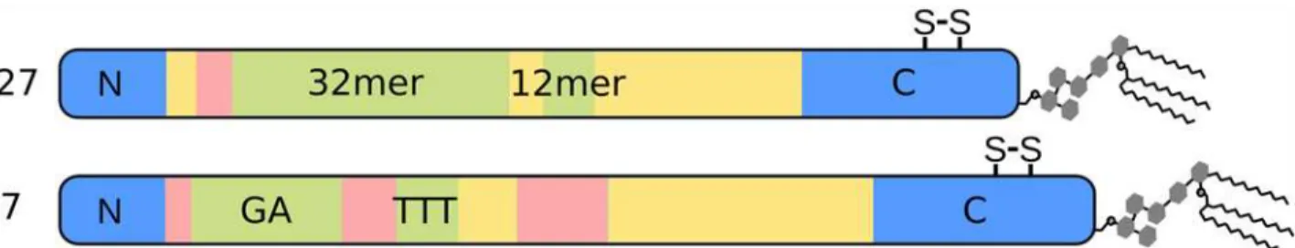
MSP2 is highly polymorphic, with conserved N- and C-terminal domains flanking a central variable region, which contains tandemly arrayed repetitive sequences [32,33]. All MSP2 alleles have been categorized into two families typified by the 3D7 and FC27 alleles, respectively, be-cause of differences in the repeats and flanking variable sequences (Fig. 1) [32,34,35]. Indeed, the sequence variability within each allelic family is limited to the repeat regions and to a few localised regions of heterogeneity within the regions flanking the repeats (green and pink in
Fig. 1).
MSP2 is a candidate for inclusion in a malaria vaccine [36], and the 3D7 allele of MSP2 was a component of a subunit vaccine that significantly reduced parasite densities in a clinical trial in Papua New Guinea [25]. This vaccine showed protective efficacy against infections with par-asites expressing the vaccine-like 3D7-type MSP2 sequence, indicating that vaccine efficacy was mediated by strain-specific responses to MSP2 [23]. Efforts to elicit protective antibodies against the conserved regions of MSP2 are complicated by the observation that MSP2 anti-bodies induced by infection withP.falciparumare largely directed against epitopes in the cen-tral variable region of the molecule [37,38], and that many conserved-region epitopes are cryptic on the parasite surface [10]. As such, the generation of a broadly effective MSP2-based vaccine is likely to require fine control of the specificity of the induced immune response. In this context, we have undertaken a detailed study of the conformational dynamics of MSP2, with the goal of establishing the extent to which these properties might contribute to the NHMRC. The Burnet Institute is supported by funding
from the NHMRC Infrastructure for Independent Research Institutes Support Scheme, and Victoria State Government Operational Infrastructure Support. The funders had no role in study design, data collection and analysis, decision to publish, or preparation of the manuscript.
observed patterns of antigenicity and immunogenicity against MSP2, and the extent to which they might be exploited to fine-tune the specificity of the antibody response against MSP2.
Methods
Materials
Untagged full-length FC27 MSP2 was expressed and purified using a strategy specific for recombinantly expressed disordered proteins, as described previously [39]. A synthetic gene encoding 3D7 MSP2, codon optimised for expression inEscherichia coli(Genescript), was cloned into pET32a (Novagen) using KpnI and NcoI. The resulting construct contains an N-terminal thioredoxin (Trx) and His6-tag for affinity purification. Bacterial cell pellets were
lysed by heating, as for FC27 MSP2 [39]. The expressed fusion protein was isolated on a His-TrapFF affinity column (GE Healthcare), eluted with imidazole and cleaved with 1% (w/w) TEV protease. The released Trx-tag and any uncleaved fusion protein were subsequently re-moved by a second passage through the His-trap column. Final purification of 3D7 MSP2 was by HPLC, using a C18 column (0.9 x 25 cm, Zorbax) and a linear acetonitrile gradient in 0.1% TFA. Isotopically enriched 3D7 and FC27 MSP2 for NMR studies was prepared by growing ex-pression cultures in M9 minimal medium, with 1 g/L15N ammonium chloride and/or 4 g/L
13C glucose as the sole nitrogen and carbon sources, respectively. The final recombinant 3D7
MSP2 has an N-terminal Gly derived from the TEV cleavage site whereas recombinant FC27 MSP2 has an N-terminal Met derived from the start codon.
NMR spectroscopy
NMR samples contained 0.4 mM 3D7 or FC27 MSP2 in 50 mM sodium acetate, pH 4.5, with 7%2H2O included for the spectrometer lock. All of the data used for resonance assignments
were acquired on a 700 MHz Bruker Avance III spectrometer equipped with the1H/13C/15N TXO cryogenic probehead with z-axis gradients at 25°C. The HNCO spectrum was acquired with spectral widths set to 9800 (aq) x 2500 (15N) x 2000 (13C’) Hz, and with maximal evolu-tion times of 80 ms (13C’) and 80 ms (15N) in the indirectly detected dimensions. The inter-scan delay was set to 1.1 s, and 4 transients per free induction decay (FID) were cumulated. The overall number of 2048 complex points was acquired in the acquisition dimension, where-as 600 hypercomplex points were randomly distributed over the indirectly-detected dimen-sions. The experiment was acquired in 3.5 h, which represents 1.9% of the time needed for a conventional experiment with similar settings. The 5D HN(CA)CONH experiment was ac-quired with the spectral widths set to 9800 (aq) x 2500 (15N) x 2000 (13C’) x 2800 (15N) x 8000 (1H) Hz [40]. The maximal acquisition times were adjusted to 15 ms for the1H indirectly-de-tected dimension, to 27 ms and 40 ms for15N dimensions, and to 30 ms for the13C’dimension. Fig 1. Schematic depiction of the primary structure of the two allelic families of MSP2.The conserved N- and C-terminal regions of MSP2 are in blue, while the allele-specific central region is composed of polymorphic repeats (green) and non-repetitive sequences (pink) as well as dimorphic regions (yellow) that differ between the allelic families but are conserved within them. The position of the conserved disulfide bond in the C-terminal regions is indicated.
The experiment was acquired with 2048 complex points in the acquisition dimension and 1750 hypercomplex points were randomly distributed over the indirectly detected dimensions. The inter-scan delay was set to 1.25 s and 4 transients per FID were collected. The experimental time of 46 h represents 0.0036% of the time needed for a similar experiment using conventional settings. The 5D HabCabCONH experiment was acquired with spectral widths set to 9800 (aq) x 2500 (15N) x 2000 (13C’) x 10000 (13Caliph) x 5000 (1Haliph) [40]. The maximal evolution times were set to 12 ms for1Haliph, 6.5 ms for13Caliph, 30 ms for13C’, and 22 ms for15N indirect dimensions. The total number of 1536 complex points was measured in the directly-detected dimension, and 1750 hypercomplex points were randomly distributed in the indirectly-detected dimensions. The experiment was acquired with 4 transients per collected FID and an interscan delay of 1.25 s. The overall experimental time of 46 h represents 0.008% of the time needed for acquisition of the conventional experiment providing similar resolution.
NMR relaxation experiments were performed on a 600 MHz Bruker Avance III NMR spec-trometer equipped with a QCI-P cryogenic probehead with z-axis gradients at 25°C. Tempera-ture was calibrated according to the chemical shift differences of pure methanol peaks. Spectral widths were set to 8370 (aq) x 1428 (15N) Hz. The overall number of 2048 complex points was acquired in the acquisition dimension and 400 complex points were acquired in the indirect di-mension for auto-relaxation rates R1, R2, cross-correlated relaxation ratesΓx,Γzand steady
state15N-1H nuclear Overhauser effect (NOE) [41]. Standard experiments were used for the measurement of R1(relaxation delays 11.2, 56, 134.4, 235.2, 380.8, 560, 896, 1344, 1848, and
2352 ms) and R2(relaxation delays 0, 14.4, 28.8, 43.2, 57.6, 72, 86.4, 115.2, and 144 ms) [42].
Asterisks denote spectra recorded twice in order to estimate experimental error. Experiments based on symmetrical reconversion were performed for determination of transverse cross-correlated relaxation ratesΓx(relaxation delays 30, 50, and 70 ms) and longitudinal cross-correlated relaxation ratesΓz(relaxation delays 100, 150, 200, and 250 ms) [43,44].
Antigenicity
Antigenicity across the MSP2 sequence was determined using sera from mice and rabbits immunised with full-length recombinant 3D7 or FC27 MSP2 (Genebank JN248383 and JN248384). Both proteins were expressed inE.coliwith C-terminal His6tags and purified by
metal-chelating, anion-exchange and reverse-phase chromatography [31]. Animals were immunised with the recombinant MSP2 formulated in Montanide ISA720. Mice (C57Bl/6) were immunised with 10μg subcutaneously and rabbits were immunised with 100μg intra-muscularly on two occasions with a four-week interval between immunisations. Serum samples used in antigenic analyses were obtained from blood samples collected two weeks after the sec-ond immunization. Immunisations were approved by the La Trobe University Animal Ethics Committee and were conducted in accord with the policies of the National Health and Medical Research Council, Australia. Reactivity to a panel of 13-residue biotinylated peptides covering the sequence of both antigens with an 8-residue overlap, was measured by ELISA, as described previously [10,19]. The panel contains one copy of the first three peptides common to both 3D7 and FC27 MSP2, but because the central variable regions of 3D7 and FC27 MSP2 are dif-ferent lengths, the two peptide sets (3D7 and FC27) extended through the conserved
pairwise comparisons of mean ELISA results for each animal. Permutation tests were used to estimate two-tailed p-values. Within each condition, each residue in MSP2 was accorded the average response of all peptides in which that residue is represented and the resulting antige-nicity profiles were normalised.
Results
Backbone resonance assignments for 3D7 MSP2
The assignment of observed spectral frequencies (chemical shifts) in an NMR spectrum to spe-cific atoms in the protein is a prerequisite for detailed structural analysis by NMR, allowing measured spectral parameters to be ascribed to specific structural features. We have previously determined near-complete backbone assignments for FC27 MSP2, but expression yields for 3D7 MSP2 were insufficient to permit the detailed analysis of that allelic form [39]. Here, we employ a new expression system, based on a thioredoxin fusion strategy, that yields ~ 10 mg 3D7 MSP2 per litre of culture medium. An assignment strategy tailored to repetitive disordered proteins and exploiting two 5D experiments, HN(CA)CONH and HabCabCONH, was em-ployed to assign the resonance frequencies of 3D7 MSP2 [45]. All the non-proline residues were successfully assigned, although residues 37–58, within the GGSA repeats, show degener-ate backbone chemical shifts, as do residues 77, 78, and 84–87 within the TTT repeats.
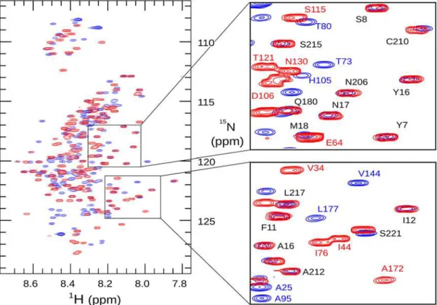
The amide chemical shifts of 3D7 MSP2 show minimal dispersion (Fig. 2), and backbone shifts are close to those expected for a disordered protein (Fig. 3). This demonstrates that 3D7 MSP2, like FC27, is extensively disordered, consistent with our previous analyses [39,46]. Com-parison of the backbone chemical shifts of the FC27 and 3D7 forms of MSP2 reveals almost perfect correspondence between the shifts of the conserved N and C-terminal regions, indicat-ing that the conformational propensities of these regions are identical in the two allelic forms (Figs.2and3). In particular, slightly elevated Cαsecondary chemical shifts are seen in the N-terminal region of both MSP2 forms, indicating a weak preference for helical conformation in this region (Fig. 3) [39,47,48]. Cβchemical shifts of the two Cys residues confirm the pres-ence of the single disulfide in MSP2 [49].
Conformational dynamics probed by
15N relaxation
NMR relaxation rates are sensitive to fast conformational dynamics [51], and as such are valu-able probes of the extent of disorder in unstructured proteins [52]. We have measured relaxa-tion rates of the backbone amides of MSP2 to determine conformarelaxa-tional dynamics at ps-ns timescales and at single-residue resolution (Fig. 4). Values of the spectral density functionJ(ω) at zero frequency and at the15N and1H Larmor frequencies were calculated from15N relaxa-tion rates (Fig. 5) [53]. These values represent the direct link between the experimental data and the conformational dynamics of the protein, with a larger value ofJ(ω) indicating a larger contribution to relaxation from dynamic processes with frequencyω. In order to identify resi-dues exhibiting dynamics atμs-ms timescales, theJ(0) andJ(ωN) values were calculated from
both auto-correlated (R1,R2, steady-state [1H]-15N NOE) and cross-correlated (Γz,Γx) relaxa-tion data [54].
under more acidic conditions [39]. Under both conditions, we observe more rapid relaxation and smaller magnitude (and in some cases small positive)1H-15N NOEs, consistent with a de-gree of conformational constraint, in the following three distinct regions: throughout the con-served N-terminus, in part of the C-terminal region coincident with the single disulfide bond in MSP2, and in part of the FC27-specific dimorphic region, between residues 140 and 150 (Figs.4and5). It should be stressed that, although these regions are more ordered than the rest of MSP2, they do not represent regions of folded regular structure as both their relaxation properties and chemical shifts are indicative of significant residual disorder, well beyond that observed in conventional structured proteins. Rather, the flexibility of these regions is weakly constrained by transient helical structure in the N-terminal region, and by the disulfide in the C-terminal region. For a few residues in the C-terminal conserved region, values ofJ(0) calcu-lated from auto-correcalcu-lated relaxation data are larger than those obtained from the cross-correlated relaxation (Fig. 5). This is suggestive of exchange contributions to the measuredR2
relaxation rates for these residues, and may imply the existence of a weakly populated meta-stable conformational state with a lifetime in theμs-ms range [55]. The repeat regions of FC27 MSP2 show somewhat variable dynamic properties, with elevated values ofJ(ωH), indicating
more extensive sub-ns dynamics than observed in the rest of the dimorphic and C-terminal re-gions, but with significant variation in the lower-frequency spectral densities across the 32-residue repeat (Fig. 5).
The variable region of 3D7 MSP2 shows greater diversity in its dynamic properties, as re-ported by relaxation measurements. The largest region of polymorphism in 3D7 MSP2, the Fig 2.1H,15N heteronuclear single-quantum correlation spectra of 3D7 (blue) and FC27 (red) MSP2.Assigned peaks are labelled in the expanded
regions, highlighting the similarity of chemical shift for these residues in the conserved N- and C-terminal regions (labelled in black type, FC27 numbering) of the two allelic forms of MSP2.
GGSA repeats, is exceptionally flexible, with relaxation properties indistinguishable from the extreme termini of the protein (Fig. 5; residues 32–63). Presumably this flexibility is a conse-quence of the uniformly small side chains in this region, and the correspondingly small steric barriers to backbone reorganisation. It is noteworthy that, despite the high levels of polymor-phism, this region is consistently rich in small residues, with Gly, Ser and Ala representing over 90% of residues seen in this region across all 3D7 MSP2 alleles characterised. In contrast, the next largest region of polymorphism, residues 103–122 appears relatively ordered, to essentially the same degree as the more ordered region in the FC27 dimorphic domain, residues 140–150. Likewise, the degree of order in the 3D7 dimorphic region (residues 123–180; yellow in Figs.4
and5) is comparable to the remainder of the FC27 dimorphic region, and the 3D7 TTT repeats are comparable to the FC27 32- and 12-residue repeats.
In contrast to the variable regions, the dynamic properties of the conserved regions of MSP2 are indistinguishable in the two allelic forms at ps-ns timescales, as indicated by identical relax-ation rates, with the regions of reduced flexibility within the N- and C-terminal regions being the most ordered regions in both alleles (Fig. 3). Together with the perfect correspondence of backbone chemical shifts, this agreement indicates that the ensembles of rapidly inter-converting conformational states sampled by these regions are identical in FC27 and 3D7 MSP2. The intervening variable regions exert no perceptible influence on these properties. Fig 3. Secondary chemical shifts of MSP2.The difference between the observed Cα(A) and HN (B) chemical shifts and those predicted for a disordered
protein by the method of Tamiola et al. [50] is plotted for 3D7 (black) and FC27 (red) MSP2. The data for FC27 are plotted on a broken axis (top) in order to correctly align the conserved regions.
More generally, this suggests that the conformational properties of MSP2 are entirely locally determined. This inference is consistent with our observation that each member of a large panel of monoclonal antibodies recognises a simple linear epitope [10].
For 3D7 MSP2, theJ(0) values calculated from auto-correlated relaxation data are never sig-nificantly larger than those obtained from cross-correlated relaxation (Fig. 5), suggesting that theμs-ms dynamics that was inferred for FC27 MSP2 C-terminal conserved region are absent in this region of 3D7 MSP2. This suggests that the variable region may influence the population or lifetime of meta-stable conformations in the conserved C-terminal domain. Remarkably, this appears to occur without affecting the ps-ns dynamics, or the overall conformational pref-erences (as reported by chemical shift) of either region.
Antigenicity is correlated with local dynamics
Potential correlations between the conformational dynamics characterised above and the anti-genicity of MSP2 have been investigated by examining the patterns of local antianti-genicity in sera of mice and rabbits immunised with recombinant 3D7 and FC27 MSP2. The reactivity of Fig 4. The15N auto-relaxation rates
R1,R2, cross-correlated relaxation ratesΓx,Γz, and steady-state15N-1H NOE for FC27 (left) and 3D7 (right) MSP2.The sequence regions of MSP2 are colour-coded as inFig. 1.
MSP2 antisera to an array of overlapping peptides covering the entire sequences of 3D7 and FC27 MSP2 was measured by ELISA [10,19]. To enable direct comparison between these re-sults and our NMR measurements, which are resolved at the level of individual residues, we adopted a scoring scheme in which each residue was scored according to the average reactivity of each of the peptides in which that residue was represented. There was good agreement across the individual mice immunised against each antigen. Pairwise comparisons of ELISA results from individual mice immunised with 3D7 MSP2 yielded average correlation coefficients of 0.7 ± 0.1 and for mice immunised with FC27 MSP2 the average correlation was 0.6 ± 0.1 (Pear-son’s r; p<10-5for all comparisons). For rabbits, there is substantially greater variation between individuals, with correlation coefficients of 0.3 ± 0.2 for each antigen (p<0.05 for seven of 12 comparisons). Nonetheless there was reasonable qualitative agreement across all antigenicity profiles for each antigen, with most regions identified to be antigenic in mice also antigenic in at least one rabbit, and vice versa (Fig. 6). The following analysis therefore considers a single average profile for each antigen in each species (Fig. 7A).
Several lines of evidence give rise to confidence that these profiles of antigenicity are robust estimations of the intrinsic immunogenicity of MSP2. First, there is excellent agreement Fig 5. Values of the15N spectral density functions for FC27 (left) and 3D7 (right) MSP2, as determined from reduced spectral density mapping from the auto-relaxation rates and steady-state15N-1H NOE (black) and cross-correlated relaxation rates (red).The sequence regions of MSP2 are
colour-coded as inFig. 1.
Fig 6. Antigenic profile of MSP2 mapped by ELISA.Four mice (top panels, black bars) and four rabbits (bottom panels, blue bars) were immunised with either FC27 (left panels) or 3D7 (right panels) MSP2. Individual immune sera were tested against a single panel of overlapping peptides covering the sequences of both 3D7 MSP2 (peptides 1–45) and FC27 MSP2 (peptides 46–84), as shown schematically above. The conserved N terminal (peptides 1–3)
and C terminal (peptides 37–45 and 77–84) regions are common to both 3D7 and FC27 MSP2 and are delineated with dashed red lines. Peptides showing
Fig 7. Comparison of experimental patterns of antigenicity, predicted antigenicity, and conformational dynamics, for FC27 (left) and 3D7 (right) MSP2.A. Antigenicity profiles of MSP2 inferred from experimental immunisation of mice (black) and rabbits (red) are plotted against the sequence. Black bars (top) denote the location of epitopes of a panel of monoclonal antibodies to MSP2. B. Conformational flexibility of MSP2 as measured by the spectral density functions derived from the15N relaxation data. Spectral density functions are plotted at zero frequency (black line) and at the15N Larmor frequency
(red line). C. Antigenicity of MSP2 as predicated using BepiPred [60] (red, right axis) and the method of Kolaskar and Tongaonkar [61] (black, left axis). The threshold for epitope prediction for both methods is denoted by the grey line.
doi:10.1371/journal.pone.0119899.g007
Table 1. Conformationally constrained peptides are more antigenic.
No. animals respondingb No. peptides with>2 animals respondingc
J(0)a mice rabbits total mice rabbits total
Constrained 42 1.06–1.88 2.2±0.4 1.1±0.2 1.7±0.3 14 4 18
Flexible 42 0.36–1.06 0.50±0.15 0.45±0.10 0.48±0.09 3 0 3
a Range of maximumJ(0) values defining each peptide class.
b The number of sera generating a background-corrected response greater than 0.3 OD (mice) or 0.05 OD (rabbits) to individual peptides, averaged (± SEM) over each class.
c Number of peptides in each class to which more than two sera respond.
between the current 3D7 profile and those derived previously from experimental immunisa-tions of mice and humans with recombinant 3D7 MSP2 [56,57]. Second, the conserved regions of MSP2 show similar patterns of antigenicity in both the FC27 and 3D7 profiles. Finally, the epitopes of an extensive panel of monoclonal antibodies to MSP2 [10,30,58,59] all coincide with peaks in the antigenicity profile (Fig. 7A).
Strikingly, both of the regions of marked conformational restriction in MSP2, the conserved N-terminal region and the region around the disulfide in the conserved C-terminal region, co-incide with peaks in the experimental antigenicity profiles of both 3D7 and FC27 MSP2, and with the epitopes of several monoclonal antibodies (Fig. 7). Likewise, antigenic regions within the repeats and dimorphic regions of FC27 correspond to those that show slightly elevated low-frequency spectral densities, indicative of conformational restriction. Although the GGSA repeat, which is the most flexible region in 3D7 MSP2, shows some antigenicity, this arises from a significant response in only a single rabbit (Fig. 6), suggesting that this very flexible re-gion is only rarely antigenic (Fig. 7). Indeed, there is a significant correlation between the anti-genicity profile and relaxation-based measures of conformational flexibility: Spearman’sρfor the comparison of the average antigenicity profiles over all mice withJ(0) are 0.35 and 0.54 for FC27 and 3D7 MSP2, respectively, and for rabbits 0.30 and 0.21 (two-tailed p<0.005 for all comparisons, by permutation). Thus, it appears that restricted conformational disorder within MSP2 may be a robust predictor of local antigenicity. To explore this further, we divided the peptides into two equal groups according to the maximum value ofJ(0) measured for the resi-dues in each peptide, representing the conformationally constrained and flexible regions of MSP2 (Table 1). The peptides from constrained regions are almost four times as likely as the peptides from flexible regions to be significantly antigenic, while 85% of peptides that show sig-nificant responses in more than two animals (of either species) are from conformationally con-strained regions of MSP2.
In contrast, sequence-based predictors of B-cell epitopes [60,61] perform poorly when ap-plied to MSP2, showing weak and in some cases negative correlation with the experimental an-tigenicity, and failing to predict monoclonal antibody epitopes (Fig. 7). The Bepipred predictor [60] predicts 80 of 84 peptides in our array to contain B-cell epitopes, when in fact only 21 pep-tides reacted significantly with more than two antisera (Fig. 6), and these 21 peptides included two of the four peptides not predicted to be epitopes by this method. The approach of Kolaskar and Tongaonkar [61] performs only slightly better, predicting 35 peptides to contain epitopes, including 10 that reacted with more than two antisera.
Discussion
conformational dynamics of MSP2, as reported by15N relaxation measurements, with local an-tigenicity as inferred from experimental animal immunisations. We find that regions of MSP2 that are most antigenic correspond to those regions in which conformational flexibility is somewhat constrained, whereas those regions that are most flexible appear to be the least antigenic.
In contrast, we find no evidence that the polymorphic regions of MSP2 are particularly anti-genic. Indeed, the most polymorphic region of MSP2, the GGSA repeats of 3D7, is also the most flexible and amongst the least antigenic regions. Other polymorphic regions (green and pink inFig. 7) are no more antigenic than are the dimorphic and conserved regions. There is evidence that the polymorphisms within these regions are selectively favoured, and although details of these selective processes are unclear, they are expected to involve host immune pres-sure [63,64]. As such, the lack of obvious antigenic bias towards these regions is surprising, and may highlight important immunogenic differences between recombinant MSP2 and the native parasite antigen [10].
Previous studies of structured antigens have established that increased epitope flexibility tends to increase antigenicity [65,66], in contrast to the current findings. An important distinc-tion is that these studies have addressed epitopes that are variably flexible loops in largely struc-tured proteins. The most flexible of these loops are unlikely to be as flexible as even the least flexible regions of MSP2. In the model structured antigen lysozyme, all residues show positive steady-state [1H]-15N NOE values greater than 0.6 [67], reflecting markedly more constrained sub-ns dynamics than is seen for any region of MSP2 (Fig 4). One possible explanation for the apparent discrepancy, therefore, may be that a moderate degree of flexibility is optimal for anti-genicity, with epitopes that are either too rigid, or too flexible, being less effective. Alternatively, the determinants of antigenicity in structured and disordered proteins may differ in a more fundamental way. For example, it has been suggested that the correlation between flexibility and antigenicity observed in structured proteins reflects accessibility, rather than flexibilityper se[68], whereas the accessibility of potential epitopes in a disordered antigen is likely to be uni-formly high. Perhaps consistent with this interpretation is our observation that epitope predic-tors, parameterised primarily on the basis of structured antigens, perform poorly for MSP2.
The consistency of the antigenic profiles we have measured here between animals and with other previous studies in mice and in humans, strongly suggests that these profiles are deter-mined by the intrinsic immunogenicity of the recombinant MSP2 antigen. As such, the correla-tion we observe between conformacorrela-tional restriccorrela-tion and antigenicity probably reflects a tendency for more flexible regions of MSP2 to be less immunogenic. The mechanistic basis un-derlying this tendency is currently unclear, though several possible explanations are worthy of consideration. It has been suggested that the unusual residue composition of disordered and re-petitive antigens may give rise to extensively cross-reactive responses, which fail to mature into high-affinity and specific antibodies [69]. Alternatively, it may be that conformational disorder itself frustrates the process by which a mature antibody response develops. Any disordered an-tigen exists in a vast ensemble of distinct conformations, but a developing antibody is likely to be limited in the range of conformations it is capable of recognising. The conformational diver-sity of disordered antigens may therefore impose a significant barrier to antibody maturation, as proposed for theStaphylococcus aureusfibronectin binding protein [70]. This effect may be viewed as a conformational analogue of the epitope dilution effect recently described in the context of a polyvalent vaccine of the polymorphic antigen apical membrane antigen
are diluted to an extent determined by the degree of disorder present in the epitope, with the result that the antibody response is biased towards more ordered epitopes.
Little is known about which MSP2 epitopes contribute to a protective immune response. Vaccine-derived protection mediated by MSP2 appears to be strain specific [23,73], suggesting that variable epitopes dominate. However this does may not be the case for the natural immune response to MSP2, where strain-specific protection has not been detected [37,74]. Nonetheless, a protective, strain-independent response is clearly desirable in the context of vaccine develop-ment. As such, our observation that conserved N- and C-terminal epitopes are amongst the most immunogenic regions of MSP2 is encouraging, although it is likely that not all of these epitopes will be accessible on the parasite surface [10].
The correlation established here begs the question of causation: is it possible to modulate the immunogenicity or antigenicity of a disordered antigen by altering its flexibility? Antigen flexibility could be modulated by directly modifying the antigen by addition of bulky residues or disulfide bonds at sites flanking a target epitope. Alternatively, simply changing the formula-tion of the antigen may have the desired effect. For example, the N-terminal region of MSP2 can be conformationally stabilised by interactions with lipid membranes, in a way that may better reflect the conformation of MSP2 on the merozoite surface [48]. These possibilities have important implications for the development of vaccines based on MSP2, where it is desirable to tune antigenicity towards epitopes that are conserved and exposed on the parasite surface [10,36].
Author Contributions
Conceived and designed the experiments: CAM JSR JN VS RFA RSN. Performed the experi-ments: MZ DA LZ. Analyzed the data: CAM MZ DA JN LZ JSR JGB. Contributed reagents/ materials/analysis tools: CAM BK RFA. Wrote the paper: CAM MZ DA BK JN LZ VS JSR JGB RFA RSN.
References
1. Uversky VN, Oldfield CJ, Dunker AK. Showing your ID: intrinsic disorder as an ID for recognition, regu-lation and cell signaling. 2005; J Mol Recognit 18: 343–384. PMID:16094605
2. Oldfield CJ, Dunker AK. Intrinsically disordered proteins and intrinsically disordered protein regions. 2014; Annu Rev Biochem 83: 553–584. doi:10.1146/annurev-biochem-072711-164947PMID: 24606139
3. Xue B, Blocquel D, Habchi J, Uversky AV, Kurgan L, Uversky VN, et al. Structural disorder in viral pro-teins. 2014; Chem Rev 114: 6880–6911. doi:10.1021/cr4005692PMID:24823319
4. Feng ZP, Zhang X, Han P, Arora N, Anders RF, Norton RS. Abundance of intrinsically unstructured pro-teins inP.falciparumand other apicomplexan parasite proteomes. 2006; Mol Biochem Parasitol 150: 256–267. PMID:17010454
5. Pavlovic MD, Jandrlic DR, Mitic NS. Epitope distribution in ordered and disordered protein regions. Part B—Ordered regions and disordered binding sites are targets of T- and B-cell immunity. 2014; J
Immu-nol Methods 407: 90–107. doi:10.1016/j.jim.2014.03.027PMID:24726865
6. Dunker AK, Brown CJ, Lawson JD, Iakoucheva LM, Obradovic Z. Intrinsic disorder and protein function. 2002; Biochemistry 41: 6573–6582. PMID:12022860
7. Kwong PD, Doyle ML, Caspar DJ, Cicala C, Leavitt SA, Majeed S, et al. HIV-1 evades antibody-mediat-ed neutralisation through conformational masking of receptor binding sites. 2002; Nature 420: 678–682. PMID:12478295
8. Stahl HD, Coppel RL, Brown GV, Saint R, Lingelbach K, Cowman AF, et al. Differential antibody screening of clonedPlasmodium falciparumsequences expressed inEscherichia coli: procedure for isolation of defined antigens and analysis of human antisera. 1984; Proc Natl Acad Sci U S A 81: 2456–2460. PMID:6371814
10. Adda CG, MacRaild CA, Reiling L, Wycherley K, Boyle MJ, Kienzle V, et al. Antigenic characterization of an intrinsically unstructured protein,Plasmodium falciparummerozoite surface protein 2. 2012; Infect Immun 80: 4177–4185. doi:10.1128/IAI.00665-12PMID:22966050
11. Raj DK, Nixon CP, Nixon CE, Dvorin JD, DiPetrillo CG, Pond-Tor S, et al. Antibodies to PfSEA-1 block parasite egress from RBCs and protect against malaria infection. 2014; Science 344: 871–877. doi:10. 1126/science.1254417PMID:24855263
12. Richards JS, Stanisic DI, Fowkes FJ, Tavul L, Dabod E, Thompson JK, et al. Association between natu-rally acquired antibodies to erythrocyte-binding antigens ofPlasmodium falciparumand protection from malaria and high-density parasitemia. 2010; Clin Infect Dis 51: e50–60. doi:10.1086/656413PMID: 20843207
13. Olugbile S, Kulangara C, Bang G, Bertholet S, Suzarte E, Villard V, et al. Vaccine potentials of an intrin-sically unstructured fragment derived from the blood stage-associatedPlasmodium falciparumprotein PFF0165c. 2009; Infect Immun 77: 5701–5709. doi:10.1128/IAI.00652-09PMID:19786562
14. Yagi M, Bang G, Tougan T, Palacpac NM, Arisue N, Aoshi T, et al. Protective epitopes of the Plasmodi-um falciparPlasmodi-umSERA5 malaria vaccine reside in intrinsically unstructured N-terminal repetitive se-quences. 2014; PLoS One 9: e98460. doi:10.1371/journal.pone.0098460PMID:24886718
15. Foquet L, Hermsen CC, van Gemert GJ, Van Braeckel E, Weening KE, Sauerwein R, et al. Vaccine-induced monoclonal antibodies targeting circumsporozoite protein preventPlasmodium falciparum in-fection. 2014; J Clin Invest 124: 140–144. PMID:24292709
16. Muster T, Steindl F, Purtscher M, Trkola A, Klima A, Himmler G, et al. A conserved neutralizing epitope on gp41 of human immunodeficiency virus type 1. 1993; J Virol 67: 6642–6647. PMID:7692082 17. Foucault M, Mayol K, Receveur-Brechot V, Bussat MC, Klinguer-Hamour C, Verrier B, et al. UV and
X-ray structural studies of a 101-residue long Tat protein from a HIV-1 primary isolate and of its mutat-ed, detoxifimutat-ed, vaccine candidate. 2010; Proteins 78: 1441–1456. doi:10.1002/prot.22661PMID: 20034112
18. Dyson HJ, Satterthwait AC, Lerner RA, Wright PE. Conformational preferences of synthetic peptides derived from the immunodominant site of the circumsporozoite protein ofPlasmodium falciparumby1H
NMR. 1990; Biochemistry 29: 7828–7837. PMID:2261440
19. Boyle MJ, Langer C, Chan JA, Hodder AN, Coppel RL, Anders RF, et al. Sequential processing of mer-ozoite surface proteins during and after erythrocyte invasion byPlasmodium falciparum. 2014; Infect Immun 82: 924–936. doi:10.1128/IAI.00866-13PMID:24218484
20. al-Yaman F, Genton B, Anders R, Taraika J, Ginny M, Mellor S, et al. Assessment of the role of the hu-moral response toPlasmodium falciparumMSP2 compared to RESA and SPf66 in protecting Papua New Guinean children from clinical malaria. 1995; Parasite Immunol 17: 493–501. PMID:8552419 21. Aucan C, Traore Y, Tall F, Nacro B, Traore-Leroux T, Fumoux F, et al. High immunoglobulin G2 (IgG2)
and low IgG4 levels are associated with human resistance toPlasmodium falciparummalaria. 2000; In-fect Immun 68: 1252–1258. PMID:10678934
22. Ekala MT, Jouin H, Lekoulou F, Mercereau-Puijalon O, Ntoumi F. Allelic family-specific humoral re-sponses to merozoite surface protein 2 (MSP2) in Gabonese residents withPlasmodium falciparum in-fections. 2002; Clin Exp Immunol 129: 326–331. PMID:12165090
23. Flück C, Smith T, Beck HP, Irion A, Betuela I, Alpers MP, et al. Strain-specific humoral response to a polymorphic malaria vaccine. 2004; Infect Immun 72: 6300–6305. PMID:15501757
24. al-Yaman F, Genton B, Anders RF, Falk M, Triglia T, Lewis D, et al. Relationship between humoral re-sponse toPlasmodium falciparummerozoite surface antig2 and malaria morbidity in a highly en-demic area of Papua New Guinea. 1994; Am J Trop Med Hyg 51: 593–602. PMID:7985752
25. Genton B, Betuela I, Felger I, Al-Yaman F, Anders RF, Saul A, et al. A recombinant blood-stage malaria vaccine reducesPlasmodium falciparumdensity and exerts selective pressure on parasite populations in a phase 1–2b trial in Papua New Guinea. 2002; J Infect Dis 185: 820–827. PMID:11920300 26. Osier FH, Feng G, Boyle MJ, Langer C, Zhou J, Richards JS, et al. Opsonic phagocytosis of
Plasmodi-um falciparPlasmodi-ummerozoites: mechanism in human immunity and a correlate of protection against malar-ia. 2014; BMC Med 12: 108. doi:10.1186/1741-7015-12-108PMID:24980799
27. Stanisic DI, Richards JS, McCallum FJ, Michon P, King CL, Schoepflin S, et al. Immunoglobulin G sub-class-specific responses againstPlasmodium falciparummerozoite antigens are associated with con-trol of parasitemia and protection from symptomatic illness. 2009; Infect Immun 77: 1165–1174. doi: 10.1128/IAI.01129-08PMID:19139189
29. Osier FH, Fegan G, Polley SD, Murungi L, Verra F, Tetteh KK, et al. Breadth and magnitude of antibody responses to multiplePlasmodium falciparummerozoite antigens are associated with protection from clinical malaria. 2008; Infect Immun 76: 2240–2248. doi:10.1128/IAI.01585-07PMID:18316390 30. Stubbs J, Olugbile S, Saidou B, Simpore J, Corradin G, Lanzavecchia A. Strain-transcending
Fc-dependent killing ofPlasmodium falciparumby merozoite surface protein 2 allele-specific human anti-bodies. 2011; Infect Immun 79: 1143–1152. doi:10.1128/IAI.01034-10PMID:21189324
31. McCarthy JS, Marjason J, Elliott S, Fahey P, Bang G, Malkin E, et al. A phase 1 trial of MSP2-C1, a blood-stage malaria vaccine containing 2 isoforms of MSP2 formulated with Montanide ISA 720. 2011; PLoS One 6: e24413. doi:10.1371/journal.pone.0024413PMID:21949716
32. Smythe JA, Coppel RL, Day KP, Martin RK, Oduola AM, Kemp DJ, et al. Structural diversity in the Plas-modium falciparummerozoite surface antigen 2. 1991; Proc Natl Acad Sci U S A 88: 1751–1755.
PMID:2000383
33. Fenton B, Clark JT, Khan CM, Robinson JV, Walliker D, Ridley R, et al. Structural and antigenic poly-morphism of the 35- to 48-kilodalton merozoite surface antigen (MSA-2) of the malaria parasite Plasmo-dium falciparum. 1991; Mol Cell Biol 11: 963–971. PMID:1990294
34. Smythe JA, Peterson MG, Coppel RL, Saul AJ, Kemp DJ, Anders RF. Structural diversity in the 45-kilo-dalton merozoite surface antigen ofPlasmodium falciparum. 1990; Mol Biochem Parasitol 39: 227–234. PMID:2181307
35. Thomas AW, Carr DA, Carter JM, Lyon JA. Sequence comparison of allelic forms of thePlasmodium falciparummerozoite surface antigen MSA2. 1990; Mol Biochem Parasitol 43: 211–220. PMID: 2090943
36. Anders RF, Adda CG, Foley M, Norton RS. Recombinant protein vaccines against the asexual blood-stages ofPlasmodium falciparum. 2010; Hum Vaccin 6: 1–15.
37. Osier FH, Murungi LM, Fegan G, Tuju J, Tetteh KK, Bull PC, et al. Allele-specific antibodies to Plasmo-dium falciparummerozoite surface protein-2 and protection against clinical malaria. 2010; Parasite Immunol 32: 193–201. doi:10.1111/j.1365-3024.2009.01178.xPMID:20398182
38. Taylor RR, Smith DB, Robinson VJ, McBride JS, Riley EM. Human antibody response toPlasmodium falciparummerozoite surface protein 2 is serogroup specific and predominantly of the immunoglobulin G3 subclass. 1995; Infect Immun 63: 4382–4388. PMID:7591074
39. Zhang X, Perugini MA, Yao S, Adda CG, Murphy VJ, Low A, et al. Solution conformation, backbone dy-namics and lipid interactions of the intrinsically unstructured malaria surface protein MSP2. 2008; J Mol Biol 379: 105–121. doi:10.1016/j.jmb.2008.03.039PMID:18440022
40. Kazimierczuk K, Zawadzka-Kazimierczuk A, Kozminski W. Non-uniform frequency domain for optimal exploitation of non-uniform sampling. 2010; J Magn Reson 205: 286–292. doi:10.1016/j.jmr.2010.05. 012PMID:20547466
41. Ferrage F, Cowburn D, Ghose R. Accurate sampling of high-frequency motions in proteins by steady-state15N-{1H} nuclear Overhauser effect measurements in the presence of cross-correlated relaxation. 2009; J Am Chem Soc 131: 6048–6049. doi:10.1021/ja809526qPMID:19358609
42. Korzhnev DM, Billeter M, Arseniev AS, Orekhov VY. NMR studies of Brownian tumbling and internal motions in proteins. 2001; Prog Nucl Magn Reson Spectrosc 38: 197–266.
43. Pelupessy P, Espallargas GM, Bodenhausen G. Symmetrical reconversion: measuring cross-correlation rates with enhanced accuracy. 2003; J Magn Reson 161: 258–264. PMID:12713978 44. Pelupessy P, Ferrage F, Bodenhausen G. Accurate measurement of longitudinal cross-relaxation rates
in nuclear magnetic resonance. 2007; J Chem Phys 126: 134508. PMID:17430048
45. Motackova V, Novacek J, Zawadzka-Kazimierczuk A, Kazimierczuk K, Zidek L, Sanderova H, et al. Strategy for complete NMR assignment of disordered proteins with highly repetitive sequences based on resolution-enhanced 5D experiments. 2010; J Biomol NMR 48: 169–177. doi: 10.1007/s10858-010-9447-3PMID:20890634
46. Adda CG, Murphy VJ, Sunde M, Waddington LJ, Schloegel J, Talbo GH, et al.Plasmodium falciparum
merozoite surface protein 2 is unstructured and forms amyloid-like fibrils. 2009; Mol Biochem Parasitol 166: 159–171. doi:10.1016/j.molbiopara.2009.03.012PMID:19450733
47. Low A, Chandrashekaran IR, Adda CG, Yao S, Sabo JK, Zhang X, et al. Merozoite surface protein 2 of
Plasmodium falciparum: expression, structure, dynamics, and fibril formation of the conserved N-termi-nal domain. 2007; Biopolymers 87: 12–22. PMID:17516503
48. MacRaild CA, Pedersen MØ, Anders RF, Norton RS. Lipid interactions of the malaria antigen merozoite surface protein 2. 2012; Biochim Biophys Acta 1818: 2572–2578. doi:10.1016/j.bbamem.2012.06.015
PMID:22749949
49. Sharma D, Rajarathnam K.13C NMR chemical shifts can predict disulfide bond formation. 2000; J
50. Tamiola K, Acar B, Mulder FAA. Sequence-specific random coil chemical shifts of intrinsically disor-dered proteins. 2010; J Am Chem Soc 132: 18000–18003. doi:10.1021/ja105656tPMID:21128621 51. Palmer AG. NMR characterization of the dynamics of biomacromolecules. 2004; Chemical Reviews
104: 3623–3640. PMID:15303831
52. Jensen MR, Zweckstetter M, Huang JR, Blackledge M. Exploring Free-Energy Landscapes of Intrinsi-cally Disordered Proteins at Atomic Resolution Using NMR Spectroscopy. 2014; Chem Rev 114: 6632–6660. doi:10.1021/cr400688uPMID:24725176
53. Farrow NA, Zhang O, Szabo A, Torchia DA, Kay LE. Spectral density function mapping using15N
relax-ation data exclusively. 1995; J Biomol NMR 6: 153–162. PMID:8589604
54. Kaderavek P, Zapletal V, Rabatinova A, Krasny L, Sklenar V, Zidek L. Spectral density mapping proto-cols for analysis of molecular motions in disordered proteins. 2014; J Biomol NMR 58: 193–207. doi: 10.1007/s10858-014-9816-4PMID:24515886
55. Mittermaier AK, Kay LE. Observing biological dynamics at atomic resolution using NMR. 2009; Trends Biochem Sci 34: 601–611. doi:10.1016/j.tibs.2009.07.004PMID:19846313
56. Lawrence N, Stowers A, Mann V, Taylor D, Saul A. Recombinant chimeric proteins generated from con-served regions ofPlasmodium falciparummerozoite surface protein 2 generate antiparasite humoral responses in mice. 2000; Parasite Immunol 22: 211–221. PMID:10792760
57. Saul A, Lawrence G, Smillie A, Rzepczyk CM, Reed C, Taylor D, et al. Human phase I vaccine trials of 3 recombinant asexual stage malaria antigens with Montanide ISA720 adjuvant. 1999; Vaccine 17: 3145–3159. PMID:10462251
58. Epping RJ, Goldstone SD, Ingram LT. An epitope recognised by inhibitory monoclonal antibodies that react with a 51 kilodalton merozoite surface antigen inPlasmodium falciparum. 1988; Mol Biochem Parasitol 28: 1–10. PMID:2453800
59. Saul A, Lord R, Jones G, Geysen HM, Gale J, Mollard R. Cross-reactivity of antibody against an epitope of thePlasmodium falciparumsecond merozoite surface antigen. 1989; Parasite Immunol 11: 593–601. PMID:2482473
60. Larsen JE, Lund O, Nielsen M. Improved method for predicting linear B-cell epitopes. 2006; Immunome Res 2: 2. PMID:16635264
61. Kolaskar AS, Tongaonkar PC. A semi-empirical method for prediction of antigenic determinants on pro-tein antigens. 1990; FEBS Lett 276: 172–174. PMID:1702393
62. Yang X, Adda CG, MacRaild CA, Low A, Zhang X, Zeng W, et al. Identification of key residues involved in fibril formation by the conserved N-terminal region ofPlasmodium falciparummerozoite surface pro-tein 2 (MSP2). 2010; Biochimie 92: 1287–1295. doi:10.1016/j.biochi.2010.06.001PMID:20542076 63. Putaporntip C, Jongwutiwes S, Hughes AL. Differential selective pressures on the merozoite surface protein 2 locus ofPlasmodium falciparumin a low endemic area. 2008; Gene 427: 51–57. doi:10. 1016/j.gene.2008.09.009PMID:18840512
64. Zilversmit MM, Volkman SK, DePristo MA, Wirth DF, Awadalla P, Hartl DL. Low-complexity regions in
Plasmodium falciparum: missing links in the evolution of an extreme genome. 2010; Mol Biol Evol 27: 2198–2209. doi:10.1093/molbev/msq108PMID:20427419
65. Ofek G, Guenaga FJ, Schief WR, Skinner J, Baker D, Wyatt R, et al. Elicitation of structure-specific anti-bodies by epitope scaffolds. 2010; Proc Natl Acad Sci U S A 107: 17880–17887. doi:10.1073/pnas. 1004728107PMID:20876137
66. Westhof E, Altschuh D, Moras D, Bloomer AC, Mondragon A, Klug A, et al. Correlation between seg-mental mobility and the location of antigenic determinants in proteins. 1984; Nature 311: 123–126.
PMID:6206398
67. Buck M, Boyd J, Redfield C, MacKenzie DA, Jeenes DJ, Archer DB, et al. Structural determinants of protein dynamics: analysis of15N NMR relaxation measurements for main-chain and side-chain nuclei
of hen egg white lysozyme. 1995; Biochemistry 34: 4041–4055. PMID:7696270
68. Novotny J, Handschumacher M, Haber E, Bruccoleri RE, Carlson WB, Fanning DW, et al. Antigenic de-terminants in proteins coincide with surface regions accessible to large probes (antibody domains). 1986; Proc Natl Acad Sci U S A 83: 226–230. PMID:2417241
69. Anders RF. Multiple cross-reactivities amongst antigens ofPlasmodium falciparumimpair the develop-ment of protective immunity against malaria. 1986; Parasite Immunol 8: 529–539. PMID:3543808 70. Penkett CJ, Redfield C, Jones JA, Dodd I, Hubbard J, Smith RA, et al. Structural and dynamical
charac-terization of a biologically active unfolded fibronectin-binding protein fromStaphylococcus aureus. 1998; Biochemistry 37: 17054–17067. PMID:9836601
membrane antigen-1. 2013; PLoS Pathog 9: e1003840. doi:10.1371/journal.ppat.1003840PMID:
24385910
72. Chaudhury S, Reifman J, Wallqvist A. Simulation of B Cell Affinity Maturation Explains Enhanced Anti-body Cross-Reactivity Induced by the Polyvalent Malaria Vaccine AMA1. 2014; J Immunol 193: 2073–2086. doi:10.4049/jimmunol.1401054PMID:25080483
73. Flück C, Schopflin S, Smith T, Genton B, Alpers MP, Beck HP, et al. Effect of the malaria vaccine Com-bination B on merozoite surface antigen 2 diversity. 2007; Infect Genet Evol 7: 44–51. PMID: 16647307