Cerebral Ischemia in Adult Rat Brain with Treadmill
Exercise
Jin-Young Chung1., Min-Wook Kim2., Moon-Suk Bang3
, Manho Kim1*
1Department of Neurology, Seoul National University Hospital, Chongno-ku, Seoul, Korea,2Department of Rehabilitation Medicine, College of Medicine, The Catholic University of Korea, Seoul, and Institute of Catholic Integrative Medicine (ICIM), Incheon St. Mary’s Hospital, Incheon, South Korea,3Department of Rehabilitation Medicine, Seoul National University Hospital, Chongno-ku, Seoul, Korea
Abstract
Neurotrophin 4 (NT-4) belongs to the family of neurotrophic factors, and it interacts with the tyrosine kinase B (trkB) receptor. NT-4 has neuroprotective effects following cerebral ischemia. Its role might be similar to brain-derived neurotrophic factor (BDNF), because both interact with trkB. Exercise also improves neural function by increasing neurotrophic factors. However, expression profiles of NT-4 in the brain during exercise are unknown. Here, we assessed the expressions of NT-4 and its receptor, trkB, following cerebral ischemia and hypothesized that exercise changes the expressions of NT-4 and trkB. Results showed that in a permanent middle cerebral artery occlusion rat model, ischemia decreased NT-4 and trkB expression. Immunohistochemistry showed their immunoreactivities around the region of the ischemic area. Treadmill exercise changed the expression of NT-4, which increased in the contralateral hemisphere in rats with ischemic injury. TrkB also showed similar patterns to its neurotophins. The change in NT-4 suggested that exercise might have primed NT4 production so that further injury causes slightly greater increases in NT4 compared with non-exercise controls.
Citation:Chung J-Y, Kim M-W, Bang M-S, Kim M (2013) Increased Expression of Neurotrophin 4 Following Focal Cerebral Ischemia in Adult Rat Brain with Treadmill Exercise. PLoS ONE 8(3): e52461. doi:10.1371/journal.pone.0052461
Editor:Stefano L. Sensi, University G. D’Annunzio, Italy
ReceivedSeptember 15, 2012;AcceptedNovember 19, 2012;PublishedMarch 18, 2013
Copyright:ß2013 Chung et al. This is an open-access article distributed under the terms of the Creative Commons Attribution License, which permits unrestricted use, distribution, and reproduction in any medium, provided the original author and source are credited.
Funding:This work was supported by grants from the Korea Health 21 R&D Project, Ministry of Health and Welfare (A092058) and WCU-Neurocytomics program and by grants from 21C Frontier Functional Proteomics Project (FPR08K1301-02210). The funders had no role in study design, data collection and analysis, decision to publish, or preparation of the manuscript.
Competing Interests:The authors have declared that no competing interests exist.
* E-mail: [email protected]
.These authors contributed equally to this work.
Introduction
Neurotrophic factors are the family of proteins that includes nerve growth factor (NGF), BDNF, NT-3, and NT-4 [1–3]. Each neurotrophic factor shows specific selective biological activity, interacting with different members of the tyrosine kinase (trk) receptors [1]. BDNF, which is one of the most active substances to stimulate neurogenesis, acts with tyrosine kinase B (trkB). Neurotrophin 4 (NT-4), which is also called neurotrophin 4 or 4/5 (NT-4 or NT-4/5), also initiates signals by binding with trkB. Since both BDNF and NT-4 bind trkB, the roles of NT-4 and BDNF might be similar. For example, NT-4 might play a role in long-term potentiation and plasticity [4,5]. BDNF has been frequently described in damaged brain or in response to physiologic stimuli [6–8]. The BDNF binding trkB also interacts with NT-4, which indicates that altered expression of trkB can possibly affect the function of NT-4. However, in comparison with BDNF, reports on NT-4 in damaged brain or in response to the physiologic stimuli are rare [9,10]. Chan et al. showed that treatment with NT-4 reduced the infarction volume in a permanent focal cerebral ischemic rat model [11], demonstrating that NT-4 is involved in ischemic brain injury.
Exercise improves functional recovery following brain injury. It also increases neurotrophic factors, stimulates neurogenesis, or
improves resistance to neuronal injury [12–14]. Moreover, exercise regenerates and develops the neural system and improves activities associated with learning ability [15–17]. Following focal cerebral ischemia, expression of BDNF/trkB and NGF/trkA have been reported, and exercise increases the expression of BDNF/ trkB and trkA; it does so more in the side contralateral to the ischemic lesion [6,18].
Cerebral injury and exercise may alter neurotrophic factors that improve neural function. However, even expression profiles of NT-4 in the ischemic brain are unknown. Therefore, we observed the changes in NT-4 and trkB expression following ischemic injury in the rat brain. We also postulated that exercise can affect the expressions of NT-4 and its receptor, trkB.
Materials and Methods
Experimental design and classification of animals
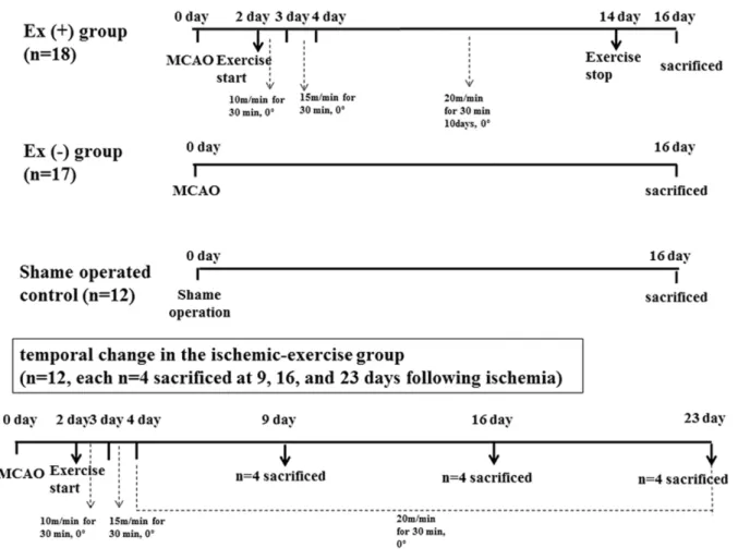
either the exercise (n = 18) or non-exercise group (n = 17). The severity was determined according to the Garcia scale as previously described [6,18]. Six items (spontaneous activity, symmetry of movements, symmetry of forelimbs, climbing the wall of wire cage, reaction to touch, and response to vibrissae touch) were measured with a total score that ranged from 3 to 18. The higher the score, the better the performance: mild (scores 12– 18), moderate (8–11), and severe degrees (3–7). Twelve adult male Sprague-Dawley rats (275–325 g) were additionally used for determination of temporal change in the ischemic-exercise group (n = 12, n = 4 each and sacrificed at 9, 16, and 23 days following ischemia) (Figure 1). Protocols for care and use of animals in this procedure were in compliance with guidelines and were approved by the Catholic University animal care committee.
Surgical procedures
For a focal cerebral ischemia model, modified Longa’s method was used, as previously described [19]. Induction was performed using a mixture of 3% isoflurane in 30% O2and 70% N2O. For maintenance, 1.5% isoflurane was used. Through a midline cervical incision, the left common carotid artery was exposed at its bifurcation. Branches from the external carotid artery were coagulated. The pterygopalatine artery was ligated with a 5.0 silk suture. A 4.0 nylon monofilament was used for the occlusion. Heating to the tip of this filament made it rounded, and then it was
inserted into the bifurcation site of the common carotid artery. The monofilament was advanced 16–18 mm into the internal carotid artery from the bifurcation site in order to occlude vessels at the location of the origins of the middle cerebral and proximal anterior cerebral artery. The monofilament was secured in place with a ligature, and the wound was closed. With this procedure, the origin of the middle cerebral artery was occluded. The animals were allowed to survive with food and water ad libitum. Body temperature was maintained at 3761uC (rectal temperature) using a thermistor-controlled heating blanket [6,18].
Treadmill exercise
For the exercise, we used the treadmill test (Columbus instruments, USA). The start of treadmill exercise was performed 2 days following the MCAO operation. This exercise training was done for 30 min every day for 12 days. The velocity of the treadmill was gradually increased, from 10 m/min on the first exercise day, 15 m/min on the second, and 20 m/min on the third and subsequent days. The tilting angle of the exercise table was maintained and set to 0u[6,18].
Immunohistochemistry
On postoperative day 16, for sacrificing, rats were anesthetized, and transcardiac perfusion with heparinized saline followed by 4% paraformaldehyde in phosphate-buffered saline (PBS) was
per-Figure 1. Experimental design.Among total of 59 rats, 35 rats underwent middle cerebral artery occlusion (MCAO), and 12 rats were used as sham-operated control. In 48 hours, the MCAO group was divided into either the exercise (n = 18) or non-exercise group (n = 17). Twelve rats were additionally used for determination of temporal change in the ischemic-exercise group (n = 12, n = 4 each and sacrificed at 9, 16, and 23 days following ischemia).
formed. Using a sliding microtome, sections were cut at 30 um thickness. Blocking was done in a mixture of 10% normal goat serum (NGS), 1% bovine serum albumin (BSA), 0.2% Triton X-100, and 1% H2O2in PBS. After washing with PBS three times, primary antibody was incubated in 10% NGS and 1% BSA for 40 h at 4uC. For detection of NT-4 and trkB immunoreactivity, anti-NT-4 (1:300, Santa Cruz, CA, USA) and anti-trkB (1:300, Santa Cruz, CA, USA) antibodies were used. Immunoperoxidase labeling was performed using a DAB kit (Dako, Carpinteria, CA), and slides were evaluated using an Olympus BX51 microscope (Olympus, Japan) [6,18].
Western blot analysis
For protein extraction, brains were dissected into right and left hemispheres and placed on ice in 10 volumes of cold homogeni-zation buffer (50 mM Tris, 120 mM NaCl, pH 7.4). Protease inhibitors (Complete Mini, Gibco, Grand Island, NY, USA) were added, and then the tissue was homogenized. Protein concentra-tions were determined by Bradford method (Bio-Rad, Richmond, CA, USA). For each well, 20mg of protein extracts were loaded and separated by sodium dodecyl sulfate-polyacrylamide gel
electrophoresis using 10% polyacrylamide with 0.05% bis-acrylamide [20]. Proteins were transferred to nitrocellulose membrane and probed with anti-NT-4 (1:300, Santa Cruz, CA, USA) and anti-trkB (1:300, Santa Cruz, CA, USA). Peroxidase anti-rabbit IgG (vector, PI-1000, 1:3000 dilution) was used as a secondary antibody.btubulin (1:300, Santa Cruz, CA, USA) was used for an internal control. Signals were detected by enhanced chemiluminescence (Supersignal, Pierce, Rockford, IN, USA) using autoradiograms exposed from 10 to 30 min [6,18]. These experiments were repeated independently in triplicate.
Statistical analysis
The Mann-Whitney test was used to compare the control and exercise groups. In addition, non-parametric test for the paired sample was also performed. SPSS ver. 12.0 was used, and a p-value below 0.05 was considered to be statistically significant. We replicated experiments more than three times to confirm the results.
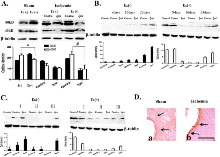
Figure 2. NT-4 expression profile.(A) Two forms, dimer (80 kDa) and monomer (40–47 kDa), were detected. Ischemia decreases monomer and dimer proteins in the ipsilateral region (Ipsi). Exercise increased monomer and dimer proteins in both hemispheres, particularly in the contralateral hemisphere in ischemia (#p,0.05, n = 7). Exercise-only increased dimer (* p,0.05, n = 7). (B) Expression of dimers increased at postinfarct day 23 in ischemia. Exercise increased dimer proteins at postinfarct as early as day 9 and particularly in the contralateral hemisphere at postinfarct day 23. (p,0.05, n = 6). (C) Dimer level is lowered in moderate to severe conditions. Exercise induces increased expression of dimers, more so in the milder condition (I: mild, II: moderate, III: severe) (p,0.05, n = 5). (D) The distribution of immunoreactivity by exercise increased adjacent to the ischemic region (b) comparing to the ischemia-only control (a).S= 100 um.
Results
Expression profile of NT-4
NT-4 exists in two forms, either as a dimer (80 kDa) or as a monomer (40–47 kDa). Both forms of proteins were decreased in the ipsilateral ischemic region at 2 weeks when compared to the non-ischemic contralateral side (Figure 2A). NT-4 was increased by treadmill exercise, more so in the contralateral hemisphere following ischemic injury. Exercise alone increased monomer and dimer forms of NT-4 proteins in the bilateral hemispheres (Figure 2A). Analysis of temporal changes in NT-4 showed that NT-4 dimer protein, the level of which was low in week 2, increased post-infarct on day 23. Treadmill exercise increased NT-4 as early as post-infarct day 9. At post-infarct day 23, this dimer protein was also increased, particularly in the contralateral hemisphere (Figure 2B). NT-4 dimer protein decreased when the ischemic severity increased. Exercise increased the expression of NT-4 dimer protein (Figure 2C). NT-4 showed that immunore-activity increased in the ischemic region, and the distribution of
immunoreactivity came out adjacent to the ischemic region after exercise (Figure 2D).
Expression profiles of trkB
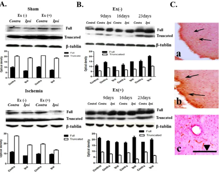
TrkB exists in two forms, either as a full-length (140 kDa) protein or as a truncated (90–95 kDa) protein. In the ischemia group, the full-length protein was decreased; however, the truncated protein was not changed. Exercise increased the full length and truncated proteins in ischemic conditions. Treadmill exercise also increased the full-length protein in both hemispheres of the sham control (Figure 3A).
Temporal changes in trkB showed that expression of the two forms of trkB increased following day 23. After exercise, expression of the full-length form was increased in both hemispheres at day 16, and the truncated form was increased by day 16, particularly in the contralateral hemisphere (Figure 3B). No relationship between the expression of trkB protein and severity of ischemia was observed.
Figure 3. TrkB expression.(A) Two forms of trkB are noted: full length form (140 kDa) and truncated form (90,95 kDa). Ischemia decreased the full-length protein in the ipsilateral region (Ipsi). Exercise increased two forms of the protein in both hemispheres, particularly contralateral (Contra) to the ischemic hemisphere (p,0.05, n = 7). (B) Expression of two forms of protein increased at day 23 after ischemia. Exercise increased the full-length form in both hemispheres at day 16 and increased the truncated form by day 16, particularly in the contralateral hemisphere (p,0.05, n = 6). (C) (a) Immunoreactivities in the ischemic region. (b) Exercise increased the immunoreactivities adjacent to the ischemic region in the ipsilateral hemisphere. (c) In the control hemisphere, exercise increased immunoreactivities, particularly in vascular structures.S= 100 um.
Immunohistochemistry showed that exercise increased immu-noreactivity in both hemispheres of the sham group, particularly in vascular structures, and exercise also increased the distribution of immunoreactivity around the ischemic region in the ipsilateral hemisphere (Figure 3C).
Discussion
In previous studies, we confirmed the expression of BDNF/trkB and NGF/trkA following focal cerebral ischemia [6,18].
In this study, we attempted to observe the changes in NT-4 and trkB expression following ischemic injury in the rat brain. We hypothesized that exercise changes expression of neurotrophic factors and their tyrosine kinase receptors. Our results showed that ischemia decreased NT-4 and trkB, a specific receptor of the NT-4 full-length protein, in the ipsilateral region; however, there were no changes in the truncated protein. Treadmill exercise altered the levels of NT-4 and trkB, increasing them more in the contralateral hemisphere. In terms of immunohistochemistry, immunoreactiv-ities of NT-4 and trkB appeared to be most predominant around the ischemic area. These staining intensities became dense and smaller following exercise. These results suggest that NT-4 altered in response to ischemic injury, and treadmill exercise plays a role in the changes of neurotrophins and their receptors.
Although NT4 is included in the neurotrophic family, NT-4 and trkB decreased, and their expressions in the ischemic brain were different. Since NT-4 has a high affinity for trkB, the function of NT-4 is supposed to be the same as BDNF, which also plays a role in long-term potentiation and plasticity [4,5]. However, BDNF in ischemic rat brain increased [6] whereas NT-4 decreased in the same experimental set in this study. These findings cannot account for the fact that the roles of NT-4 and BDNF are identical.
Effects of exercise include a better functional outcome [6,18], exercise may increase neurotrophic factors, neurogenesis, or have neuroprotective effects [12–14]. Duration and intensity of exercise are factors for promoting plasticity and enhancement of perfor-mance. Compared with voluntary exercise, progressive treadmill exercise was intense and lasted long enough to improve brain function [6,8,14,21,22]. As a result, treadmill exercise enhanced NT-4 in the contralateral hemisphere in an ischemic model and
even in control sham-operated rats. This supports idea that increasing neurotrophic factors contribute to functional recovery [12,13]. Immunohistochemistry showed more NT-4 immunore-activity in the ischemic area compared to the non-ischemic region. Exercise concentrated the area of immunoreactivities in our experiment. It has been reported that exercise reduces brain damage in ischemic rats [23], suggesting one possibility that accounts for the concentrating area of immunoreactivities. Immunohistochemistry also showed that exercise increased trkB immunoreactivity, particularly in vascular structures. Exercise is known to be associated with regional angiogenesis [23]. There is no direct evidence to show that trk receptors increased in vascular structures.
Trials for treatment with neurotrophic factors involving direct administration under pathologic conditions have been conducted [5,24,25]. Among types of brain injury, stroke is the most common cause that leads to death [26]. Studies of exercise as a rehabilitation program show that it can also change neurotrophic factors and trk receptors in the damaged brain [6,14,27]. Under experimental cerebral ischemia and exercise conditions, the expression profiles of NT4/trkB as well as NGF/trkA and BDNF/trkB are changed [6,18]. Taken together, these findings suggest that functional recovery in cerebral ischemia is associated with not only BDNF or NGF, but it can also be mediated by NT-4 and other tyrosine kinase receptors.
Conclusions
Overall, ischemia decreased NT-4 and trkB expressions in a permanent middle cerebral artery occlusion rat model. However, treadmill exercise changed expressions of NT-4 and trkB. Altered expression profiles in ischemic brain indicate that NT-4 and trkB might participate in the recovery process in rats with brain damage.
Author Contributions
Conceived and designed the overall study: JYC MWK MK MSB. Performed the experiments: JYC MWK MK MSB. Analyzed the data: JYC MWK MK MSB.
References
1. Persson H, Ibanez CF (1993) Role and expression of neurotrophins and the trk family of tyrosine kinase receptors in neural growth and rescue after injury. Curr Opin Neurol Neurosurg 6: 11–18.
2. Thoenen H (1991) The changing scene of neurotrophic factors. Trends Neurosci 14: 165–170.
3. Thoenen H (1995) Neurotrophins and neuronal plasticity. Science 270: 593– 598.
4. Ferrer I, Krupinski J, Goutan E, Marti E, Ambrosio S, et al. (2001) Brain-derived neurotrophic factor reduces cortical cell death by ischemia after middle cerebral artery occlusion in the rat. Acta Neuropathol 101: 229–238. 5. Klintsova AY, Greenough WT (1999) Synaptic plasticity in cortical systems.
Curr Opin Neurobiol 9: 203–208.
6. Kim MW, Bang MS, Han TR, Ko YJ, Yoon BW, et al. (2005) Exercise increased BDNF and trkB in the contralateral hemisphere of the ischemic rat brain. Brain Res 1052: 16–21.
7. Goutan E, Marti E, Ferrer I (1998) BDNF, and full length and truncated TrkB expression in the hippocampus of the rat following kainic acid excitotoxic damage. Evidence of complex time-dependent and cell-specific responses. Brain Res Mol Brain Res 59: 154–164.
8. Widenfalk J, Olson L, Thoren P (1999) Deprived of habitual running, rats downregulate BDNF and TrkB messages in the brain. Neurosci Res 34: 125– 132.
9. Burette A, Jalenques I, Romand R (1997) Neurotrophin receptor immunostain-ing in the rat ventral cochlear nucleus. Brain Res 776: 10–23.
10. Abe K (2000) Therapeutic potential of neurotrophic factors and neural stem cells against ischemic brain injury. J Cereb Blood Flow Metab 20: 1393–1408.
11. Chan KM, Lam DT, Pong K, Widmer HR, Hefti F (1996) Neurotrophin-4/5 treatment reduces infarct size in rats with middle cerebral artery occlusion. Neurochem Res 21: 763–767.
12. Johansson BB, Zhao L, Mattsson B (1999) Environmental influence on gene expression and recovery from cerebral ischemia. Acta Neurochir Suppl 73: 51– 55.
13. Keyvani K, Sachser N, Witte OW, Paulus W (2004) Gene expression profiling in the intact and injured brain following environmental enrichment. J Neuropathol Exp Neurol 63: 598–609.
14. Ploughman M, Attwood Z, White N, Dore JJ, Corbett D (2007) Endurance exercise facilitates relearning of forelimb motor skill after focal ischemia. Eur J Neurosci 25: 3453–3460.
15. Cotman CW, Berchtold NC (2002) Exercise: a behavioral intervention to enhance brain health and plasticity. Trends Neurosci 25: 295–301.
16. Hicks RR, Zhang L, Atkinson A, Stevenon M, Veneracion M, et al. (2002) Environmental enrichment attenuates cognitive deficits, but does not alter neurotrophin gene expression in the hippocampus following lateral fluid percussion brain injury. Neuroscience 112: 631–637.
17. Russo-Neustadt A, Ha T, Ramirez R, Kesslak JP (2001) Physical activity-antidepressant treatment combination: impact on brain-derived neurotrophic factor and behavior in an animal model. Behav Brain Res 120: 87–95. 18. Chung JY, Kim MW, Bang MS, Kim M (2010) The effect of exercise on trkA in
the contralateral hemisphere of the ischemic rat brain. Brain Res 1353: 187– 193.
20. DiFiglia M, Sapp E, Chase K, Schwarz C, Meloni A, et al. (1995) Huntingtin is a cytoplasmic protein associated with vesicles in human and rat brain neurons. Neuron 14: 1075–1081.
21. Chang HC, Yang YR, Wang SG, Wang RY (2009) Effects of treadmill training on motor performance and extracellular glutamate level in striatum in rats with or without transient middle cerebral artery occlusion. Behav Brain Res 205: 450–455.
22. Tong L, Shen H, Perreau VM, Balazs R, Cotman CW (2001) Effects of exercise on gene-expression profile in the rat hippocampus. Neurobiol Dis 8: 1046–1056. 23. Ding Y, Li J, Luan X, Ding YH, Lai Q, et al. (2004) Exercise pre-conditioning reduces brain damage in ischemic rats that may be associated with regional angiogenesis and cellular overexpression of neurotrophin. Neuroscience 124: 583–591.
24. Zhang WR, Hayashi T, Wang JM, Sasaki C, Sakai K, et al. (1999) Reduction of tyrosine kinase B and tyrosine kinase C inductions by treatment with neurotrophin-3 after transient middle cerebral artery occlusion in rat. Neurosci Lett 276: 161–164.
25. Semkova I, Krieglstein J (1999) Neuroprotection mediated via neurotrophic factors and induction of neurotrophic factors. Brain Res Brain Res Rev 30: 176– 188.
26. Bang OY (2009) Multimodal MRI for ischemic stroke: from acute therapy to preventive strategies. J Clin Neurol 5: 107–119.