271
Rev Bras Oftalmol. 2013; 72 (4): 271-3
C
ASER
EPORTOclusão de ramo arterial retiniano bilateral
Luiz Guilherme Azevedo de Freitas
1, David Leonardo Cruvinel Isaac
2, Luiz Alexandre Rassi Gabriel
3,
Lívia Carla de Souza Nassar Bianchi
4, Marcos Pereira de Ávila
5.
1Retina and Vitreous Department, Santa Luzia Eye Hospital/Santa Luzia Foundation — Recife/PE, Brazil.
2Retina and Vitreous Unit, Reference Centre for Ophthalmology, University Hospital of the Federal University of Goiás (UFG) — Goiânia - GO, Brazil. 3Ocular Genetics Unit, Federal University of Goiás (UFG), Goiânia/GO, Brazil.
4Retina and Vitreous Unit, Reference Centre for Ophthalmology, University Hospital of the Federal University of Goiás (UFG) — Goiânia/GO, Brazil. 5Reference Centre for Ophthalmology, University Hospital of the Federal University of Goiás (UFG) — Goiânia/GO, Brazil.
Institution:
Work conducted at the Reference Centre for Ophthalmology, University Hospital of UFG.
The authors declare no conflict of interest.
Received for publication: 26/10/2011 - Accepted for publication: 27/3/2012
A
BSTRACTThe authors report a case of a patient who presented bilateral branch retinal artery occlusion without any meaningful systemic underlying conditions.
Keyworks: Retina; Vascular diseases; Retinal artery occlusion; Retinal vasculitis; Fuorescein angiography; Case reports
Bilateral branch retinal artery occlusion
R
ESUMOOs autores relatam o caso de uma paciente que apresentou quadro bilateral de oclusão de ramos arteriais da retina sem causas sistêmicas identificáveis para o aparecimento da doença.
272
I
NTRODUCTIONT
he first report of central retinal artery occlusion (CRAO) was by von Graefe in 1859, on a patient with endocarditis and multiple systemic emboli. The histopathological findings of CRAO were subsequently described by Sweiger(1,2).Central retinal artery occlusion is a severe eye disease with a poor prognosis. Its incidence is 8.5 per 100,000 people, and it most often affects men around 60 years of age with systemic diseases such as atherosclerosis and hypertension(3).
Occlusion of the central retinal artery or one of its branches is usually caused by an embolism from atherosclerotic deposits in the carotid artery. Other sources of occlusion can be valvular heart disease, rheumatic vegetations, and atrial myxoma. Emboli may be made of cholesterol (Hollenhorst plaques), platelets, and calcium, of which the former is the most common(4). In young
persons, the occlusion is most commonly due to vascular spasms in conditions such as migraine(5).
Bilateral involvement occurs in 1-2% of cases. When both eyes are affected simultaneously by central retinal artery occlusion, valvular heart disease, giant cell arteritis, or vasculitis should be strongly considered(6).
The central retinal artery is a terminal vessel without collateral vessels; for this reason, its occlusion quickly triggers retinal ischemia, followed by abrupt unilateral loss of visual acuity. In such cases, final visual acuity is usually equal to or less than counting fingers, while a small percentage of patients with occlusion of the central artery or macular branches may retain better vision(7,8). Currently, there is no treatment of choice shown
to produce a beneficial effect to patients with the condition.
C
ASER
EPORTSCB, a 54-year-old white female patient, was referred for ophthalmic evaluation complaining of non-simultaneous episodes of visual impairment in both eyes (BE), with an interval of approximately 24 hours between events. She had no family history of eye disease.
On examination, visual acuity with best correction was 20/ 400 in both eyes. Slit lamp examination showed the presence of relative afferent pupillary defect, and intraocular pressure was 18 mmHg in BE. Fundus examination showed pale retinal oedema in the macular region and papillomacular bundle in both eyes, with epiretinal haemorrhage nasal to the macula and increased vascular tortuosity.
Fluorescein angiography found retinal ischemia with late staining of retinal arterioles and capillaries, capillary filling defect, and bilateral macular ischemia on the path of the occlusion (Fi-gure 1). The patient was prescribed 0.2% brimonidine tartrate every 8 hours and intramuscular betamethasone (1 ml/3mg).
The following tests were also requested: antinuclear antibodies (ANA), erythrocyte sedimentation rate (ESR), anticardiolipin antibodies (IgG and IgM), lupus anticoagulant, and serum protein electrophoresis, all of which were within nor-mal ranges.
MRI of the carotid arteries, resting electrocardiogram, stress testing, transesophageal Doppler echocardiography, and Doppler echocardiography with colour flow mapping were also normal. Colour duplex scan of the carotids found a fibrotic plaque with 10-20% stenosis of the left carotid bulb.
On another visit two months later the patient had a corrected VA of 20/200 in the right eye (RE) and 20/50 in the left eye (LE). Fundus examination showed occlusion of peripheral
Freitas LGA, Isaac DLC, Gabriel LAR, Bianchi LCSN, Ávila MP
vessels with haemorrhages. The patient was kept on 0.2% brimonidine tartrate every 8 hours and monitoring of symptoms. Given these findings, the patient underwent periodic observation, and the condition remained stable in both eyes.
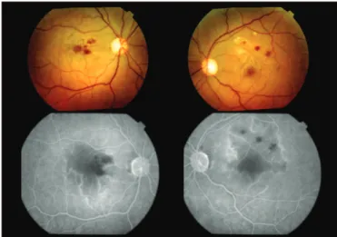
Ophthalmic examination 92 months after the first visit showed a corrected VA of 20/400 in the RE and 20/25 in the LE. Biomicroscopy showed early-stage nuclear cataract in BE and intraocular pressure of 16 mmHg in BE. Monochrome and colour fundus photography showed an attached retina, optic nerve with temporal pallor, epiretinal haemorrhages temporal to the macula, silver wire vessels, and dilated capillaries temporal to the macula in BE, as well as cotton wool spots in the inferior temporal retina in the RE and the superior retina in the LE. Fluorescein angiography found in BE: areas of retinal nonperfusion tem-poral-superior to macula, isolated hyperfluorescence spots that remained throughout the test, and temporal capillary telangiectasias (Figure 2)
D
ISCUSSIONThis paper presents a rare case of retinal artery branch occlusion occurring almost simultaneously in both eyes. The aetiological factor was not clearly determined, and visual acuity was eventually preserved in one eye.
Male patients and patients over 60 years of age with systemic diseases such as hypertension, valvular heart disease, and carotid atherosclerosis are more likely to present some kind of occlusion of the retinal artery. The condition is generally caused by an embolism originating from the carotid arteries. We presented the case of a patient under 60 years of age who had no history of systemic diseases.
Systemic investigation found normal ANA, VHS, anticardiolipin antibodies (IgG and IgM), lupus anticoagulant, and serum protein electrophoresis, ruling out any risk factors that might increase the likelihood of occlusive phenomena.
Thrombotic occlusion is usually related to changes in the vascular endothelium or blood flow velocity and coagulation disorders. The main conditions associated with such causes are: vascular disorders (atherosclerosis, hypertension), haematological disorders (haemoglobinopathy, polycythemia vera), inflammation triggered by vasculitis (Behçet’s disease, syphilis, temporal arteritis), and collagen diseases (systemic lupus erythematosus with or without antiphospholipid antibody syndrome).
We did not investigate trombophilia, which could have been done by assessing protein S deficiency, protein C deficiency, protein C resistance, hyperhomocysteinemia, dysfibrinogenemia, factor V Leiden, antithrombin III, and antiphospholipid AC syndrome.
Vasculitis of undetermined cause was found in the vascular arcades in both eyes. Such vasculitis could have been triggered as a reaction to the occlusive process, however it is not possible to determine the cause of inflammation, or whether the inflammation was responsible for the occlusion.
The retinal artery branch occlusion was bilateral, but the visual outcome was very different in each eye. One eye (left) regained good visual acuity, which is very uncommon according to literature, while the other suffered significant vision loss. We believe that the left eye retained better visual acuity because its macula was less affected by the ischemia; also, its telangectesias were smaller and more distant from the centre of the fovea (Figure 2).
273
Figure 2: Area of retinal nonperfusion greater than the temporal macula and temporal telangectásicas dilatation of capillaries Bilateral Retinal Artery Branch Occlusion
Figure 1: Bilateral retinal and macular ischemia due to capillary filling defect
One suggested therapy for the condition consists in reducing the intraocular pressure using topical and systemic hypotensive agents, in an attempt to improve retinal perfusion. Our patient was prescribed brimonidine tartrate, a topical hypotensive agent, to good effect. Interestingly, it has recently been suggested that the drug may also have a neuroprotective effect. Given the presence of vasculitis, systemic betamethasone may have reduced vascular inflammation, thus improving perfusion in some of the affected branches.
The course of the disease in this patient and the fact that no further episodes were noted in the following eight years make us consider the possibility of an isolated occlusive event without further ocular or systemic abnormalities. The precise aetiology of the case remains elusive. A limitation of this report is that thrombophilia was not investigated at the time, as this group of diseases could possibly lead to such a clinical presentation.
This was a rare case that deserves to be reported, so that patients with similar presentations can undergo investigation for all types of diseases that may present with vascular occlusion. This may help prevent occlusive processes, avoiding ischemic damage to susceptible organs such as the heart, kidneys, and brain.
C
ONCLUSIONRetinal occlusions can have different presentations, not limited to those found in the literature. Ophthalmologists should always be prepared to face rare disease presentations and should be familiar with their management.
R
EFERENCES1. Von Graefe A. Ueber Embolie der arteria centralis retinae als Urscahe plotzlicher Erblingdung. Arch fur Ophthalmol. 1859; 5:136-57.
2. Duke-Elder S, Dobree H. System of ophthalmology. St. Louis: Mosby; 1967. Diseases of the retina. p. 66-97.
3. Rumelt S, Dorenboim Y, Rehany U. Aggressive systematic treat-ment for central retinal artery occlusion. Am J Ophthalmol. 1999;128(6):733-8. Erratum in Am J Ophthalmol. 2000;130(6): 908. 4. Abujamra S, Ávila M, Barsante C, Farah ME, Gonçalves JO, Lavinsky J, et al. Retina e vítreo: clínica e cirurgia. São Paulo: Roca; 2000. 5. Hykin PG, Gartry D, Brazier DJ, Graham E. Bilateral cilio-retinal
artery occlusion in classic migraine. Postgrad Med J. 1991;67(785):282-4.
6. BrownGC, Shields JA. Cilioretinal arteries and retinal arterial occlusion. Arch Ophthalmol. 1979;97(1):84-92.
7. Augsburger JJ, Magargal LE. Visual prognosis following treat-ment of acute central retinal artery obstruction. Br J Ophthalmol. 1980;64(12):913-7.
8. Brown GC, Magargal LE. Central retinal artery obstruction and visual acuity. Ophthalmology. 1982;89(1):14-9.
9. Ávila M, Lavinsky J, Moreira Jr CA. Retina e vítreo. Rio de Janeiro: Guanabara Koogan, Cultura Médica; 2008. p. 243-8.
Corresponding author:
Luiz Guilherme Azevedo de Freitas Hospital de Olhos Santa Luzia
Estrada do Encanamento 909 - Casa Forte - Recife – Pernambuco, Brazil - CEP: 52070-000
Telephone: +5581 2121 9100 /+5581 7811 8559 Fax: (81) 2121-9100
E-mail: [email protected]