C
ASER
EPORT501
Takayasu's arteritis with renal artery stenosis diagnosed in a
patient with 65 years old
Authors
Ellen Simionato Valente 1 Rafael de Almeida 1 Alexander Gonçalves Sacco 2
Mauricio Costa Lazzarin 1
André Melchiades da Silva 1
Marcos Andreazza 2
1 Universidade Federal de Pelotas-RS.
2 Hospital Santa Casa de Pelotas-RS.
Submitted on: 05/04/2015. Approved on: 07/16/2015.
Correspondence to:
Ellen Simionato Valente. Universidade Federal de Pelotas-RS. Avenida Duque de Caxias, 336, Bloco E, Apto 202, Fragata, Pelotas, RS, Brasil. CEP: 96030-000
E-mail: [email protected]
I
NTRODUCTIONTakayasu arteritis (TA) is a rare chronic granulomatous inflammatory arterial disease of unknown etiology that may affect the aorta and its main branches.1-4 It is an uncommon disease, which mainly affects women within their reproductive years; more commonly seen in Southeast Asia.5-8 According to data from North America, the annual incidence is 2.6 cases per one million people.5,6
Initial clinical manifestations can be insidious, with nonspecific signs and systemic symptoms, varying according to the affected arterial sites, and the left subclavian artery is the DOI: 10.5935/0101-2800.20150079
Takayasu arteritis is a rare disease of unknown etiology that affects the aorta and its main branches. It is a condition, geographically more common in Southeast Asia, which mainly affects women of reproductive age. The clinical presentation is nonspecific, with signs and symptoms that vary according to the affected arterial segment. The most commonly affected vessel is the subclavian artery, while renal artery stenosis is relatively uncommon. Cardiac involvement and association with other diseases may also be present. We present in this report the case of an elderly patient with late diagnosis of Takayasu's arteritis and various comorbidities or related complications.
ABSTRACT
Keywords: aged; chronic, kidney failure; takayasu arteritis.
one most often involved, followed by the aorta, the common carotid, the renal and the vertebral artery.1,4,7
Renal artery injury associated with TA is reported in 30-35% of cases, with reports mainly from Asia.6,9 Stenosis occurs in 23-31% of affected renal arteries, whereas other lesions (occlusion, dilation and aneurysm) are often more common.6 TA-induced renal artery stenosis can result in malignant hypertension, severe renal impairment, cardiac decompensation and premature death.3
C
ASEREPORTA sixty-five year-old female, born and living in Pelotas, a former smoker, with a previous history of mitral valve disease due to rheumatic fever sequelae, withs replacement by bioprosthesis for 9 years. Since then, the bearer of heart failure, atrial fibrillation, and dyslipidemia, without a diagnosis of hypertension (SAH).
J Bras Nefrol 2015;37(4):501-504
Takayasu arteritis with renal artery stenosis
502
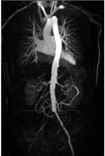
the legs. Her laboratory tests did not stand out, with creatinine of 1.05 mg/dL and urea of 17 mg/dL.We ordered a magnetic resonance angiography (MRA) of her thoracic aorta and her abdominal and iliac arteries (Figure 1), which results showed signs suggestive of inflammatory parietal artery disease involving the thoracoabdominal aorta and its branches, characterized by occlusion of the left subclavian artery, the right common iliac artery and also the superior mesenteric artery, moderate diffuse reduction of the innominate artery gauge (around 50%), critical stenosis at the origin of the celiac trunk (70-90%) and moderate stenosis in the middle third of the right renal artery (between 60- 70%). These findings were consistent with the diagnosis of Takayasu arteritis. The patient was then submitted to an angioplasty with a stent placement in the right iliac artery with complete resolution of the right lower limb injury. She was started on a drug treatment for vasculitis with deflazacort at 30 mg/day.
The patient continued treatment with corticosteroids for three months only, after which he stopped and did not return to the angiologist. She continued under medical care at a basic healthcare unit in her own district, where she used to present nonspecific symptoms such as fatigue, diffuse arthralgia, dizziness, intermittent claudication, diffuse myalgia, tachycardia, and depressive symptoms. In one of these visits, she complained of recent oliguria, slowness, drowsiness and hair loss. Her laboratory tests showed TSH 66.09 mIU/L; free T4 of 0.53 ng/dL; creatinine of 5.58 mg/dL and urea of 172 mg/dL. She was then referred to the nephrology clinic, where she was admitted for investigation. During hospitalization, the patient was treated for hypothyroidism and was submitted to an abdominal ultrasound scan, which showed topical kidney, with normal morphology and echogenicity, with a slight reduction in the right kidney size, measuring 8.8 cm in its longest axis, and the left kidney measured 10.3 cm. The patient improved in renal function with only intravenous hydration and was discharged with a creatinine of 2.60 mg/dL for outpatient monitoring.
Figure 1. Coronal MRA showed an occlusion of the left subclavian artery, of the right common iliac artery and also of the superior mesenteric artery with moderate stenosis in the middle third of the right renal artery.
J Bras Nefrol 2015;37(4):501-504
Takayasu arteritis with renal artery stenosis
503
and more rarely: acute vascular events.7 The nonspecific nature of the symptoms upon presentation, combined with the absence of physical symptoms, typically results in late diagnosis and a fail to install proper treatment early in the course of the disease.4 According to Kerr et al.,11 20% of the patients are not diagnosed for three years after the onset of symptoms. This demonstrates the difficulty in diagnosis, and corroborates the fact that several recent studies have shown a high prevalence in the elderly.7
Cardiac involvement occurs in up to 50% of patients with TA and may involve any heart structure.1 This association between rheumatic fever and TA, as described in our patient, is infrequent and, according to Vale et al., this combination raises the possibility of a common immunological basis in the pathogenesis of both diseases.1,12 In advanced cases, the occlusion of arterial vessels of the limbs may result in ischemic ulcers or gangrene in a minority of cases.11 Our patient only sought specialized medical care after she developed an erosive lesion in the right lower limb, which ultimately led to the diagnosis of TA, showing the importance of dermatological aspects, albeit unusual. The involvement of the subclavian artery is frequent, present in up to 93% of cases of TA.13 The stenosis of this vessel can lead to neurological symptoms related to the phenomenon called the Subclavian Steal Syndrome, with impaired cerebral blood flow due to the reverse flow of the vertebral artery.14 In our case, we associated the partial improvement of the symptoms with the early hypothyroidism - the subclavian steal syndrome, as the patient remained with dizziness and slowness of thought even after a state of euthyroidism.
According to surveys, renal artery stenosis induced by TA is seen in 23-31% of patients.6 Atherosclerosis and fibromuscular dysplasia account for the vast majority of cases of renal artery stenosis, so it is very rare to have this stenosis as a manifestation of systemic vasculitis.13 Usually, most TA studies are focused started treatment with prednisone 40 mg/day for
her vasculitis.
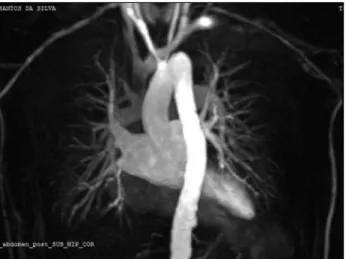
Figure 2. Coronal MRA shows an atypical aortic arch, with occlusion of the proximal segment of the left subclavian artery from its origin all the way to the emergence of the vertebral artery, with a filling of the distal subclavian bed. Brachiocephalic trunk and left common artery with smooth walls and moderate diffuse reduction in gauge of around 50%.
D
ISCUSSIONChronic and slow-developing TA -the clinical setting that is the most frequent, causes vascular lesion characterized by thickening of the adventitia and cellular infiltration of the tunica media, with local destruction of smooth muscle cells and elastin of the vessels.4 The intimal hyperplasia results from
the proliferation of myofibroblasts, followed by fibrosis of the media and intima, leading to stenosis, and occasionally overlapping atherosclerosis of the affected arterial segment.3,4
The diagnostic criteria for TA, defined by the American Rheumatology College, include: age of onset < 40, claudication of the limbs, decreased pulse of the brachial artery, difference in systolic blood pressure > 10 mmHg between the two arms, murmurs of the subclavian arteries or the aorta, and abnormal arteriography. Having three or more of these criteria yields a sensitivity of 90.5% and specificity of 97.8%.10 In this case, the patient had four criteria, with an unknown age of onset and late diagnosis.
J Bras Nefrol 2015;37(4):501-504
Takayasu arteritis with renal artery stenosis
504
on brachiocephalic involvement, with no reports on the exact consequence of renal stenosis, but it is known that it can result in hypertension and fatal cardiovascular events.3,6 In 33-83% of TA cases there is arterial hypertension (AH), and in 20-38% of these patients, renal artery stenosis contributes to this factor.7 However, AH can often be hidden, since the BP measured in the upper limbs may underestimate the true central pressure as a result of the subclavian artery involvement.4 In our case, besides the occlusion of the left subclavian artery, there was a diffuse reduction in the diameter of the innominate artery, suggesting lower BP values, reason why the renovascular hypertension was probably not diagnosed.
In addition to the considerable morbidity, TA-related mortality rates have been described as above 35% in 5 years.4 This arterial disease remains a clinical challenge at all stages, and treatment decisions are hampered by a shortage of evidence pro and against targeted therapies. In about 50% of cases, steroid treatment alone is not sufficient to prevent vasculitis progression, requiring the addition of immunosuppressive therapy.7 The difficulty in dealing with TA is a reflection of the nature of the disease and its rarity, which limits the feasibility of future clinical trials.4
R
EFERENCES1. Gormezano NWS, Santos MC, Okuda EM, Catani EH, Sacchet-ti SB. Associação entre febre reumáSacchet-tica e arterite de Takayasu - relato de caso. Rev Bras Reumatol 2014;epub ahead of print.
2. Gotway MB, Araoz PA, Macedo TA, Stanson AW, Higgins CB, Ring EJ, et al. Imaging findings in Takayasu's arteritis. AJR Am J Roentgenol 2005;184:1945-50. PMID: 15908559 DOI: http://dx.doi.org/10.2214/ajr.184.6.01841945
3. Weaver FA, Kumar SR, Yellin AE, Anderson S, Hood DB, Rowe VL, et al. Renal revascularization in Takayasu arteritis-induced renal artery stenosis. J Vasc Surg 2004;39:749-57. PMID: 15071436 DOI: http://dx.doi.org/10.1016/j.jvs.2003.12.022 4. Andrews J, Mason JC. Takayasu's arteritis-recent advances in
imaging offer promise. Rheumatology (Oxford) 2007;46:6-15. DOI:http://dx.doi.org/10.1093/rheumatology/kel323
5. Lee TH, Chen IM, Chen WY, Weng CF, Hsu CP, Shih CC. Ear-ly endovascular experience for treatments of Takayasu's arte-ritis. J Chin Med Assoc 2013;76:83-7. PMID: 23351418 DOI: http://dx.doi.org/10.1016/j.jcma.2012.10.006
6. Agarwal G, Vats HS, Raval AN, Yevzlin AS, Chan MR, Gime-lli G. Chronic total occlusion and successful drug-eluting stent placement in Takayasu arteritis-induced renal artery stenosis. Clin Med Res 2013;11:233-6. DOI: http://dx.doi.org/10.3121/ cmr.2013.1132
7. Neves BD, Raimundo A, Figueira TA, Machado FP, Roquette J, Sá J. Arterite de Takayasu: a propósito de um caso clínico. Rev Port Cardiol 2015;34:215.e1-215.e4.
8. Tso E, Flamm SD, White RD, Schvartzman PR, Mascha E, Hoffman GS. Takayasu arteritis: utility and limitations of magnetic resonance imaging in diagnosis and treatment. Ar-thritis Rheum 2002;46:1634-42. PMID: 12115196 DOI:http:// dx.doi.org/10.1002/art.10251
9. Lahaxe L, Cailleux N, Plissonier D, Levesque H, Marie I. Right renal artery stenosis complicating Takayasu disease. QJM 2008;101:589. PMID: 18388155 DOI: http://dx.doi. org/10.1093/qjmed/hcn041
10. Davarpasand T, Hosseinsabet A, Sotudeh Anvary M. Mitral--aortic intervalvular fibrosa involvement by takayasu' arteritis. Int Cardiovasc Res J 2014;8:181-3.
11. Kerr GS, Hallahan CW, Giordano J, Leavitt RY, Fauci AS, Rot-tem M, et al. Takayasu arteritis. Ann Intern Med 1994;120:919-29. PMID:7909656 DOI: http://dx.doi.org/10.7326/0003-4819-120-11-199406010-00004
12. Vale TC, Maciel RO, Maia D, Beato R, Cardoso F. Takayasu's Arteritis in a Patient with Sydenham's Chorea: is There an As-sociation? Tremor Other Hyperkinet Mov (N Y) 2012;2. pii: tre-02-94-542-1.
13. Delles C, Weidner S, Schobel HP, Rupprecht HD. Renal-artery stenosis in a patient with Takayasu's arteritis. Nephrol Dial Transplant 2002;17:1339-41. DOI: http://dx.doi.org/10.1093/ ndt/17.7.1339