PARENCHYMAL ABNORMALITIES IN CEREBRAL VENOUS
THROMBOSIS: FINDINGS OF MAGNETIC RESONANCE IMAGING
AND MAGNETIC RESONANCE ANGIOGRAPHY*
Clécia Santos Ferreira1
,Marcos Pellini2
, Edson Boasquevisque3
, Luís Alberto M. de Souza4
OBJECTIVE: To determine the frequency and localization of parenchymal abnormalities in cerebral venous thrombosis on magnetic resonance imaging and magnetic resonance angiography as well as their correlation with the territory and affected venous drainage. MATERIALS AND METHODS: Retrospective analysis (1996 to 2004) of 21 patients (3 male and 18 female) age range between 3 and 82 years (mean 40 years, median 36 years) with clinical and radiological diagnosis of cerebral venous thrombosis on magnetic resonance imaging and magnetic resonance angiography in 2D PC, 3D PC and contrast-enhanced 3D TOF sequences. The sta-tistical analysis was performed with the qui-square test. Four patients had follow-up exams and three pa-tients underwent digital subtraction angiography. RESULTS: Main predisposing factors were: infection, use of oral contraceptives, hormone replacement therapy and collagenosis. Predominant symptoms included: focal deficit, headache, alteration of consciousness level and seizures. Most frequent parenchymal manifes-tations were: cortical/subcortical edema or infarct, venous congestion and collateral circulation, meningeal enhancement and thalamic and basal ganglia edema or infarct. Occlusion occurred mainly in superior sagit-tal, left transverse, left sigmoid and straight sinuses. Cavernous sinus and cortical veins thrombosis are uncommon events. CONCLUSION: Cerebral venous thrombosis is an uncommon cause of stroke, with fa-vorable prognosisbecause of its reversibility. Diagnosis is highly dependent on the radiologist capacity to recognize the presentations of this disease, principally in cases where the diagnosis is suggested by paren-chymal abnormalities rather than necessarily by visualization of the thrombus itself. An accurate and rapid diagnosis allows an immediate treatment, reducing the morbidity and mortality rates.
Keywords: Magnetic resonance; Cerebral venous thrombosis; Cerebral infarct; Intracranial hemorrhage.
Alterações parenquimatosas na trombose venosa cerebral: aspectos da ressonância magnética e da angior-ressonância.
OBJETIVO: Determinar a freqüência e localização das alterações parenquimatosas da trombose venosa ce-rebral nos exames de ressonância magnética e de angiorressonância, bem como a correlação com o territó-rio e a drenagem venosa comprometida. MATERIAIS E MÉTODOS: Foram analisados exames de 21 pacien-tes realizados entre 1996 e 2004, com diagnóstico clínico e radiológico de trombose venosa cerebral em exames de ressonância magnética e de angiorressonância nas seqüências 2D PC, 3D PC e 3D TOF com contraste paramagnético. Análise estatística foi realizada com o teste do qui quadrado. Quatro pacientes tinham exames de controle e três realizaram angiografia por subtração digital. RESULTADOS: Dos 21 pa-cientes, 18 eram mulheres, todos com idade entre três e 82 anos (média de 40 anos e mediana de 36 anos). Os principais fatores etiológicos foram infecção, uso de contraceptivos orais, reposição hormonal e colage-noses. Predominaram os sintomas de déficit focal, cefaléia, alteração do nível de consciência e convulsões. Por freqüência, as manifestações parenquimatosas foram: edema/infarto de distribuição cortical e/ou sub-cortical, congestão venosa e circulação colateral, realce meníngeo e infarto ou edema dos tálamos e núcleos da base. Os principais seios comprometidos foram o sagital superior, o transverso esquerdo, o sigmóide es-querdo e o seio reto, sendo incomum o acometimento dos seios cavernosos e de veias corticais. CONCLU-SÃO: A trombose venosa cerebral é causa incomum de acidente vascular encefálico, com prognóstico favo-rável pelo caráter reversível das lesões. Seu diagnóstico depende fundamentalmente da capacidade do radio-logista reconhecer suas formas de apresentação, principalmente nos casos em que ele é sugerido pelas al-terações parenquimatosas e não necessariamente pela visualização do trombo. A precisão e a rapidez no diagnóstico permitem o pronto tratamento, reduzindo a morbi-mortalidade da doença.
Unitermos: Ressonância magnética; Trombose venosa cerebral; Infarto cerebral; Hemorragia intracraniana.
Abstract
Resumo
* Study developed at Department of Diagnostic Imaging of Hospital da Beneficência Portuguesa do Rio de Janeiro and at Department of Radiology of Hospital Universitário Clementino Fraga Filho da Universidade Federal do Rio de Janeiro, Rio de Janeiro, RJ, Brazil.
1. MD, Master degree student at Department of Radiology, Faculdade de Medicina da Universidade Federal do Rio de Janeiro. 2. Adjunct Professor at Department of Radiology, Faculdade de Medicina da Universidade Federal do Rio de Janeiro.
INTRODUCTION
Cerebral venous thrombosis, contrarily to vascular accidents of arterial origin(1), is an uncommon, potentially fatal entity, cor-responding to less than 1–2% of cases of
3. Adjunct Professor at Department of Pathology, Faculdade de Ciências Médicas da Universidade do Estado do Rio de Janeiro. 4. Chief for Magnetic Resonance Section of Imaging Service at Hospital da Beneficência Portuguesa do Rio de Janeiro.
Mailing address: Dra. Clécia Santos Ferreira. Rua Mariz e Barros, 1025/605, Bl. B. Rio de Janeiro, RJ, Brazil 20270-004. E-mail: [email protected]
studies have reported a mortality rate of 5– 15%(3,4). It is a subdiagnosed condition because of the low rate of clinical suspect, considering non-specificity of signs and symptoms(5–8), which usually may impede an accurate and early diagnosis as well as na early treatment aiming at avoiding se-quels for the patient. Therefore, it is essen-tial that the radiologist has the capacity to recognize the parenchymal manifestations of this disease to proceed with the diagnos-tic investigation.
Currently, magnetic resonance angiog-raphy (MRA) is the method of choice for the diagnosis of cerebral venous thrombo-sis(4,7) although it detects only direct signs of thrombosis and is not capable of dem-onstrating the related parenchymal abnor-malities. Due the existence of cerebral venous system anatomical variations like agenesia and hypoplasia, the correlation between MRA and MR images becomes is essential for the diagnosis confirmation.
The objective of this study was the de-termination of frequency and localization of parenchymal abnormalities on MRI and MRA in cases of cerebral venous thrombo-sis, as well as its correlation with the terri-tory and the affected venous drainage.
MATERIALS AND METHODS
In the period between 1996 and 2004, we have retrospectively analyzed MRI and MRA studies of 21 patients in three differ-ent institutions (Hospital da Beneficência Portuguesa do Rio de Janeiro, Hospital Quinta D’Or and Hospital São Vicente de Paulo), with clinical and radiological diag-nosis of cerebral venous thrombus. Our sample consisted of three men and 18 women, with ages ranging between three and 82 years (mean age 40 years, median 36 years).
In the three institutions, 0.5 T and 1.0T equipment with similar protocols were uti-lized, including, at least: gadolinium con-trast- and non-concon-trast-enhanced axial SE (spin echo) T1-weighted images; axial, coronal and sagittal FSE (fast spin echo) T2-weighted images; and axial Flair (fast fluid-attenuated inversion recovery) im-ages with slices thickness ranging between 5 mm and 7 mm. In one patient, an axial
sequence* was performed.
2D PC MRA were performed in 18 pa-tients, contrast-enhanced 3D TOF se-quences in five, and 3D PC sese-quences in two patients. The PC sequences velocity was 20 cm/s. For images reconstruction, the maximum intensity projection (MIP) technique was employed.
Four patients had follow-up MRI and MRA studies. Three patients underwent digital subtraction angiography (DSA).
MRI and MRA studies were indepen-dently analyzed by two specialists, and the cases where there was disagreement were reviewed by one of them and included in the casuistic, provided no doubt remained regarding the interpretation of cerebral venous thrombosis signs on the images. The final diagnoses were confirmed by means of MRA studies.
Each patient MRI radiological finding was classified on the basis of the identifi-cation of direct signs of the cerebral venous system occlusion as well as indirect signs related to parenchymal and circulatory re-percussions in the affected territory, and correlated with epidemiological and predis-posing factors, time of disease evolution and clinical manifestations in patients by the time of diagnosis.
The qui-square test was employed for statistical analysis of secondary radiologi-cal findings, with a significance degree of
p < 0.05.
RESULTS
The mean time of symptoms evolution was determined for 16 of 21 patients. Three of them presented symptoms with less than 48 hours, nine patients, between two and 30 days and four, more than 30 days.
Distribution by sex and age range is shown in Figures 1 and 2.
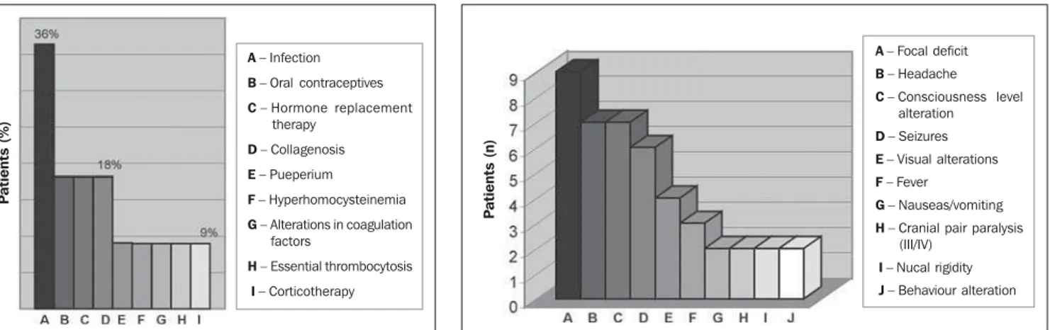
Factors related to the development of cerebral venous thrombosis (Figure 3) were identified in ten of 21 patients with data available in dossiers. Amongst the cases of infection, there were two of otitis, one of sinusitis and one of tonsillitis. Systemic infectious processes included gastroenteri-tis, pneumonia and urinary tract infection. Seventeen of 21 patients presented the clinical feature found by the time of
diag-nosis, as per Figure 4, focal deficit, head-aches and consciousness level alterations being the most frequent symptoms. For the remaining patients, this information was not available.
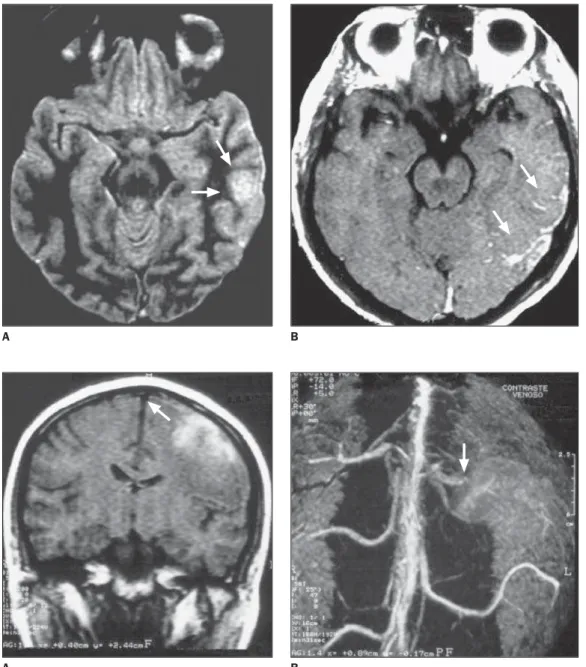
Cerebral venous thrombosis sites are demonstrated in Figure 5. The secondary radiological findings are described in Table 1. The main parenchymal manifestation was cortical and/or subcortical infarct or edema (Figures 6 and 7) in 16 patients (78%), eight (39%) of them presenting a hemorrhagic component (Figure 8). In four of 21 patients, hemorrhagic manifestations also were observed as intraparenchymal he-matoma with perilesional edema (19%).
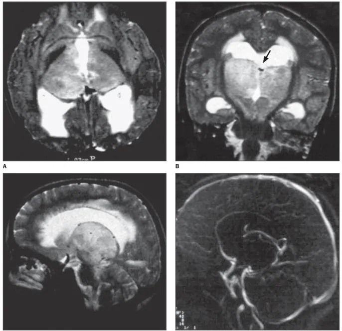
In five of 21 patients (24%) there was a thalamic-nuclear infarct or edema (Figure 9), four of them resulting from the straight sinus thrombosis and one from right trans-verse and sigmoid sinuses or ipsilateral jugular vein thrombosis.
In cases of straight sinus occlusion, the thrombus was evidenced at MRI of three patients, and in others, the thrombus was detected by MRA.
DISCUSSION
Cerebral venous thrombosis is a rela-tively rare condition when compared with vascular accidents of arterial origin(1). Al-though it may affect individuals in any age
Figure 1. Distribution of patients by sex.
Men (n = 3) Women (n = 18)
Figure 2. Distribution of age ranges – Female pa-tients (n = 18).
Figure 3. Frequency of predisposing factors (n = 10). Figure 4. Frequency of clinical manifestation (n = 17).
Figure 5. Venous thrombosis localization.
Table 1 MRI and MRA: secondary findings statistical analysis.
Findings
Cortical and/or subcortical edema or infarct Congestion/collateral circulation
Meningeal enhancement Thalamic/nuclear edema or infarct
Intraparenchymal hematoma White matter lesion
Hydrocephaly
Intraventricular hemorrhage
Subarachnoid hemorrhage
Total
Positive
16 13
5 5
4 3
1 1
1
49
Negative
5 8
16 16
17 18
20 20
20
140
p
0.01 0.001
0.82 0.82
0.34 0.16
0.08 0.08
0.08 Results
Figure 6. Flair MRI, axial view (A) showing bilateral, asymmetrical cortical alterations on frontal lobes, compatible with edema or infarcts (arrow) and presence of subarachnoid hemorrhage (arrowhead). Sagittal sinus presenting signal void inside suggesting flow patency (asterisk). 2D TOF MRA (B), showing signal intensity irregularities and alterations in the superior sagittal sinus, compatible with thrombosis and partial recanalization.
B A
A– Infection
B– Oral contraceptives
C– Hormone replacement therapy
D– Collagenosis
E– Pueperium
F– Hyperhomocysteinemia
G– Alterations in coagulation factors
H– Essential thrombocytosis
I– Corticotherapy
P
a
ti
e
n
ts
(%)
A– Focal deficit
B– Headache
C– Consciousness level alteration
D– Seizures
E– Visual alterations
F– Fever
G– Nauseas/vomiting
H– Cranial pair paralysis (III/IV)
I– Nucal rigidity
J– Behaviour alteration
P
a
ti
e
n
ts
(n
)
P
a
ti
e
n
ts
(n
)
A– Superior sagittal sinus
B– Left transverse sinus
C– Left sigmoid sinus
D– Straight sinus
E – Right transverse sinus
F– Right sigmoid sinus
G– Left jugular vein
H– Right jugular vein
I– Cortical veins
J– Cavernous sinuses (bilat-eral)
range, in the present study, only one child was affected. There was a prevalence in women between 16 and 36 years of age, differently from the male group (n = 3) were ages ranged between 46 and 59 years. The female prevalence corroborates the results reported in the literature.
Terazzi et al.(8) describe the duration time of the cerebral venous thrombosis, ac-cording the classification of Bousser et al., in acute (< 48 hours), subacute (between 48 hours and 30 days) and chronic (more than 30 days). According this classification, three patients presented an acute clinical picture, nine, subacute and four, chronic. The other could not be classified due the
absence of clinical data in the patients dos-siers.
The predisposing factors and clinical manifestation identified in the present sample were similar to those described in the literature(3,4,7,9,10).
At a lower rate, hematological alter-ations like essential thrombocytosis, hyperhomocysteinemia and other non-specified alterations of coagulation factors. Cases of venous thrombosis in the su-perficial system predominated, and just in eight (38.0%) of 21 cases the deep venous system was affected. In agreement with the literature, the present study found rare cases of cavernous sinus and cortical veins
thrombosis, principally isolated cases(7,10). Venous thrombosis and parenchymal hemorrhage, either in the form of hemor-rhagic infarct or parenchymal hematoma, occurred in 58% of cases. This finding has been described by several authors and is a result of lesion in vein walls, with rupture of the hematoencephalic barrier due intra-vascular hypertension in the occluded ter-ritory causing from petechial hemorrhages to intraparenchymal hematomas(3–7,10,11). Additionally, in one of these patients, a T2-weighted GRE* sequence demonstrated a large area of hemorrhage within a volumi-nous infarct and that, on a SE T1-weighted sequence was visualized just as discreet
Figure 7. T2-weighted MRI (A), axial view showing a small area of cortical hypersignal on the left temporal lobe, compatible with edema or infarct (arrows). Con-trast-enhanced T1-weighted MRI (B), axial view showing dural en-hancement and signs of adjacent venous congestion adjacent to the lesion (arrows).
A B
Figure 8. Non-contrast-enhanced T1-weighted MRI, coronal view (A) showing cortical-subcortical hem-orrhagic infarct on the left frontal lobe. The superior sagittal sinus presents normal signal (arrow). Gadolinium-enhanced 2D TOF MRA (B) shows the cortical vein signal interruption at left (arrow), compatible with thrombosis. Patent superior sagittal sinus.
foci of hypersignal. This finding was in agreement with the literature that reports a higher sensitivity of GRE sequences in re-lation to conventional SE and Fair se-quences for detection of parenchymal of parenchymal hemorrhages - even the small ones -, and in the identification of sub-arachnoid hemorrhage(7,10).
In the present study, one has not ob-served parenchymal abnormalities produc-ing a mass effect (characterized by fadproduc-ing of cortical and compression effect over the
ventricular system), without parenchymal signal alteration at the T2-weighted se-quence, according studies by Tsai et al.(6) and Yuh et al.(9).
Evidences of infarct or edema affecting the thalamus and basal nuclei were ob-served in five patients, this finding being related principally to straight sinus throm-bosis in four patients. In only one patient it was a result of right transverse and sig-moid sinuses and ipsilateral jugular vein thrombosis.
The second most frequent abnormality was the circulatory one, present in 62% of cases, and depicted by signs of venous con-gestion and collateral circulation diagnosed at MRI as intravascular enhancement after paramagnetic contrast agent injection(2).
Another frequent manifestation, also re-lated to circulatory alterations resulting from cerebral venous thrombosis was the presence of dural enhancement adjacent to parenchymal lesions (infarct or hematoma). This is explained by the fact that the dura
Figure 9. T2 weighted MRI, axial (A), coronal (B) and sagittal (C) views. Bilateral edema in thalami and basal nuclei, causing supratentorial hydrocephaly with signs of transependymal transudation. The internal cerebral veins present normal flow signal (arrow). Straight sinus with absent signal inside, possibly corre-sponding to hypointense thrombus. 2D TOF MRA (D). Absence of flow signal on the straight sinus, indicating thrombosis. Internal cerebral veins with preserved flow.
A B
venous thrombosis and slowing of the blood flow, enhance due the venous con-gestion in the affected territory(2).
A regression of parenchymal alterations was observed in the four patients with fol-low-up studies and there was just one case of death of a patient presenting left trans-verse and straight sinuses thrombosis, pro-gressing to a condition of intracranial hy-pertension and progressive decay of the neurological picture.
Thrombosis of the deep venous system is an uncommon condition with high mor-bidity and mortality rates, and the absence of typical clinical findings results in diag-nostic difficulty, mistakes and delayed treatment start(12,13).
In the present study, thrombosis of the deep venous system involving isolatedly the straight sinus, occurred in only one pa-tient. In all the other seven cases, thrombo-sis occurred concomitantly in structures of the superficial venous system. This pattern of involvement of multiple structures of both superficial and deep venous systems also has been reported by Lafitte et al.(5),
Ming et al.(12)and Crawford et al.(13), and has been attributed to a trend towards ret-rograde progression of the thrombus present in the superficial venous system.
In most of our cases, the main signs of the presence of thrombosis of the deep venous system were due to the localization of the secondary parenchymal manifesta-tions which were found in all of the patients with cerebral venous thrombosis, similar to data reported by Lafitte et al.(5). In half of cases (four patients) of the present study, there was a thalamic-nuclear infarct or edema, and in the other patients one has observed hemorrhagic infarct of varied dis-tribution in three cases, and diffuse alter-ation of the white matter in one patient.
The deep venous system present a more constant anatomy and parenchymal alter-ations occur in a more predictable pattern and may involve the thalami, basal nuclei and the superior portion of the cerebellum and mesencephalon(5,10). In cases where parenchymal alterations consist basically of bilateral or, more rarely, unilateral sig-nal alterations on the thalami, the deep venous thrombosis and infarct of the basi-lar artery and thalamus-perforating arteries
ies of Percheron) are included in the differ-ential diagnosis, especially if the symptoms are recent and with fast progression(14,15).
When alterations are cortical and/or subcortical, the diagnosis of infarct of ar-terial etiology should be the first hypoth-esis to be considered, since arterial throm-boembolism is much more frequent than cerebral venous thrombosis. However, ar-terial infarcts are characterized by well defined anatomical territory, which does not occur in venous infarcts involving the superficial system. For this reason, in cases of cortical and/or subcortical infarcts in atypical sites one should suspect cerebral venous thrombosis(4).
The superior sagittal sinus thrombosis should be taken into consideration in the presence of parenchymal alterations near the midline, involving frontal, parietal and occipital lobes. When bilateral, usually they are asymmetrical (3). An alteration of signal in the posterior region of the temporal lobe should raise suspect of an infarct by trans-verse sinus thrombosis on the same side of the lesion(3). The differential diagnosis of lesions in these sites includes arterial inf-arct, contusion and encephalitis(3).
In the present study, white matter signal alterations were not frequent and were identified in only three of the 21 patients evaluated (14,3%). In one of these cases, a diffuse alteration of the periventricular white matter signal attributed to a vasogenic edema in a 36-year old patient with a history of nephropathy and systemic arterial hypertension presenting signs of venous congestion and extensive collateral circulation observed at MRI, the dural si-nuses presenting absent signal. The MRA was inconclusive and the final diagnosis was achieved only by means of cerebral angiography that showed a diffuse throm-bosis of dural sinuses and deep venous system with an intense superficial and deep collateral circulation. Despite the scarcity of data in the literature on this subject, the significance of this finding is in the revers-ible character of these lesions which was observed in follow-up studies of this pa-tient after diagnosis and an appropriate treatment.
Only one patient presented hydroceph-aly attributed to accentuate edema in
sion over the third ventricle and supraten-torial hydrocephaly similar to those de-scribed in studies of Crombé et al.(16) and Crawford et al.(13). Although the cerebral venous thrombosis physiopathology indi-cates the possibility of ventricular system dilatation during the progression of the disease, this was not what we found in the present study(9).
Pitfalls were observed on MR images of three patients. In two of them there was a signal void inside the thrombosed dural sinus mimetizing flow patency. These pa-tients presented a period of symptoms de-velopment longer than two months and the radiological investigation occurred belat-edly.
In another patient, despite the presence of cortical infarcts in the frontal lobes, there was no superior sagittal sinus signal alter-ation, with signal void on all the weighting images. The final diagnosis of cerebral venous thrombosis was achieved by means of MRA and cerebral angiography compat-ible with thrombosis with partial recanali-zation of this sinus.
Both Lafitte et al.(5) and Zaiqiang et
al.(17) emphasize the importance of a radio-logical investigation to be performed within up to two weeks from the first clini-cal manifestations, a period where the thrombus may be iso- or hyperintense on T1-weighted sequences. During the chronic phase there is a progressive loss of signal intensity on T1- and T2-weighted se-quences and the thrombus presents a poorly defined aspect and may be partial or com-pletely recanalized, resulting in flow-void. This may generate hyposignal on MR im-ages, and is an acknowledged cause of false-negative results which reduces the method sensitiveness.
MRI provides superior anatomical de-tail when compared with CT, but is limited as regards differentiation between the hypersignal generated by the slowed blood flow and the presence of thrombosis, and, besides, the signal intensities change along time, with possible false-negative results both in hyperacute and chronic cases (18).
edema, or the existence of venous conges-tion and collateral circulaconges-tion demonstrated statistical significance (Table 1). It is pos-sible that these findings indicate a higher clinical and radiological suspicion of cere-bral venous thrombosis. Especially in these cases, the assessment by means of MRA is indicated to define the certainty diagnosis. An investigation for cerebral venous thrombosis should start with an evaluation of the parenchyma through conventional MRI with a protocol including T1- and T2-weighted sequences in at least two mutu-ally orthogonal views to avoid possible artifacts. The MRA is a non-invasive method that allows visualization of both deep and superficial systems, confirming the diagnosis in suspect or inconclusive cases diagnosed by MRI.
CONCLUSION
Cerebral venous thrombosis is an un-common cause of encephalic vascular ac-cidents, with a favorable prognosis because of the lesions reversibility. However, its diagnosis is highly dependent on the radi-ologist capacity to recognize the disease presentations, principally in cases where the diagnosis is suggested by parenchymal alterations rather than necessarily by
visu-alization of the thrombus itself. An accu-rate and rapid diagnosis allows an imme-diate treatment, reducing the morbidity and mortality rates.
REFERENCES
1. Ferro JM, Canhão P, Stam J, Bousser MG, Barina-garrementeria. Prognosis of cerebral vein and dural sinus thrombosis: results of the International Study on Cerebral Vein and Dural Sinus Throm-bosis (ISCVT). Stroke 2004;35:664–670. 2. Provenzale JM, Joseph GJ, Barboriak DP. Dural
sinus thrombosis: findings on CT and MR imag-ing and diagnostic pitfalls. AJR Am J Roentgenol 1998;170:777–783.
3. Connor SEJ, Jarosz JM. Magnetic resonance im-aging of cerebral venous sinus thrombosis. Clin Radiol 2002;57:449–461.
4. Lee SK, terBrugge KG. Cerebral venous throm-bosis in adults: the role of imaging evaluation and management. Neuroimaging Clin N Am 2003;13: 139–152.
5. Lafitte F, Boukobza M, Guichard JP, Reizine D, Woimant F, Merland JJ. Deep cerebral venous thrombosis: imaging in eight cases. Neuroradiol-ogy 1999;41:410–418.
6. Tsai FY, Wang AM, Matovich VB, et al. MR stag-ing of acute cerebral thrombosis: correlation with venous pressure measurements and implications for treatment and prognosis. AJR Am J Roent-genol 1995;16:1021–1029.
7. Mathews PV, Whitlow WD, Bryan RN. Cerebral ischemia and infarction. In: Atlas SW, editor. Mag-netic resonance of the brain and spine. 2nded. Philadelphia: Lippincott-Raven, 2002:537–601. 8. Terazzi E, Mittino R, Cerrato P, et al. Cerebral venous thrombosis: a retrospective multicentre study of 48 patients. Neurol Sci 2005;25:311– 315.
9. Yuh WTC, Simonson TM, Wang AM, et al. Ve-nous occlusive disease: MR findings. AJNR Am J Neuroradiol 1994;15:309–316.
10. Osborn AG. Acidentes vasculares. In: Osborn AG, editor. Diagnóstico neurorradiológico. Rio de Janeiro: Revinter, 1999;385–395.
11. Bianchi D, Maeder P, Bogousslavsky J, Schnyder P, Meuli RA. Diagnosis of cerebral thrombosis with routine magnetic resonance: an update. Eur Neurol 1998;40:179–190.
12. Shuhong M, Zengfei QI, Luning W, Ke Z. Deep cerebral thrombosis in adults. Chin Med J 2002; 115:395–397.
13. Crawford SC, Digre KB, Palmer CA, Bell DA, Osborn AG. Thrombosis of deep venous drainage of the brain in adults: analysis of seven cases with review of the literature. Arch Neurol 1995;52: 1101–1108.
14. Bell DA, Davis LW, Osborn AG, Harnsberger HR. Bithalamic hiperintensity on T2 weighted MR: vascular causes and evaluation with MR angiog-raphy. AJNR Am J Neuroradiol 1994;15:893– 899.
15. Herrmann KA, Sporer B, Yousay TA. Thrombo-sis of the internal cerebral vein associated with transient unilateral thalamic edema: a case report and review of the literature. AJNR Am J Neuro-radiol 2004;25:1351–1355.
16. Crombé D, Haven F, Gille M. Isolated deep ve-nous thrombosis diagnosed on CT and MR im-aging. A case study and literature review. JBR-BTR 2003;86:257–261.
17. Zaiqiang Z, Long J, Li W. Cerebral venous throm-bosis: a clinical study of 23 cases. Clin Med J 2000;113:1043–1045.