Copyright © 2008 by Sociedade Brasileira de Pediatria
O
RIGINALA
RTICLEFunctional capacity assessment during exercise in children
and adolescents with post-infectious bronchiolitis
obliterans
Rita Mattiello,1 Edgar E. Sarria,2 Ricardo Stein,3 Gilberto Bueno Fischer,4
Helena Teresinha Mocelin,5 Sergio Saldanha Menna Barreto,4
João Antônio Bonfadini Lima,6 Diego Brandenburg7
Abstract
Objective:To assess functional capacity during exercise in children and adolescents with post-infectious bronchiolitis obliterans (PIBO).
Methods:20 children with PIBO, aged 8-16 years old, and in follow-up at an outpatient clinic carried out cardiopulmonary exercise testing (CPET), a 6-minute walk test (6MWT) and pulmonary function tests (PFT), according to American Thoracic Society (ATS), European Respiratory Society (ERS) and American College of Chest Physicians (ACCP) guidelines. Results were expressed as percentages of predicted reference values: Armstrong’s for CPET, Geiger’s for 6MWT, Knudson’s for spirometry, and Zapletal’s for plethysmography.
Results:Mean age (± SD) was 11.4±2.2 years; 70% were boys; mean weight: 36.8±12.3 kg; mean height: 143.8±15.2 cm. When compared to reference values, PFT detected lower airflows (spirometry) and higher volumes (plethysmography). Eleven patients had reduced peak VO2values in CPET (< 84% predicted). The mean distance
walked (6MWT) was 77.0±15.7% of predicted (512±102 m). Peak VO2was not correlated with 6MWT, but it was
correlated with FVC (L) (r = 0.90/p=0.00), with FEV1(L) (r = 0.86/p = 0.00) and with RV/TLC, both in absolute values
(r = -0.71/p = 0.02) and as percentages of predicted values (r = -0.63/p = 0.00).
Conclusions:The majority of these post-infectious bronchiolitis obliterans patients exhibited reduced functional capacity, exhibited during both CPET and the 6MWT. Due to its greater feasibility, 6MWT could be an alternative where CPET is not available.
J Pediatr (Rio J). 2008;84(4):337-343:Bronchiolitis obliterans, children, exercise.
Introduction
Post-infectious bronchiolitis obliterans (PIBO) is a chronic lung disease that results from some type of aggression to the lower respiratory tract in previously healthy children. From the pathological perspective, PIBO is characterized by
obstruction of the lumen with granulation tissue, inflamma-tion and fibrosis with obliterainflamma-tion of small airways and bronchiectasis.1
Functional assessment of PIBO patients at rest demon-strate airflow limitation, characterizing an obstructive venti-latory disorder that is generally severe and irreversible.2This
1. MSc. Doctoral Student in Pediatrics, Universidade Federal do Rio Grande do Sul (UFRGS), Porto Alegre, RS, Brazil. 2. PhD. Research assistant, Universidad de Santiago de Chile, Chile.
3. PhD. Professor, Cardiology Postgraduate Course, UFRGS, Porto Alegre, RS, Brazil. 4. PhD. Professor, Pediatrics Postgraduate Course, UFRGS, Porto Alegre, RS, Brazil.
5. PhD. Pediatric Pulmonology Section, Hospital da Criança Santo Antônio, Porto Alegre, RS, Brazil. 6. MSc. Doctoral Student in Pulmonology, UFRGS, Porto Alegre, RS, Brazil.
7. MD. Pediatric Pulmonology Section, Hospital da Criança Santo Antônio, Porto Alegre, RS, Brazil.
No conflicts of interest declared concerning the publication of this article.
Suggested citation:Mattiello R, Sarria EE, Stein R, Fischer GB, Mocelin HT, Barreto SS, et al. Functional capacity assessment during exercise in children and adolescents with post-infectious bronchiolitis obliterans. J Pediatr (Rio J). 2008;84(4):337-343.
Manuscript received Feb 12 2008, accepted for publication Apr 23 2008.
doi:10.2223/JPED.1807
airflow restriction can limit physical activity levels (PAL) among these individuals.
Exercise capacity testing is considered part of the multi-dimensional assessment of patients with chronic lung dis-ease, since it objectively evaluates cardiac, respiratory, muscular, and metabolic systems.3,4In this context, cardiop-ulmonary exercise testing (CPET) is considered the gold stan-dard for investigating the causes of exercise intolerance.3,5
There are few studies evaluating the pulmonary function of PIBO patients during exercise. Considering the importance of PAL in children and adolescents, particularly those with res-piratory problems, and in view of the lack of scientific knowl-edge on the functional behavior of these individuals during exercise, the aim of this study is to assess their functional capacity during exercise.
Methods
Children and adolescents aged 8 to 16 years old and with previous diagnosis of PIBO were enrolled. They were in follow-up at the pediatric pulmonology outpatient clinic from one of two hospitals, Hospital Materno Infantil Presidente Var-gas (HMIPV) or Hospital da Criança Santo Antônio (HCSA), both in Porto Alegre, RS, Brazil. Diagnosis was based on the following combination of clinical, radiological and functional criteria:1,21) history of acute pulmonary infection in a previ-ously healthy child less than 2 years old; 2) permanent respi-ratory signs and symptoms (e.g. wheezing, crepitations, coughing) 4 weeks after the initial event; 3) high resolution computerized tomography with abnormalities characteristic of bronchiolitis obliterans (BO), such as mosaic pattern, bron-chiectasis and atelectasis; 3) airflow limitation demonstrated by pulmonary function tests; and 4) exclusion of other chronic lung conditions with a clinical course involving persistent res-piratory symptoms, such as severe asthma, cystic fibrosis, alpha-1-antitrypsin deficiency and immunodeficiencies, among others.
Patients were excluded if they had: 1) cognitive or motor limitations, or any other condition that could compromise per-formance of the tests; 2) pulmonary or systemic hyperten-sion and electrocardiographic findings suggestive of heart disease (rhythm/conduction disorders and/or ST segment abnormalities); 3) deterioration of their respiratory status during the 30 days prior to the tests, denoted by worsening of signs and symptoms (coughing, wheezing, expectoration, dyspnea) or infection.
Patients were instructed not to drink coffee, tea or soft drinks, nor to eat chocolate or to perform exercise during the 2 hours prior to the tests and also not to use bronchodilators prior to testing, 8 hours for short acting and 24 hours for long acting.
This study was approved by the Research Ethics Commit-tees at the HMIPV and the Hospital de Clínicas de Porto Alegre.
The children indicated their willingness to participate ver-bally and their guardians signed free and informed consent forms.
Three appointments were made for each participant to be tested. At the first visit, patient history was taken, a physical examination carried out, nutritional status and PAL were assessed and the patient performed a 6-minute walk test (6MWT).6The pulmonary function tests were performed at the second visit and the patient underwent CPET at the third appointment. All interviews were conducted by the same interviewer, and all clinical assessments were carried out by a single pediatric pulmonologist. The minimum interval between appointments was 2 days, and the maximum was 14 days.
Nutritional assessment
Weight and height were measured for nutritional assess-ment. These were then used to calculate body mass index (kg/ m2), which was classified according to the Centers for Disease Control (CDC) reference values, by age and sex.7All measure-ments were taken by the same examiner, with instrumeasure-ments properly calibrated and employing standardized measure-ment techniques.8
Pulmonary function
American Thoracic Society (ATS) technical procedures as well as acceptability and reproducibility criteria were used for pulmonary function tests.9,10Spirometry and plethysmogra-phy were carried out using a Master-Screen equipment (Jae-ger, Germany), and the following parameters were recorded: 1) spirometry: forced vital capacity (FVC), forced expiratory volume in 1 second (FEV1), FEV1to FVC ratio (FEV1/FVC), forced expiratory flow between 25-75% of FVC (FEF25-75%); and 2) plethysmography: total lung capacity (TLC), intratho-racic gas volume (ITGV), residual volume (RV), RV to TLC ratio (RV/TLC). These data are given as percentage of predicted values according to Knudson11for spirometry, and Zapletal12 for plethysmography.
Physical Activity Questionnaire
The full version of the International Physical Activity Ques-tionnaire (IPAQ) was applied by interview. The time spent per-forming the different IPAQ domains was calculated from the product of the duration (minutes/day) and the frequency (days/week) patients reported for each activity type and then, from these results, they were categorized as having low, mod-erate or intense PAL.6
Submaximal exercise test
(Nonin’s WristOx®3100, USA), peak expiratory flow (PEF) and the Borg scale (perceived exertion scale for lower limbs and dyspnea). Peak expiratory flow was assessed at 3, 5, 10, 15 and 20 minutes after the test. A fall in PEF was considered significant if there was a variation of ≥ 15% between post and pre measurement [((PEF post - PEF pre)/PFE post) x 100]. Total distance walked was also recorded. Each child was asked to walk as far as possible in 6 minutes, along a 30 m corridor. Once a minute they were encouraged verbally, using phrases recommended by the ATS. The following were criteria for halt-ing the test: intense tiredness and dyspnea as expressed by the patient; SaO2≤ 80% or refusal to continue the test. The reference values published by Geiger et al. were used to cal-culate the percentage of predicted distance that was actually walked.14
In order to assess maximum heart rate during the 6MWT and CPET, an estimated heart rate formula was employed, based on patient’s age [205 - (0.5 x age in years)].15
Maximal exercise test
Cardiopulmonary exercise testing was conducted by a car-diologist proficient in the method, with the aid of a pediatric pulmonologist and a physiotherapist. Tests were carried out in accordance with ATS and the American College of Chest Physicians recommendations.5All tests were performed dur-ing the morndur-ing, at room temperature of 22 to 24 ºC and rela-tive humidity of aproximately 60%.
Maximal cardiopulmonary testing was carried out using a computerized system (Metalyzer 3B, Cortex, Germany) and a treadmill (Inbramed®KT 10200, Brazil) with velocity in the range of 0 to 16 km/h (0 to 10 mph) and a ramp angle ranging from 0 to 26%.
At the start of the test, the subject walked for around two minutes to become accustomed to the treadmill. The proto-col began at a velocity of 2.4 km/h with a 2% incline. Velocity was increased every 20 s (0.1 to 0.2 km/h), and inclination was increased every 60 s (0.1 to 0.2%). During the test patients were encouraged by the same person to maintain their rhythm until exhaustion or until limiting symptoms appeared. The exercise intensity was calculated with the intention that the test lasted approximately 8 to 10 minutes.
The following variables were analyzed (breath by breath) using a previously validated system:16oxygen consumption (VO2), in mL/min, STPD (Standard Temperature and Pres-sure, Dry); carbon dioxide output (VCO2), in mL/min, STPD; respiratory exchange coefficient (R); minute volume (VE), in L/min, BTPS (Body Temperature, Pressure Saturated); respi-ratory rate (RR) and heart rate (HR). Peak VO2was defined as the highest value observed during the last 20 s of exercise and we used reference values published by Armstrong et al..17
Cardiopulmonary parameters were recorded before and after the test pulmonary and cardiac auscultation, SaO2, RR, HR, blood pressure, PEF and Borg). Oxygen saturation and
electrocardiographic tracing were continuously monitored, using, respectively, a pulse oximeter (Nonin’s WristOx®3100, United States) and a cardiac monitor (Nikon Kohden Corpo-ration®, Japan). Peak expiratory flow was measured at 3, 5, 10, 15 and 20 minutes after the test. Blood pressure (BP) was measured using an aneroid sphygmomanometer (Tycos®, United States).
Statistical analysis
Data were analyzed using SPSS version 14.0 (SPSS Inc, United States). The distribution of variables was analyzed using the Kolmogorov-Smirnov test. Continuous variables are presented in the form of means and standard deviations or medians and interquartile ranges; categorical measures are given as absolute and relative frequencies. The results of pul-monary function and exercise tests are given as percentages of predicted values. Student’sttest was used for compari-sons between means of variables with normal distribution, while the Wilcoxon test was used for measures that exhibited asymmetrical distribution. Correlations between pulmonary function and exercise test variables were assessed using Pear-son’s correlation coefficient (r). Based on a sample of 20 patients, accepting an alpha error of 5% and a beta error of 20%, correlations were accepted at r ≥ 0.6.
Results
A total of 73 children with PIBO were in follow-up at the two institutions. Of these, 20 met the inclusion criteria and took part in the study. Their mean age was 11 years, and 70% were male. In terms of nutritional status, the majority were well-nourished (16 patients), three were classified as mal-nourished and one as overweight.
The IPAQ results classified 17 patients (85%) as active (four with intense PAL), three (15%) were classified as not very active and none of the patients were classified as seden-tary. No correlations were observed between PAL and peak oxygen consumption (r = 0.14/p = 0.57).
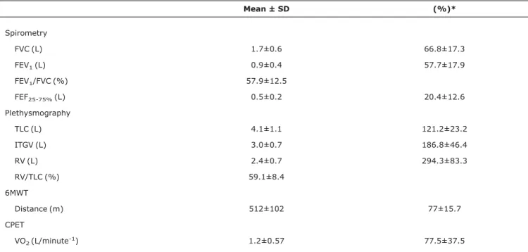
When compared to the reference population, these patients exhibited reduced forced flows on spirometry, and increased volumes on plethysmography (Table 1).11,12
Three patients did not perform the CPET because they did not attend their appointments. Three of the 17 who did undergo CPET exhibited dyspnea and an important fall in satu-ration at the end of the test. None of the subjects exhibited abnormalities on electrocardiogram before, during or after exercising, and the respiratory quotient (R) reached a mean of 1.0±0.6. Mean peak VO2(absolute value) was reduced in comparison with reference values (1.2±0.5 vs. 1.7±0.6; p = 0.00). This reduction is more evident when mean percentage of the predicted value is compared with the cutoff point of nor-mality (≥ 84%)17(Table 1).
(512±102 vs. 665±33.5 m; p = 0.000), which, in terms of percentage of predicted, was lower than the value consid-ered normal (≥ 80%).14Distance was not correlated with peak oxygen consumption, either when evaluated as absolute fig-ures, (r = -0.28/p = 0.29) or as percentages of the predicted distance (r = -0.50/p = 0.85).
When we analyzed HR, RR and the Borg scale after the exercise test, we observed that these parameters were sig-nificantly higher in the CPET than in the 6MWT. There was a transitory drop in saturation of more than 4% in three patients in the 6MWT and in 12 patients in the CPET. A reduction in PEF
(> 15%) was observed in three individuals in the 6MWT and three individuals during the CPET. On average, for the CPET versus the 6MWT, patients reached 90versus60% of maxi-mum heart rate (Table 2).
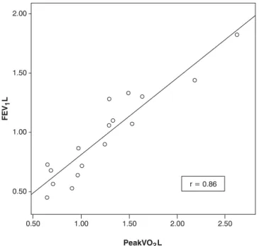
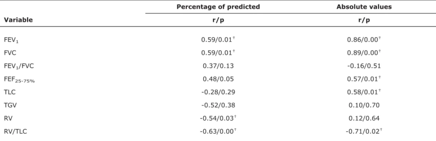
Regarding correlations between CPET and pulmonary function tests at rest (Table 3), peak VO2observed during CPET correlated with absolute volumes of FVC (r = 0.90/p = 0.00), FEV1(r = 0.86/p = 0.00) and RV/TLC (r = -0.71/p = 0.02) (Figure 1). When peak VO2was treated as percentage of predicted, the only parameter with which it correlated was RV/TLC (-0.63/p = 0.00) (Table 3).
Table 1- Characteristics of pulmonary function at rest in patients with post-infectious bronchiolitis obliterans
Mean ± SD (%)*
Spirometry
FVC (L) 1.7±0.6 66.8±17.3
FEV1(L) 0.9±0.4 57.7±17.9
FEV1/FVC (%) 57.9±12.5
FEF25-75%(L) 0.5±0.2 20.4±12.6
Plethysmography
TLC (L) 4.1±1.1 121.2±23.2
ITGV (L) 3.0±0.7 186.8±46.4
RV (L) 2.4±0.7 294.3±83.3
RV/TLC (%) 59.1±8.4
6MWT
Distance (m) 512±102 77±15.7
CPET
VO2(L/minute-1) 1.2±0.57 77.5±37.5
6MWT = 6-minute walk test; CPET = cardiopulmonary exercise testing; FEF25-75%= forced expiratory flow between 25-75% of FVC; FEV1= forced
expiratory volume in 1 second; IFVC = forced vital capacity; RV = residual volume; SD = standard deviation; TGV = intrathoracic gas volume; TLC = total lung capacity; VO2= peak oxygen consumption.
* Percentage of predicted.
Table 2- Comparison of variables for the 6-minute walk test and the cardiopulmonary exercise test
6-minute walk test Cardiopulmonary exercise test
Mean ± SD Mean ± SD p
Respiratory rate (PE) 30.0±3.7 57.4±9.0 0.000*
Heart rate (PE) 124.6±8.7 182.5±11.6 0.000*
Heart rate (MP) 62.5±9.4 91.6±5.8 0.000*
Minimum SaO2 94.1±3.2 90.2±4.6 0.001*
Borg (PE) Median (quartiles) Median (quartiles)
Fatigue in legs 0 (0-3) 3 (1-7) 0.005*
Dyspnea 1 (0-3) 2 (1-6) 0.047*
MP = predicted maximum heart rate; PE = post-exercise; SaO2min = lowest oxygen saturation during exercise; SD = standard deviation.
Discussion
The majority of patients exhibited a reduced functional capacity during exercise when submitted to CPET and 6MWT, demonstrated by their both reduced peak oxygen consump-tion and total distance covered when compared to reference values in healthy population.17
Research in pediatric patients with chronic lung condi-tions has reported contradictory findings on the physical per-formance of this population.4,18-20Previous studies have reported that a significant number of asthmatic children exhibit a limited capacity for exercise, characterized by reduced peak oxygen consumption.21,22Fanelli et al. carried out a study on 38 children with moderate to severe asthma and found that 24 patients had lower exercise capacity than healthy population.23 However, other studies that also included patients with moderate to severe asthma have found that these patients can have a normal exercise capacity.4,24
Studies in children and adolescents with cystic fibrosis, a limited exercise capacity is generally associated with worse pulmonary function. Although these parameters correlate to each other, these correlations vary greatly between them, par-ticularly between peak VO2and FEV1, two indicators that showed a good correlation in our study (Figure 1). In children and adolescents, this variability can be justified by the exist-ence of other factors related to exercise capacity, such as height, weight, puberal stage and PAL. Consequently, it is not possible to predict physical fitness solely by means of pulmo-nary function tests at rest.3-5,25
The functional characteristics of our patients, at rest, are not different from those found in other studies with PIBO, that is, characteristics of an obstructive respiratory disorder.2,26,27 Correlations found in this study between peak VO2and FEV1 and FVC with RV/TLC suggests that, in patients with PIBO, the greater the air trapping, the greater the compromise of pulmonary capacity during exercise. Nevertheless, other studies done with respiratory patients have not demon-strated the association between physical capacity and pulmo-nary function findings.4,28-30
Mocelin et al. applied the 6MWT as a submaximal test, using the same protocol we used here, to assess exercise capacity in a sample of 19 patients with PIBO.31The most important findings of that study were that 37% of the patients exhibited a fall in oxygen saturation during the test and that the total distance walked did not correlate with spirometric data. In our study, just 15% of the patients exhibited a reduc-tion in SaO2during the 6MWT. Despite the operational advan-tages of the 6MWT, it does not offer the possibility of measuring the intensity needed to perform prolonged exer-cise, nor of quantifying its limiting factors or defining co-existing pathophysiologic mechanisms; for these pur-poses, CPET is the method of choice.3,5,32
As would be expected, when submitted to CPET, our patients exhibited significantly higher HR, RR and Borg val-ues, and also a greater drop in saturation compared to the 6MWT. These findings are similar to what can be found in other studies using incremental testing in patients with chronic res-piratory conditions. Therefore, it appears to us that maximal testing (CPET with progressive increase of the workload) can induce higher results in cardiorespiratory variables and, con-sequently, provide a wider range of clinically relevant data when compared with a submaximal test such as the 6MWT.33
Some authors recommend assessing PAL using standard-ized questionnaires.34,35However, there is conflicting evi-dence in the literature on the association between oxygen consumption and reported habitual PAL.17,20Added to this is the scarcity of this type of instruments in Brazil, particularly for younger population.36In this situation, researchers have to use questionnaires designed for adults, like the IPAQ. In our study, the IPAQ results patients with PIBO did not corre-late with exercise test results. We believe that the absence of specific domains for activities that children take part of and the resulting under/overestimation of other activities is the main cause of these results.
The principal limitation of our study is the lack of a control group with which to compare the exercise testing results. This would also compensate for the relatively small number of patients, which limits plausible inferences that could be made based on the results, if we considering the variability of param-eters included in the study. Nevertheless, since this is not a very prevalent disease, the rather homogenous group of
r = Pearson’s correlation coefficient; FEV1(L) = forced expiratory volume in 1
second (Liters); VO2(L) = oxygem consumption
patients studied offers an important initial view of their patho-physiologic behavior when submitted to exercise. We can con-clude, based on our results, that the majority of patients with PIBO studied exhibit reduced capacity for exercise, both in CPET and the 6MWT. Due to the ease of application of the 6MWT, it can be used as an alternative for the initial evalua-tion of patients at places that do not have access to CPET equipment.
Acknowledgements
We would like to thank Globalmed for one of their oxime-ters to monitor patients through all the field study period. We are also grateful to Vânia Hirakata for supervising the statis-tical analysis and Dr. Maria Ângela Moreira for helping us with the logistics in the pulmonary function testing.
References
1. Kim CK, Kim SW, Kim JS, Koh YY, Cohen AH, Deterding RR, et al.Bronchiolitis obliterans in the 1990s in Korea and the United States.Chest. 2001;120:1101-6.
2. Colom AJ, Teper AM, Vollmer WM, Diette GB.Risk factors for the development of bronchiolitis obliterans in children with bronchiolitis.Thorax. 2006;61:503-6.
3. ERS Task Force, Palange P, Ward SA, Carlsen KH, Casaburi R, Gallagher CG, et al.Recommendations on the use of exercise testing in clinical practice. Eur Respir J. 2007;29:185-209.
4. Nixon PA.Role of exercise in the evaluation and management of pulmonary disease in children and youth.Med Sci Sports Exerc. 1996;28:414-20.
5. American Thoracic Society; American College of Chest Physicians.ATS/ACCP Statement on cardiopulmonary exercise testing.Am J Resp Crit Care Med. 2003;167:211-77.
6. Craig CL, Marshall AL, Sjostrom M, Bauma AE, Booth TH, Ainsworth BE, et al.International physical activity questionnaire: 12-country reliability and validity. Med Sci. Sports Exerc. 2003; 35:1381-95.
7. Kuczmarski RJ, Ogden CL, Guo SS, Grummer-Strawn LM, Flegal KM, Mei Z, et al. 2000 CDC Growth Charts for the United States: methods and development. Vital Health Stat 11. 2002;246: 1-190.
8. Accioly E, Saunders C, Lacerda AL. Nutrição em obstetrícia e pediatria. Rio de Janeiro: Cultura Médica; 2003.
9. Miller MR, Hankinson J, Brusasco V, Burgos F, Casaburi R, Coates A.Standardisation of spirometry.Eur Respir J. 2005;26: 319-38.
10. Wanger J, Clausen JL, Coates A, Pedersen OF, Brusasco V, Burgos F, et al.Standardisation of the measurement of lung volumes.Eur Respir J. 2005;26:511-22.
11. Knudson RJ, Lebowitz MD, Holberg CJ, Burrows B.Changes in the normal maximal expiratory flow-volume curve with growth and aging.Am Rev Respir Dis. 1983;127:725-34.
12. Zapletal A, Motoyama EK, Van De Woestijne KP, Hunt VR, Bouhuys A.Maximum expiratory flow-volume curves and airway conductance in children and adolescents. J Appl Physiol. 1969; 26:308-16.
13. ATS Committee on Proficiency Standards for Clinical Pulmonary Function Laboratories.ATS statement: guidelines for the six-minute walk test.Am J Resp Crit Care Med. 2002;166: 111-7.
14. Geiger R, Strasak A, Treml B, Gasser K, Kleinsasser A, Fischer V, et al.Six-minute walk test in children and adolescents.J Pediatr. 2007;150:395-9.
15. Hammond HK, Froelicher VF.Normal and abnormal heart rate responses to exercise.Prog Cardiovasc Dis. 1985;27:271-96.
16. Meyer T, Georg T, Becker C, Kindermann W.Reliability of gas exchange measurements from two different spiroergometry systems.Int J Sports Med. 2001;22:593-7.
Table 3- Correlations between peak oxygen consumption and pulmonary function variables at rest*
Percentage of predicted Absolute values
Variable r/p r/p
FEV1 0.59/0.01† 0.86/0.00†
FVC 0.59/0.01† 0.89/0.00†
FEV1/FVC 0.37/0.13 -0.16/0.51
FEF25-75% 0.48/0.05 0.57/0.01†
TLC -0.28/0.29 0.58/0.01†
TGV -0.52/0.38 0.10/0.70
RV -0.54/0.03† 0.12/0.64
RV/TLC -0.63/0.00† -0.71/0.02†
FEF25-75%= forced expiratory flow between 25-75% of FVC; FEV1= forced expiratory volume in 1 second; FVC = forced vital capacity; RV = residual
volume; TGV = intrathoracic gas volume; TLC = total lung capacity.
* Correlations between variables in absolute values and as percentages of predicted (Knudson: spirometry, Zapletal: plethysmography, Armstrong: peak oxygen consumption).
r = Pearson’s correlation coefficient.
17. Armstrong N, Welsman JR.Assessment and interpretation of aerobic fitness in children and adolescents.Exerc Sport Sci Rev. 1994;22:435-76.
18. Santuz P, Baraldi E, Filippone M, Zacchello F. Exercise performance in children with asthma: is it different from that of healthy controls?Eur Respir J. 1997;10:1254-60.
19. Selvadurai HC, McKay KO, Blimkie CJ, Cooper PJ, Mellis CM, Van Asperen PP.The relationship between genotype and exercise tolerance in children with cystic fibrosis.Am J Resp Crit Care Med. 2002;165:762-5.
20. Pianosi PT, Davis HS.Determinants of physical fitness in children with asthma. Pediatrics. 2004;113:e225-9.
21. Clark CJ, Cochrane LM.Assessment of work performance in asthma for determination of cardiorespiratory fitness and training capacity. Thorax 1988;43:745-9.
22. Strunk RC, Rubin D, Kelly L, Sherman B, Fukuhara J. Determination of fitness in children with asthma. Use of standardized tests for functional endurance, body fat composition, flexibility, and abdominal strength.Am J Dis Child. 1988;142:940-4.
23. Fanelli A, Cabral AL, Neder JA, Martins MA, Carvalho CR.Exercise training on disease control and quality of life in asthmatic children. Med Sci. Sports Exerc. 2007;39:1474-80.
24. Voy RO. The U.S. Olympic Committee experience with exercise-induced bronchospasm, 1984. Med Sci. Sports Exerc. 1986;18:328-30.
25. Pianosi P, LeBlanc J, Almudevar A.Relationship between FEV1 and peak oxygen uptake in children with cystic fibrosis.Pediatr Pulmonol. 2005;40:324-9.
26. Zhang L, Silva FA.Bronquiolite obliterante em crianças.J Pediatr (Rio J). 2000;76:185-92.
27. Teper A, Fischer GB, Jones MH.Seqüelas respiratórias de doenças virais: do diagnóstico ao tratamento.J Pediatr (Rio J). 2002;78:187-94.
28. Celli BR, Cote CG, Marin JM, Casanova C, Mendes de Oca M, Mendez RA, et al.The body-mass index, airflow obstruction, dyspnea, and exercise capacity index in chronic obstructive pulmonary disease.N Engl J Med. 2004;350:1005-12.
29. Garfinkel SK, Kesten S, Chapman KR, Rebuck AS.Physiologic and nonphysiologic determinants of aerobic fitness in mild to moderate asthma.Am Rev Respir Dis. 1992;145:741-5.
30. Neder JA, Fernandes AL, Silva AC, Cabral AL, Nery LD. Relationship between aerobic fitness and clinical indicators of asthma severity in children. J Pneumologia. 1998;24:1-8.
31. Mocelin HT, Fischer GP, Iriar KL, Cunha LS. Evaluación clínica y funcional de niños con bronquiolitis obliterante post-infecciosa con seguimiento a largo plaz. Rev Chil Pediatr. 2004;75:12-7.
32. Roca J, Rabinovich R.Clinical exercise testing. Eur Respir J. 2005; 31:146-65.
33. Rosa FW, Camelier A, Mayer A, Jardim JR.Evaluating physical capacity in patients with chronic obstructive pulmonary disease: comparing the shuttle walk test with the encouraged 6-minute walk test.J Bras Pneumol. 2006;32:106-13.
34. Nixon PA, Orenstein DM, Kelsey SF.Habitual physical activity in children and adolescents with cystic fibrosis.Med Sci Sports Exerc. 2001;33:30-5.
35. Rogers D, Prasad SA, Doull I.Exercise testing in children with cystic fibrosis. J R Soc Med. 2003;96 Suppl 43:23-9.
36. Guedes DP, Lopes CC, Guedes JE.Reprodutibilidade e validade do questionário internacional de atividade física em adolescentes. Rev Bras Med Esporte 2005;11:151-8.
Correspondence: Rita Mattiello Rua Machadinho, 465