Inverse Correlation between Testosterone and Ventricle Ejection
Fraction, Hemodynamics and Exercise Capacity in Heart Failure
Patients with Erectile Dysfunction
Edimar A. Bocchi, Vitor O. Carvalho, Guilherme V. Guimaraes
Laboratory of Heart Failure and Transplantation, Heart Institute, Incor, University of Sao Paulo, SP, Brazil
ABSTRACT
Background: Neurohormonal activation and abnormalities in growth hormone and testosterone concentrations have been reported in heart failure (HF). Erectile dysfunction(ED) is common in these patients and contributes to a low quality of life. No data are known regarding the correlation between testosterone and hemodynamics, exercise capacity and cardiac function in HF patients with ED, a marker of endothelial dysfunction. The aim of this study was to correlate testosterone levels with cardiac function, hemodynamic and exercise capacity in HF patients with ED.
Materials and Methods: Fifteen HF patients underwent a six-minute treadmill cardiopulmonary walking test (6’CWT) and, ten minutes later, a maximum cardiopulmonary exercise test. Also, testosterone and other hormones were determined at rest.
Results:Among hemodynamic variables only diastolic blood pressure on 6’CWT was correlated with testosterone levels(r =- 0.66, p = 0.007). The variables on exercise tests, VE/VCO2 slope and oxygen consumption did not show any correlation, except the distance at 6’CWT (r = 0.50, p = 0,047). Right and left ventricle ejection fraction showed inverse correlation with testosterone (r =- 0.55, p = 0.03 and r =- 0.69, p = 0.004 respectively).
Conclusion:Testosterone levels correlated directly with distance at six-minute cardiopulmonary walk test and inversely with diastolic blood pressure, right and left ventricle ejection fraction in heart failure patients with erectile dysfunction. Further elucidation of mechanisms as regards testosterone action in these patients is warranted.
Key words: heart failure; hemodynamics; physical activity; testosterone; erectile dysfunction Int Braz J Urol. 2008; 34: 302-12
INTRODUCTION
Heart failure (HF) can be considered as the
ODVW VWDJH RI KHDUW GLVHDVH DQG D VLJQL¿FDQW FDXVH
of mortality and morbidity worldwide (1). The left ventricular systolic dysfunction and limited exercise capacity manifested by breathlessness and fatigue are determinants of mortality and clinical events in the follow-up of HF patients. (2,3). Multiple mechanisms
have been reported to be related to exercise capac-ity including diastolic and systolic cardiac function,
UHÀH[ PHWDEROLF YDVFXODU DQG PXVFXODU UHVSRQVH
(3). Sexual satisfaction is an important component
WKDWLQÀXHQFHVTXDOLW\RIOLIHLQ+)SDWLHQWV(UHFWLOH
In the physiopathology of chronic HF, the neurohormonal hypothesis for progression of heart failure has been considered of greater interest than the original hemodynamic mechanisms (5). Activation of sympathetic nervous system, renin-angiotensin-aldo-sterone system, arginine vasopressin, and endothelins are considered as neurohormonal targets in the treat-ment of HF. However, other hormonal abnormalities
KDYHEHHQUHSRUWHGLQ+)VXFKDVVLJQL¿FDQWGHFUHDVH
in growth hormones, insulin-like growth factor I and testosterone concentrations (6). The hormonal and cytokine activation contributes to peripheral muscle tissue wasting as well as anabolic/catabolic imbal-ance (7). In men, testosterone seems to play a role
LQGHWHUPLQLQJDQDEROLFIXQFWLRQDQWLLQÀDPPDWRU\
and vasodilator processes. No data has been reported as regards testosterone’s effect on hemodynamics, cardiac function and exercise capacity in HF patients with ED.
The aim of this study was to evaluate the cor-relation between serum testosterone levels and cardiac function, hemodynamics, and exercise capacity in HF patients with ED. In addition, we determined a HF
KRUPRQHSUR¿OHVHUXPOHYHOVRISURODFWLQOXWHLQ -izing hormone, follicle-stimulating hormone, resting norepinephrine, and thyroid hormones.
MATERIALS AND METHODS
Studied Population
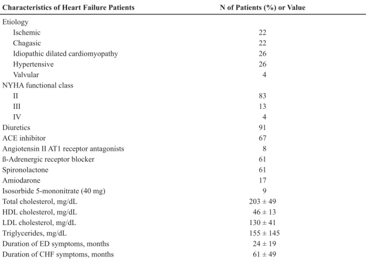
Fifteen randomized chronic male HF pa-tients, 50 ± 10 years with ED (Table-1) for at least four months, in a steady relationship and presenting
LQWHUHVWLQVH[ZHUHLQFOXGHG('ZDVGH¿QHGDVWKH
inability to achieve or maintain a durable erection to permit satisfactory sexual intercourse (the 5th-item of the International Index of Erectile Dysfunction) (8,9). Patients were in stable clinical condition for three months without testosterone replacement or drugs
WKDWFRXOGKDYHDIIHFWHGWHVWRVWHURQHOHYHOVDV¿QDV -teride, opiates, glucocorticoids and anticonvulsants. All patients were sedentary and did not have heavy alcohol consumption, nephrotic syndrome or liver cirrhosis history. All patients were evaluated for ED
HWLRORJ\VXFKDVDUWHULDOLQVXI¿FLHQF\YHQRXVOHDNDJH RUSHQLOH¿EURVLV1RHWLRORJ\ZDVIRXQGH[FHSW+)
syndrome. The protocol was approved by the Ethical Committee of the Heart Institute. Subjects provided written informed consent before participation.
Exclu-VLRQFULWHULD('VHFRQGDU\WRFDXVHVRWKHUWKDQ+)
previous ED therapy, recent use of phosphodiesterase inhibitors, psychiatric or psychological disorders, unstable angina or recent myocardial infarction, syn-cope, high-risk arrhythmias, disease with limitation for exercise except HF, and symptomatic hypotension or systolic blood pressure (SBP) < 85 mm Hg.
Exercise Protocol
We measured systolic blood pressure (SBP) and diastolic blood pressure (DBP) with the patient in an upright position immediately before each exercise test, at the last minute of the six-minute walking test (6’WT), at maximum exercise peak, and at 1-minute recovery (10). Electrocardiography was continuously monitored. Pulmonary ventilation and gas exchange data were determined on a breath-by-breath basis with a computerized system (model Vmax 229 Sensormedics). The six-minute walking test (6’WT) was performed using a programmable treadmill without inclination and with patient-con-trolled speed (Series 2000, Marquette Electronics) at least 2 hours after a light meal and with controlled room temperature (21°C to 23°C). The patients were oriented to walk according to Borg’s scale, with exertion level ranging from light to somewhat hard, from 11 to 13. After return of heart rate (HR), SBP, DBP, and symptoms to basal condition, patients underwent a progressive exercise test using a
modi-¿HG1DXJKWRQSURWRFRO7KH\ZHUHHQFRXUDJHGWR
perform maximum exercise until exhaustion or the onset of non tolerated symptoms occurred and the respiratory exchange ratio exceeded 1.0. The peak oxygen consumption (peak VO2) was considered the maximum reached VO2 value.
Cardiac Function and Hormonal Determinations
The right ventricular and left ventricular ejection fraction (RVEF and LVEF, respectively), as a percentage, were determined by echocardiogram. Hormonal dosage at rest before the exercise test included determination of serum total testosterone
hormone, norepinephrine and follicle stimulating hor-mone. The normal value of testosterone in this study
ZDV QJG/ /LSLG SUR¿OH DVVHVVPHQW ZDV
total cholesterol, HDL cholesterol, LDL cholesterol, Triglycerides. Due to hormonal circadian rhythms, all these tests were performed in the same period (early morning).
Statistical Analysis
The descriptive analysis was presented as mean and standard deviation. The variables studied underwent the non-parametric Spearman test for correlation, considering p < 0.05 (SPSS Statistical
Software for Windows version 11.5. Inc., Chicago, IL, USA).
RESULTS
All patients had total testosterone serum levels in the range for normal subjects (200-950 ng/dL). Four patients had prolactin serum levels below the normal range (2.5-11.5 ng/mL). One patient had hypothyroid-ism and the other hyperthyroidhypothyroid-ism. The cardiac func-tion, left ventricular diameters, and hormonal values of patients are reported in Table-2. Table-3 shows the hemodynamic and exercise variables.
Table 1 – Characteristics of patients with erectile dysfunction.
Characteristics of Heart Failure Patients N of Patients (%) or Value
Etiology
Ischemic 22
Chagasic 22
Idiopathic dilated cardiomyopathy 26
Hypertensive 26
Valvular 4
NYHA functional class
II 83
III 13
IV 4
Diuretics 91
ACE inhibitor 67
Angiotensin II AT1 receptor antagonists 8
ß-Adrenergic receptor blocker 61
Spironolactone 61
Amiodarone 17
Isosorbide 5-mononitrate (40 mg) 9
Total cholesterol, mg/dL 203 ± 49
HDL cholesterol, mg/dL 46 ± 13
LDL cholesterol, mg/dL 130 ± 41
Triglycerides, mg/dL 155 ± 145
Duration of ED symptoms, months 24 ± 19
Duration of CHF symptoms, months 61 ± 49
Correlation between Testosterone Serum Levels and Cardiac Diameters, Cardiac Function, and Hemodynamic Data
Left ventricle ejection fraction (r =- 0.69, p = 0.004) (Figure-1) and right ventricle ejection fraction (r =- 0.55, p = 0.03) (Figure-1) showed inverse cor-relation with testosterone levels. SBP, DBP and heart rate during MCT (maximum cardiopulmonary test) did not show any correlation with testosterone levels or any hormonal serum levels except for DBP during the 6´WT that correlated with testosterone (r =-0.66, p = 0.007) (Figure-2). Left ventricular end diastolic diameter and left ventricular end systolic diameter did not show any correlation with testosterone levels either (Table-4)
Correlation between Hormonal Levels and Exercise Capacity Data
Exercise capacity variables on 6´CWT, VE/VCO2 slope at six minutes, oxygen consump-tion at six minutes did not any show correlaconsump-tion with testosterone levels or hormonal serum levels. Only maximum distance at 6´CWT showed correlation (r = 0.50, p = 0.047) with testosterone levels. Also dur-ing the MCT, maximum VE/VCO2 slope, maximum oxygen consumption and time of maximum test did not show correlation either (Table-4)
Variables of Heart Failure Patients Number of Patients or Value
LV ejection fraction (echo) % 23 ± 7
RV ejection Fraction (echo) % 28 ± 8
LV end-diastolic diameter (echo), mm 72 ± 16
LV end systolic diameter (echo), mm 58 ± 55
Hypothyroidism/hyperthyroidism 2 patients
Serum total testosterone, ng/dL (normal 200-950) 604 ± 203
Prolactin, ng/mL (normal 2.5-11.5) 4.2 ± 2.3
Luteinizing hormone, IU/L (normal 1.4-9.2) 3.6 ± 1.9 Follicle-stimulating hormone, IU/L (normal 1-12) 4.2 ± 2.8 Resting norepinephrine, pg/mL (normal 40-268) 448 ± 214
Values are mean ± SD or n (%); LV= left ventricular; RV= right ventricular.
Table 2 – Cardiac function, left ventricular diameters and hormonal values in patients with erectile dysfunction.
Table 3 – Hemodynamic and exercise variables.
Variable p Value
HR 6m 111 ± 105 bpm
SBP 6m 141 ± 133 mmHg
DBP 6m 74 ± 71 mmHg
HR max 134 ± 127 bpm
SBP max 138 ± 128 mmHg
DBP max 78 ± 74 mmHg
Distance 6m 0.20 ± 0.03 miles
Slope 6m 31.7 ± 7
VO26m 11.9 ± 2.9 mL/Kg/min
Slope Max 34.3 ± 8.7
VO2Max 17.8 ± 3.9 mL/Kg/min
Time Max 12.3 ± 8 min
Figure 1 – A) Regression linear plots in all patients between total testosterone and right ventricular ejection fraction (%, by echog-raphy). B) Regression linear plots in all patients between total testosterone serum levels and left ventricular ejection fraction (%, by echography).
levels, except diastolic blood pressure and distance on 6’CWT. Other hormones abnormalities can be found in heart failure patients with erectile dysfunc-tion.
Low testosterone levels were reported in se-vere stroke and acute myocardial infarction (11). In HF, the testosterone serum levels may be low or in the normal range depending of the severity of the disease (11). Also, it has been reported that plasma levels of dehydroepiandrosterone sulfate decreased in patients with chronic HF in proportion to the severity evalu-ated by marker of cardiac function (12). However,
WRRXUNQRZOHGJHWKLVLVWKH¿UVWWLPHWKDWDQLQYHUVH
correlation was demonstrated between left and right systolic cardiac function and total testosterone serum levels in selected patients with HF and ED (6,13,14). It is also partially discordant that, after testosterone administration, serum total levels remained in the
QRUPDOSK\VLRORJLFDOUDQJHDQGQRVLJQL¿FDQWFKDQJHV
were found in BNP, TNF-D or LVEF. Also, there was also a positive correlation between testosterone and cardiac output (15). However, it was concordant with worsening of left ventricular remodeling with testos-terone administration. The mechanisms to explain
RXU¿QGLQJVDUHQRWFOHDU(IIHFWVRIWHVWRVWHURQHLQ
the heart are controversial and alternative hypotheses could be proposed.
7KH¿UVWK\SRWKHVLVLVWKDWWKHWRWDOWHVWRV -terone serum blood levels could play a role in patho-physiology of HF, worsening cardiac function. This is concordant with the concept that anabolic steroids were considered as having cardiac toxicity with al-terations of cellular pathology and organ physiology similar to those seen with heart failure and cardiomy-opathy (16). In addition, testosterone treatment in very high supra-physiological doses causes myocardial hypertrophy and stiffening (17). Investigators admin-istrated physiologic doses of testosterone and found an increase in the left ventricular diameters, but did
QRW¿QGDQ\FKDQJHLQWKH/9()
The second hypothesis, in contrast is that
WHVWRVWHURQHVHUXPOHYHOVFRXOGKDYHEHQH¿FLDOHI -fects on HF, and a resistance for testosterone could be proposed in selected HF patients (6). In rats, androgen therapy has been reported to improve coronary blood
ÀRZDQGLQFUHDVHGERWKIUDFWLRQDOVKRUWHQLQJWKHUHE\
improving cardiac function. In addition, animal stud-COMMENTS
Ours results demonstrated that there was an inverse correlation between right and left ventricular function and total testosterone serum levels in patients with ED. Other hemodynamic and exercise variables did not show a correlation with testosterone serum
Table 4 – Linear regression results between total testos-terone serum levels and cardiac function, and exercise variables.
Variable r p Value
LVEF (in %) - 0.69 0.004
RVEF (in %) - 0.55 0.03
LVEDD (in mm) - 0.02 ns
LVESD (in mm) 0.04 ns
HR 6m 0.10 ns
SBP 6m 0.16 ns
DBP 6m - 0.66 0.007
HR max - 0.04 ns
SBP max 0.16 ns
DBP max - 0.14 ns
Distance 6m 0.50 0.047
Slope 6m - 0.11 ns
VO26m 0.24 ns
Slope Max - 0.21 ns
VO2Max 0.02 ns
Time Max 0.00 ns
renin-angiotensin system (10,28). Also, vascular ef-fects could be dose-dependent manner, with opposite effects according to the dose.
Despite previous reported improvement in New York Heart Association functional class and exercise capacity, the potential prescription of tes-tosterone for HF should be evaluated. This potential prescription should consider our results and previous study that showed the increment of left diameters in HF patients after testosterone administration (17). Further investigations should be performed concern-ing elucidation of its action in the heart and to de-termine if it is safe. Acute hemodynamics, exercise
DQGIXQFWLRQDOEHQH¿FLDOHIIHFWVFDQQRWJXDUDQWHH D VKRUW DQG ORQJWHUP EHQH¿W LQ FDUGLDF IXQFWLRQ
and primary endpoints for heart failure treatment (17,27).
Although this study is limited by the number of patients and did not include other markers of HF, nevertheless cardiac function could be considered as one of the main surrogate endpoints in heart failure. A better elucidation of testosterone mechanisms and action is warranted in a larger patient population.
CONCLUSION
Testosterone levels correlated directly with distance at six-minute cardiopulmonary walk test and inversely with diastolic blood pressure, right and left ventricle ejection fraction in heart failure patients with erectile dysfunction.
CONFLICT OF INTEREST
None declared.
REFERENCES
1. Bocchi EA, Vilas-Boas F, Perrone S, Caamaño AG, Clausell N, Moreira M da C, Thierer J, et
DO,/DWLQ$PHULFDQ*XLGHOLQHVIRUWKH$VVHVV -ment and Manage-ment of Decompensated Heart
)DLOXUH$UT%UDV&DUGLRO6XSSO
49-94. ies demonstrated vasodilator effects of androgens with
SRWHQWLDOEHQH¿FLDOHIIHFWVLQHQGRWKHOLDOG\VIXQFWLRQ
(19), increment in IGF-1 levels with reduction in hy-perinsulinemia and insulin resistance (20). In addition, resistance to growth hormone (GH) was also proposed in severe HF patients based on higher GH concentra-tions in proportion with low IGF-1 concentration (21). However, the hypothesis of testosterone resistance needs to be proved in future investigations including mediators, nongenomic and genomic androgen action mechanisms (10).
Our method to include these patients in this
VWXG\FRXOGKDYHLQÀXHQFHGRXUUHVXOWVEHFDXVHRI VSHFL¿FIDFWRUVUHODWHGWRHUHFWLOHG\VIXQFWLRQLQ+)
and higher severity of our patients in comparison with other studies (18). Patients with HF may experience erectile dysfunction for similar reasons to the general population, however, there are social, psychological, physiological, and drug-related consequences
spe-FL¿FWR+)+RZHYHUWKHLQÀXHQFHRI('LQRXU
patients should be considered if the concept that this symptom is a marker of severity of HF and endothelial dysfunction is accepted (23). Endothelial dysfunction appears to affect all cardiac and peripheral circulation.
6LJQL¿FDQWUHODWLRQVKLSEHWZHHQVH[XDOSHUIRUPDQFH
and functional class and six-minute walk test has been reported (24). Moreover, as suggested for GH resistance in more severe patients, our results could
KDYHEHHQLQÀXHQFHGE\WKHVHIDFWRUV
The correlation between total testosterone and exercise capacity is concordant with previous random-ized, double blind, placebo-controlled parallel trial of testosterone replacement therapy. Exercise capacity
VLJQL¿FDQWO\ LPSURYHG ZLWK WHVWRVWHURQH WKHUDS\
compared with placebo (18). There are evidences in animal studies that anabolic androgens attenuate muscle fatigue in response to exercise, although the
PHFKDQLVPKDVQRWEHHQLGHQWL¿HG,QDGGLWLRQ
androgens may have different effects on heart and peripheral muscle (15).
2. Olsson LG, Swedberg K, Ducharme A, Granger
&%0LFKHOVRQ(/0F0XUUD\--HWDO$WULDO ¿EULOODWLRQDQGULVNRIFOLQLFDOHYHQWVLQFKURQLF
heart failure with and without left ventricular
V\VWROLFG\VIXQFWLRQUHVXOWVIURPWKH&DQGHVDU -tan in Heart failure-Assessment of Reduction in Mortality and morbidity (CHARM) program. J
$P&ROO&DUGLRO
3. Harrington D, Anker SD, Chua TP, Webb-Peploe
.0 3RQLNRZVNL 33 3RROH:LOVRQ 3$ HW DO
Skeletal muscle function and its relation to exer-cise tolerance in chronic heart failure. J Am Coll
&DUGLRO
-DDUVPD7'UDFXS.:DOGHQ-6WHYHQVRQ/:
Sexual function in patients with advanced heart
IDLOXUH+HDUW/XQJ
5. R Ferrara, F Mastrorilli, G Pasanisi, S Censi, N
'¶DLHOOR$)XFLOLHWDO1HXURKRUPRQDOPRGXOD -tion in chronic heart failure. Eur Heart J. 2002; 4
VXSSO'''
6. Kontoleon PE, Anastasiou-Nana MI, Papapetrou
3'$OH[RSRXORV*.WHQDV95DSWL$&HWDO +RUPRQDOSUR¿OHLQSDWLHQWVZLWKFRQJHVWLYHKHDUW IDLOXUH,QW-&DUGLRO %HUU\&&ODUN$/f&DWDEROLVPLQFKURQLFKHDUW :DJQHU*6DHQ]GH7HMDGD,8SGDWHRQPDOH
HUHFWLOHG\VIXQFWLRQ%0-9. Rosen RC, Cappelleri JC, Smith MD, Lipsky
-3HxD%0'HYHORSPHQWDQGHYDOXDWLRQRIDQ
abridged, 5-item version of the International Index of Erectile Function (IIEF-5) as a diagnostic tool for erectile dysfunction. Int J Impot Res. 1999;
10. Bocchi EA, Guimarães G, Mocelin A, Bacal
)%HOORWWL*5DPLUHV-)6LOGHQD¿OHIIHFWVRQ
exercise, neurohormonal activation, and erectile
G\VIXQFWLRQLQFRQJHVWLYHKHDUWIDLOXUHDGRXEOH
blind, placebo-controlled, randomized study followed by a prospective treatment for erectile
G\VIXQFWLRQ&LUFXODWLRQ /LX3<'HDWK$.+DQGHOVPDQ'-$QGURJHQV
and cardiovascular disease. Endocr Rev. 2003;
12. Moriyama Y, Yasue H, Yoshimura M, Mizuno Y,
1LVKL\DPD.7VXQRGD5HWDO7KHSODVPDOHYHOV
of dehydroepiandrosterone sulfate are decreased
in patients with chronic heart failure in proportion to the severity. J Clin Endocrinol Metab. 2000;
13. Woolf PD, Hamill RW, McDonald JV, Lee LA,
.HOO\07UDQVLHQWK\SRJRQDGRWURSLFK\SRJR -nadism caused by critical illness. J Clin
Endo-FULQRO0HWDE
14. Jeppesen LL, Jørgensen HS, Nakayama H,
Raas-FKRX+22OVHQ76:LQWKHU.'HFUHDVHGVHUXP
testosterone in men with acute ischemic stroke.
$UWHULRVFOHU7KURPE9DVF%LRO
54.
7DSSOHU%.DW]03LWXLWDU\JRQDGDOG\VIXQFWLRQ
in low-output cardiac failure. Clin Endocrinol
2[I
16. Sullivan ML, Martinez CM, Gennis P, Gallagher
(-7KHFDUGLDFWR[LFLW\RIDQDEROLFVWHURLGV3URJ &DUGLRYDVF'LV
17. Karila TA, Karjalainen JE, Mäntysaari MJ,
Vii-WDVDOR 07 6HSSlOl7$$QDEROLF DQGURJHQLF
steroids produce dose-dependant increase in left ventricular mass in power atheletes, and this ef-fect is potentiated by concomitant use of growth
KRUPRQH,QW-6SRUWV0HG
18. Malkin CJ, Pugh PJ, West JN, van Beek EJ, Jones
7+&KDQQHU.67HVWRVWHURQHWKHUDS\LQPHQ ZLWKPRGHUDWHVHYHULW\KHDUWIDLOXUHDGRXEOH
blind randomized placebo controlled trial. Eur
3XJK3-(QJOLVK.0-RQHV7+&KDQQHU.6 7HVWRVWHURQHDQDWXUDOWRQLFIRUWKHIDLOLQJKHDUW" 4-0
+REEV&-3O\PDWH 655RVHQ&-$GOHU5$
Testosterone administration increases insulin-like growth factor-I levels in normal men. J Clin
(QGRFULQRO0HWDE
21. Bocchi E, Moura L, Guimarães G, Conceição
6RX]D*(5DPLUHV-$%HQH¿FLDOHIIHFWVRIKLJK
doses of growth hormone in the introduction and optimization of medical treatment in decompen-sated congestive heart failure. Int J Cardiol. 2006;
5DVWRJL65RGULJXH]--.DSXU96FKZDU](5
Why do patients with heart failure suffer from
HUHFWLOHG\VIXQFWLRQ"$FULWLFDOUHYLHZDQGVXJ -gestions on how to approach this problem. Int J
23. Maguire SM, Nugent AG, McGurk C, Johnston
*'1LFKROOV'3$EQRUPDOYDVFXODUUHVSRQVHV
in human chronic cardiac failure are both endo-thelium dependent and endoendo-thelium independent.
+HDUW
-DDUVPD7'UDFXS.:DOGHQ-6WHYHQVRQ/:
Sexual function in patients with advanced heart
IDLOXUH+HDUW/XQJ
25. Tamaki T, Uchiyama S, Uchiyama Y, Akatsuka A,
5R\55(GJHUWRQ95$QDEROLFVWHURLGVLQFUHDVH
exercise tolerance. Am J Physiol Endocrinol
0HWDE(
&UHZV-..KDOLO5$$QWDJRQLVWLFHIIHFWVRI
beta-estradiol, progesterone, and testosterone on Ca2+ entry mechanisms of coronary
vasoconstric-WLRQ$UWHULRVFOHU7KURPE9DVF%LRO
1034-40.
3XJK3--RQHV7+&KDQQHU.6$FXWHKDHPRG\ -namic effects of testosterone in men with chronic
.KDOLO5$6H[KRUPRQHVDVSRWHQWLDOPRGXODWRUV
of vascular function in hypertension.
Hyperten-VLRQ
Accepted after revision: April 20, 2008
Correspondence address:
Dr. Vitor Oliveira Carvalho Laboratório de ICC-Transplante INCOR, HCFMUSP
Av. Dr. Enéas de Carvalho Aguiar 05403-000, Sao Paulo, SP, Brazil
)D[ (PDLOYLWRUFDUYDOKR#XVSEU
EDITORIAL COMMENT
Decreased testosterone level was reported throughout a 4-year follow-up in elderly patients with erectile dysfunction (ED) in addition to the association
ZLWKDQDGYHUVHPHWDEROLFSUR¿OH7HVWRVWHURQHGH
-¿FLHQF\LVDFRPPRQRFFXUUHQFHLQPHQZLWKFKURQLF
heart failure (CHF) and may underpin features of advanced disease, including reduced skeletal muscle mass and fatigue (2). Testosterone is known to act as a vasodilator in systemic, coronary and pulmonary vascular beds, as well as having anabolic properties (3). This effect could potentially lead to increased cardiac output and improved cardiovascular function
WKDWPD\FRQWULEXWHWRWKHFOLQLFDOEHQH¿W7KHUHIRUH
administration of testosterone to men with chronic congestive heart failure may lead to hemodynamic
DOWHUDWLRQV)XUWKHUPRUHWHVWRVWHURQHGH¿FLHQF\LV
positively correlated with cardiac output and exercise
FDSDFLW\LQSDWLHQWVZLWK&+)ZKHUHDVDVLJQL¿FDQW
improvement in both these parameters has been ob-served following testosterone replacement therapy. Testosterone therapy has also been shown to reduce
FLUFXODWLQJOHYHOVRILQÀDPPDWRU\PDUNHUV71)Į DQG,/ȕLQSDWLHQWVZLWKHVWDEOLVKHGFRURQDU\DUWHU\ GLVHDVHDQGWHVWRVWHURQHGH¿FLHQF\
shown to be positively correlated with the increase in serum testosterone level and was accompanied by a small increase in internal left ventricular length (2). As testosterone is a vasodilator, this could explain its anti-ischemic effects on cardiac function during exercise. However, it is currently unknown whether
WKHYDVRGLODWRU\HIIHFWVRIWHVWRVWHURQHFDQLQÀXHQFH
the fatigability of skeletal muscle in a similar fashion. Therefore, adjunctive testosterone therapy might aug-ment the positive effects of exercise rehabilitation on these clinical outcomes in hypogonadal males with stable CHF.
Currently, the main drawbacks in the design of clinical protocols and the inclusion of patients for the study of hormonal alteration lie in establishing
WKHEHVWDVVD\IRU7PHDVXUHPHQWDQGLQGH¿QLQJD
baseline T cut off level. Although some limitations of the current study have been mentioned by the authors, the important feature of this study is that it addresses one timely and important issue, which is the correla-tion between serum testosterone levels and cardiac function, hemodynamic, and exercise capacity in HF patients with ED.
REFERENCES
(O6DNND$,+DVVRED+0$JHUHODWHGWHVWRVWHURQH
depletion in patients with erectile dysfunction. J Urol.
2. Malkin CJ, Pugh PJ, West JN, van Beek EJ, Jones
7+&KDQQHU.67HVWRVWHURQHWKHUDS\LQPHQZLWK
PRGHUDWHVHYHULW\KHDUWIDLOXUHDGRXEOHEOLQGUDQ
-domized placebo controlled trial. Eur Heart J. 2006;
3. English KM, Jones RD, Jones TH, Morice AH,
Chan-QHU.67HVWRVWHURQHDFWVDVDFRURQDU\YDVRGLODWRU
by a calcium antagonistic action. J Endocrinol Invest.
4. Malkin CJ, Pugh PJ, Jones RD, Kapoor D, Channer
.6-RQHV7+7KHHIIHFWRIWHVWRVWHURQHUHSODFHPHQW
RQHQGRJHQRXVLQÀDPPDWRU\F\WRNLQHVDQGOLSLGSUR
-¿OHVLQK\SRJRQDGDOPHQ-&OLQ(QGRFULQRO0HWDE
Dr. Ahmed I. El-Sakka
Suez Canal University School of Medicine Ismailia, Egypt E-mail: [email protected]
Dr. Farid Saad
Bayer Schering Pharma
6FLHQWL¿F$IIDLUV0HQ¶V+HDOWKFDUH
Berlin, Germany
EDITORIAL COMMENT
In this study Bocchi, and his colleagues documented that testosterone (TT) levels correlated directly with distance at six-minute cardiopulmonary walk test and inversely with diastolic blood pressure, right and left ventricle ejection fraction in heart failure patients with erectile dysfunction (ED). Erectile dysfunction is considered an early sign of endothelial dysfunction and hence cardiovascular disorders while low TT levels are related to ED.
TT level is considered a marker of vascular reactivity and non-traditional risk factor (Bio Marker)
for coronary artery disease and peripheral arterial disease. It is also related to low arrhythmia threshold and prolonged QT intervals.
Low TT level is a predictor of cardiovascular mortality, and marker of low exercise tolerance and poor quality of life. So if used as an adjuvant in
treat-PHQWRIKHDUWIDLOXUHLWPD\VLJQL¿FDQWO\LPSURYHWKH
but also to the cardiologist and general physicians. However, 61% of men included in this study were taking spironolactone, which is an anti-androgen
UHQGHULQJLQWHUSUHWDWLRQRIWKHGDWDDOLWWOHELWGLI¿FXOW
Understanding the correlation between TT and cardiac
IXQFWLRQVZLOOKDYHDJUHDWEHQH¿FLDOHIIHFWRQ('
patients with heart failure.
Further studies using cardiac speckle tech-nique and tissue Doppler imaging can provide more accurate assessment of cardiac function and perhaps demonstrate a correlation between TT levels and cardiac function.
Dr. Ahmed Asaad
Cardiology Consultant National Heart Institute Cairo, Egypt
Dr. Wael Zohdy
Department of Andrology Cairo University