ABSTRACT
Objective: To determine the proportional distribution of endobronchial tuberculosis (EBTB) subtypes and to evaluate the types of bronchoscopic diagnostic procedures that can prove granulomatous inlammation. Methods: This was a retrospective study of 18 HIV-negative patients with biopsy-proven EBTB treated between 2010 and 2014. Results: The most common EBTB subtypes, as classiied by the bronchoscopic features, were tumorous and granular (in 22.2% for both). Sputum smear microscopy was performed in 11 patients and was positive for AFB in 4 (36.3%). Sputum culture was also performed in 11 patients and was positive for Mycobacterium tuberculosis in 10 (90.9%). Smear microscopy of BAL luid (BALF) was performed in 16 patients and was positive for AFB in 10 (62.5%). Culture of BALF was also performed in 16 patients and was positive for M. tuberculosis in 15 (93.7%). Culture of BALF was positive for M. tuberculosis in 93.7% of the 16 patients tested. Among the 18 patients with EBTB, granulomatous inlammation was proven by the following bronchoscopic diagnostic procedures: bronchial mucosal biopsy, in 8 (44.4%); bronchial brushing, in 7 (38.8%); ine-needle aspiration biopsy, in 2 (11.1%); and BAL, in 2 (11.1%). Bronchial anthracoibrosis was observed in 5 (27.7%) of the 18 cases evaluated. Conclusions: In our sample of EBTB patients, the most common subtypes were the tumorous and granular subtypes. We recommend that sputum samples and BALF samples be evaluated by smear microscopy for AFB and by culture for M. tuberculosis, which could increase the rates of early diagnosis of EBTB. We also recommend that bronchial brushing be employed together with other bronchoscopic diagnostic procedures in patients suspected of having EBTB.
Keywords: Tuberculosis, pulmonary; Mycobacterium tuberculosis; Diagnostic techniques and procedures; Bronchoscopy.
Bronchoscopic diagnostic procedures and
microbiological examinations in proving
endobronchial tuberculosis
Abdullah Şimşek1, İlhami Yapıcı1, Mesiha Babalık1, Zekiye Şimşek2, Mustafa Kolsuz1
Correspondence to:
Abdullah Şimşek. Department of Chest Diseases, Prof. Dr. Türkan Akyol Chest Diseases Hospital, Çamlıca Mah Gümüşyıldız Sit, 48/A, Nilüfer/Bursa, Turkey. Tel. 90 505 7130294. E-mail: [email protected]
Financial support: None.
INTRODUCTION
Pulmonary tuberculosis is one of the major health problems worldwide. There has recently been a resurgence of pulmonary tuberculosis, and that resurgence is related to the HIV epidemic, the emergence of multidrug-resistant strains of Mycobacterium tuberculosis, poverty, and immi-gration, as well as to a lack of resources in the prevention and treatment system.(1-3) Endobronchial tuberculosis
(EBTB) is deined as tuberculosis of the tracheobronchial
tree with microbial and histopathological evidence, with or without parenchymal involvement.(4) EBTB is a special
form of pulmonary tuberculosis. Previous studies have reported that 10-40% of patients with active pulmonary tuberculosis have EBTB.(5,6) EBTB can mimic a variety of
pulmonary diseases such as bronchogenic carcinoma, pneumonia, and bronchial asthma. The diagnosis of
typical pulmonary tuberculosis is easily conirmed by
bacteriological means and on the basis of radiological
indings. However, EBTB is more dificult to diagnose
because of its variable clinical manifestations. Chung
et al.(7) divided EBTB into seven subtypes according to
the features observed during bronchoscopy: actively
caseating, ibrostenotic, edematous-hyperemic, tumorous, ulcerative, granular, and nonspeciic. Other authors have found that classiication system to be valuable in
predicting the therapeutic outcome of EBTB.(8) In the
present study, we aimed to determine the proportional distribution of the EBTB subtypes, to evaluate the types of bronchoscopic diagnostic procedures that can
prove granulomatous inlammation, and to compare
bronchoscopic features with positivity for M. tuberculosis
in BAL luid and sputum samples. Thus, we wanted to
show what kinds of procedures are especially needed
in order to prove granulomatous inlammation and to deine the relationship between bronchoscopic features
and positivity for M. tuberculosis.
METHODS
The Türkan Akyol Chest Diseases Public Hospital, in the city of Bursa, Turkey, is one of several referral
1. Department of Chest Diseases, Prof. Dr. Türkan Akyol Chest Diseases Hospital, Bursa, Turkey 2. Department of Radiology, Bursa
Çekirge Public Hospital, Bursa, Turkey.
Submitted: 13 June 2015. Accepted: 14 March 2016.
hospitals for tuberculosis in the country. This was a retrospective study of 18 HIV-negative patients with biopsy-proven EBTB treated at the hospital between
2010 and 2014. The diagnosis of EBTB was conirmed
histopathologically in all 18 of the patients. Some patients were initially unable to expectorate sputum and others were sputum smear-negative according to AFB staining (sputum induction tests with hypertonic saline were not used at our hospital during the study period). Fiberoptic bronchoscopy was performed in the case of suspected tuberculosis or for the differential diagnosis of tuberculosis. Some patients expectorated
sputum after iberoptic bronchoscopy. A lexible
bronchoscope was inserted through the nasal passage. Forceps were advanced through the bronchoscope and airway to obtain biopsies from bronchial lesions. The characteristics of the patients, including demographic data, as well as radiological, bronchoscopic, and microbiological features, were reviewed, evaluated, and recorded retrospectively, as were the types of bronchoscopic diagnostic procedures employed in
order to prove granulomatous inlammation, such as bronchial mucosal biopsy, ine-needle aspiration biopsy, bronchial brushing, and BAL. Bronchoscopic indings were categorized according to the classiication system
devised by Chung et al.(7) Bronchial anthracoibrosis
was also recorded as a bronchoscopic inding. The
results are presented as means ± standard deviations or as absolute and relative frequencies.
RESULTS
Between 2010 and 2014, a total of 1,380 patients were diagnosed with pulmonary tuberculosis at our hospital. Among those 1,380 patients, endobronchial lesions were observed in 34 (2.46%), of whom 18 (52.9%) were histopathologically diagnosed with EBTB. During the study period, 3,325 patients had
been examined with iberoptic bronchoscopy because
of suspicion of pulmonary tuberculosis. Among the patients with EBTB, the female-to-male ratio was 1.57:1; ages ranged from 16 to 83 years; the mean age was 53.1 ± 20.1 years; and 38.8% of the patients were under 45 years of age.
Anatomically, the bronchoscopic findings were located primarily in the right upper lobe bronchus, in 5 (27.8%) of the 18 patients with EBTB, followed by the right lower lobe bronchus, in 4 (22.2%). Bilateral pulmonary involvement was observed in 5 patients (27.8%), right middle lobe involvement was observed in 1 (5.6%), right middle/upper lobe involvement was observed in 1 (5.6%), left upper lobe involvement was observed in 1 (5.6%), and left main bronchus involvement was observed in 1 (5.6%). Bronchoscopic
features, as classiied with the Chung et al. system,(7)
are listed in Table 1.
Radiologic alterations were observed in 14 patients:
heterogeneous iniltration in 7 cases (50%); nodular iniltration in 7 cases (50%); ground-glass appearance
in 5 cases (35.7%); consolidation in 5 cases (35.7%); atelectasis in 5 cases (35.7%); mass lesion in 4 cases (27.5%); lymphadenopathy in 3 cases (21.4%); pleural effusions in 2 cases (14.3%); and cavitary
iniltration in 1 case (7.1%). Middle lobe syndrome
was seen in 1 case (7.1%), and miliary tuberculosis was seen in 2 cases (14.3%). The lesions were mostly unilateral, being found in the right lung in 11 cases
(78.5%). Multilobar involvement was observed in 9
cases (64.3%).
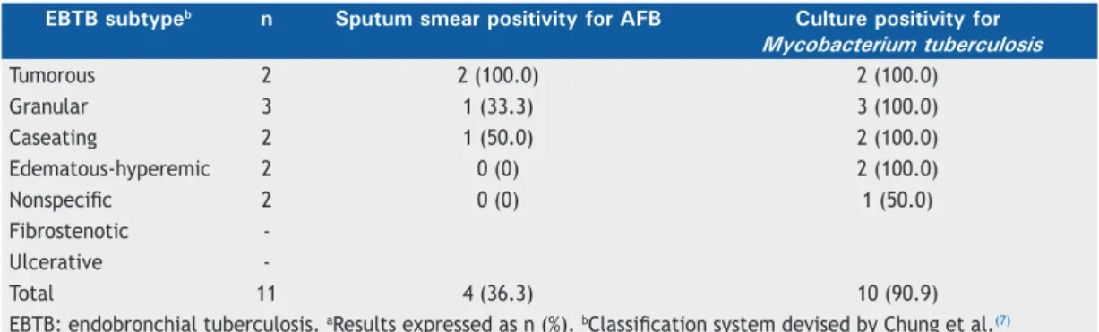
Table 2 shows the results of the microbiological and smear examinations of sputum samples evaluated in 11 cases. In some cases, the patients were unable to produce sputum, and some sputum samples were
collected after iberoptic bronchoscopy. Of the 11
samples submitted to staining, 4 (36.3%) were positive for AFB. The highest smear positivity for AFB (100%) was found in the patients with tumorous EBTB. None of the patients with edematous-hyperemic EBTB or
nonspeciic EBTB were sputum smear-positive for AFB.
Ten (90.9%) of the 11 patients had a positive sputum culture for M. tuberculosis, and the remaining patient
had nonspeciic EBTB.
In two cases (one case of caseating EBTB and one
case of nonspeciic EBTB), the BAL luid had not been
sent for AFB staining. Therefore, microbiological and
smear examinations of BAL luid for M. tuberculosis
were evaluated in only 16 patients (Table 3). Of those
16 patients, 10 (62.5%) were positive for AFB from BAL. The highest BAL smear positivity for AFB (100%) was found in the patients with tumorous EBTB. The
BAL luid culture was positive for M. tuberculosis in 15 (93.7%) of the patients, and the remaining patient
had ibrostenotic EBTB.
Among the 18 patients with EBTB, granulomatous
inlammation was proven by the following bronchoscopic
diagnostic procedures (Table 4): bronchial mucosal biopsy, in 8 cases (44.4%); bronchial brushing, in
7 cases (38.8%); ine-needle aspiration biopsy, in 2
cases (11.1%); and BAL, in 2 cases (11.1%). In one
case of nonspeciic EBTB, ine-needle aspiration biopsy
and bronchial brushing both revealed granulomatous
inlammation. Bronchial anthracoibrosis was identiied
in 5 cases (27.7%): in 2 patients with tumorous EBTB (11.1%); in 2 with granular EBTB (11.1%); and in 1 with caseating EBTB (5.5%).
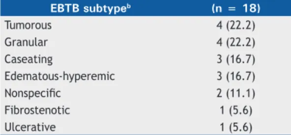
Table 1. Classiication of endobronchial tuberculosis, by bronchoscopic features.a
EBTB subtypeb (n = 18)
Tumorous 4 (22.2)
Granular 4 (22.2)
Caseating 3 (16.7)
Edematous-hyperemic 3 (16.7)
Nonspeciic 2 (11.1)
Fibrostenotic 1 (5.6)
Ulcerative 1 (5.6)
DISCUSSION
In present study, 18 patients were histopathologically diagnosed with EBTB. The lesions were mostly unilateral (in the right lung) and multilobar. The most common
radiological indings were heterogeneous iniltration (in 50%) and nodular iniltration (in 50%). The most
common EBTB subtypes were the tumorous subtype (in 22.2%) and the granular subtype (in 22.2%). Sputum smear microscopy was positive for AFB in
36.3% of the patients. The BAL luid was positive for
AFB in 62.5% of the patients. Among the diagnostic procedures employed in order to prove granulomatous
inlammation, bronchial mucosal biopsy and bronchial
brushing were the most effective. Bronchial
anthra-coibrosis was found in 27.7% of the cases.
Although the reasons are unclear, EBTB is more often observed in female patients. Possible explanations for
that include the fact that females do not expectorate sputum as well as do males, because women have thinner bronchial lumina, as well as because there are sociocultural and aesthetic proscriptions against women expectorating. In our sample of patients with EBTB, the female-to-male ratio was 1.57:1, which is consistent with the preponderance of females reported in other studies of EBTB.(8-11) EBTB usually affects
adults, although younger and elderly patients can be affected, EBTB patient ages ranging from 14 to 81 years.(12) In the present study, patients ranged from
16 to 83 years of age. The mean age was 53.1 years, and 38.8% of the patients were under 45 years of age, which is also in keeping with data in the literature.(8-11)
In the present study, the most common EBTB subtype was actively caseating EBTB, as has been reported in some previous studies.(8,11) However, in a
study conducted by Qingliang et al.,(10) the granular Table 2. Results of microbiological examination of sputum, by endobronchial tuberculosis subtype.a
EBTB subtypeb n Sputum smear positivity for AFB Culture positivity for
Mycobacterium tuberculosis
Tumorous 2 2 (100.0) 2 (100.0)
Granular 3 1 (33.3) 3 (100.0)
Caseating 2 1 (50.0) 2 (100.0)
Edematous-hyperemic 2 0 (0) 2 (100.0)
Nonspeciic 2 0 (0) 1 (50.0)
Fibrostenotic -Ulcerative
-Total 11 4 (36.3) 10 (90.9)
EBTB: endobronchial tuberculosis. aResults expressed as n (%). bClassiication system devised by Chung et al.(7)
Table 3. Results of microbiological examination of BAL luid, by endobronchial tuberculosis subtype.a
EBTB subtypeb n Smear positivity for AFB Culture positivity for
Mycobacterium tuberculosis
Tumorous 4 4 (100.0) 4 (100.0)
Granular 4 2 (50.0) 4 (100.0)
Caseating 2 1 (50.0) 2 (100.0)
Edematous-hyperemic 3 2 (66.9) 3 (100.0)
Nonspeciic 1 0 (0) 1 (100.0)
Fibrostenotic 1 0 (0) 0 (0)
Ulcerative 1 1 (100.0) 1 (100.0)
Total 16 10 (62.5) 15 (93.7)
EBTB: endobronchial tuberculosis. aResults expressed as n (%). bClassiication system devised by Chung et al.(7)
Table 4. Detection of granulomatous inlammation, by bronchoscopic diagnostic procedure employed.a
EBTB subtypeb BAL Bronchial mucosal
biopsy
Fine-needle aspiration biopsy
Bronchial brushing
Tumorous 4
Granular 3 1
Caseating 1 2
Edematous-hyperemic 1 1 1
Nonspeciic 1 1 1
Fibrostenotic 1
Ulcerative 1
Total 2 8 2 7
EBTB: endobronchial tuberculosis. aResults expressed as n of cases in which granulomatous inlammation was
subtype was the most common (in 31.8% of the patients), whereas the edematous-hyperemic type was the most common (in 34.7% of the patients) in
a study conducted by Ozkaya et al.(9) In our study, the
tumorous and granular subtypes were both seen in 22.2% of the patients, which differs from that reported in other studies.(10) The ulcerative and ibrostenotic
subtypes were the least common in our study, both being seen in 5.6% of the patients.
The yield of sputum smear microscopy for AFB is not as high in EBTB as it is in parenchymal involvement, even in an optimal laboratory set up for meticulous sputum examination. In recent studies, sputum positivity in EBTB has been demonstrated to range from 16.0% to 53.3%.(7,13,14) In an even more recent study,(9) sputum
smear microscopy for AFB was negative in all patients. In the present study, sputum smear microscopy for AFB was positive in 36.3% of the patients and the
M. tuberculosis culture positivity rate was very high (90.9%). Therefore it can be said that, when it is possible to collect sputum samples (before or after bronchoscopy), it is worthwhile to send those samples for microbiological examination for AFB.
Direct sputum smear microscopy remains a funda-mental tool in the diagnosis of tuberculosis. Alternative methods of obtaining sputum specimens, including sputum induction, BAL, and gastric lavage, are frequently called for in patients with radiological suspicion of tuberculosis who are unable to expectorate or are
smear-negative. In a study conducted by McWilliams et
al.,(15) the yield of induced sputum (96.3%) was superior
to that of BAL luid (51.9%) and the overall cost of BAL
was found to be three times that of performing sputum induction. In comparison with BAL, sputum induction has several advantages(16): it is less invasive; it has
a higher diagnostic yield; it provides greater patient comfort and safety; it is a low-cost procedure; there is no age restriction on its use; it does not require patient fasting; it is an outpatient procedure; it can be performed without the involvement of an expert; and it is less time-consuming. Unfortunately, sputum induction was not employed at our hospital during the study period.
The second step in the clinical evaluation of EBTB is bronchoscopy, in order to examine bronchial structures and obtain specimens for diagnosis. In the
study conducted by Ozkaya et al.,(9) the BAL luid was
positive for AFB in 26.0% of the cases and the BAL
luid culture was positive for M. tuberculosis in 39.1%. The authors found that positivity for AFB was highest (75.0%) among the patients with the granular subtype of EBTB. They also found mycobacterial culture positivity to be highest (also 75.0%) among the patients with the granular subtype.(9) In our study, microbiological
and smear examinations of BAL luid for AFB were
both positive in 62.5% of the patients. Cultures of
BAL luid for M. tuberculosis were positive in 93.7% of our patients. According to our data, microbiological
and smear examinations of BAL luid have high rates
of positivity of staining for AFB and culture for M.
tuberculosis. It is therefore worthwhile to send BAL
luid samples for AFB analysis in order to facilitate the
early diagnosis of EBTB.
Various bronchoscopic specimens, including those obtained through biopsy, bronchial brushing, or BAL, can be evaluated.(12) A bronchoscopic biopsy is the most
reliable method for diagnosing EBTB, because a needle aspiration biopsy sample can provide only a cytological diagnosis. The reported rate of positivity in bronchial biopsy samples ranges from 30% to 84%.(13,17) We
found it surprising that, in our study, the diagnostic yield of bronchial brushing was nearly equal to that of bronchial mucosal biopsy in detecting granulomas (38.8% and 44.4%, respectively). In a clinical analysis of 90 cases of EBTB in China,(13) bronchial brushing
yielded variable results, ranging from 10% to 85%. In the present study, bronchial mucosal biopsy was especially effective in diagnosing EBTB in patients with the tumorous or granular subtypes (positivity rate of 100% and 75%, respectively), bronchial brushing proving diagnostic in patients with any of the other subtypes.
Tuberculosis is one of the most common diseases associated with bronchial anthracofibrosis.(18,19)
Bronchial anthracoibrosis is typically induced by the
long-term inhalation of biomass smoke.(18) Previous
studies have reported high rates of tuberculosis in
patients with bronchial anthracoibrosis.(18,20-25) In the
present study, bronchial anthracoibrosis was found in
5 cases (27.7%): in 2 patients with tumorous EBTB (11.1%); in 2 with granular EBTB (11.1%); and in 1
with caseating EBTB (5.5%). These indings differ from
those reported by Kim et al.,(26) who found actively
caseating, edematous-hyperemic, and ulcerative EBTB to be the most common EBTB subtypes, respectively occurring in 49%, 21%, and 20% of their patients.
The results of the present study show the value of staining for AFB and culture for M. tuberculosis in sputum
and BAL luid samples for the early diagnosis of EBTB.
In addition, because of its high diagnostic power, we can state that bronchial brushing is a recommended bronchoscopic diagnostic procedure in patients with suspected EBTB.
Our study has certain limitations. Primarily, due to
the retrospective nature of the study, we relied on electronic medical records as our source of patient data.
In conclusion, the tumorous and granular subtypes were the EBTB subtypes most commonly seen in our study. Because of the high positivity rates, we
recom-mend that sputum samples and BAL luid samples be
evaluated by smear microscopy for AFB and by culture for M. tuberculosis, which could collectively increase the
rates of early diagnosis of EBTB. On the basis of our indings, we also recommend that bronchial brushing be
REFERENCES
1. Millard PS, Cegielski JP, Wing S, Silver A. Rurality and tuberculosis incidence trends in North and South Carolina, 1980 to 1992. J Rural Health. 1994;10(4): 226-36. http://dx.doi. org/10.1111/j.1748-0361.1994.tb00236.x
2. Glynn JR. Resurgence of tuberculosis and the impact of HIV infection. Br Med Bull. 1998;54(3):579-93. http://dx.doi.org/10.1093/ oxfordjournals.bmb.a011712
3. Lerner BH. Catching patients: tuberculosis and detention in the 1990s. Chest. 1999;115(1):236-41. http://dx.doi.org/10.1378/ chest.115.1.236
4. Lee JH, Park SS, Lee DH, Shin DH, Yang SC, Yoo BM. Endobronchial tuberculosis. Clinical and bronchoscopic feature in 121 cases. Chest. 1992;102(4): 990-4. http://dx.doi.org/10.1378/chest.102.4.990
5. Calpe JL, Chiner E, Larramendi CH. Endobronchial tuberculosis in HIV-infected patients. AIDS. 1995;9(10):1159-64. http://dx.doi. org/10.1097/00002030-199510000-00007
6. Han JK, Im JG, Park JH, Han MC, Kim YW, Shim YS. Bronchial stenosis due to endobronchial tuberculosis: successful treatment with self-expanding metallic stent. AJR Am J Roentgenol. 1992;159(5):971-2. http://dx.doi.org/10.2214/ajr.159.5.1414809
7. Chung HS, Lee JH, Han SK, Shim YS, Kim KY, Han YC, et al. Classiication of endobronchial tuberculosis by the bronchoscopic features. Tuberc Respir Dis. 1991;38:108-15.
8. Chung HS, Lee JH. Bronchoscopic assessment of the evolution of endobronchial tuberculosis. Chest. 2000;117(2):385-92. http://dx.doi. org/10.1378/chest.117.2.385
9. Ozkaya S, Bilgin S, Findik S, Kök HC, Yuksel C, Atıcı AG. Endobronchial tuberculosis: histopathological subsets and microbiological results. Multidiscip Respir Med. 2012;7(1):34. http://dx.doi.org/10.1186/2049-6958-7-34
10. Qingliang X, Jianxin W. Investigation of endobronchial tuberculosis diagnoses in 22 cases. Eur J Med Res. 2010;15:309-13. http://dx.doi. org/10.1186/2047-783X-15-7-309
11. Sahin F, Yıldız P. Characteristics of endobronchial tuberculosis patients with negative sputum acid-fast bacillus. J Thorac Dis. 2013;5(6):764-70.
12. Kashyap S, Mohapatra PR, Saini V. Endobronchial tuberculosis. Indian J Chest Dis Allied Sci. 2003;45(4):247-56.
13. Yu W, Rong Z. Clinical analysis of 90 cases with endobronchial tuberculosis [Article in Chinese]. Zhonghua Jie He He Hu Xi Za Zhi. 1999;22(27):396-8.
14. Aggarwal AN, Gupta D, Joshi K, Behera D, Jindal SK. Endobronchial
involvement in tuberculosis: a report of 24 cases diagnosed by ibreoptic bronchoscopy. J Bronchol. 1999;6:247-50. http://dx.doi. org/10.1097/00128594-199910000-00004
15. McWilliams T, Wells AU, Harrison AC, Lindstrom S, Cameron RJ, Foskin E. Induced sputum and bronchoscopy in the diagnosis of pulmonary tuberculosis. Thorax. 2002;57(12):1010-4. http://dx.doi. org/10.1136/thorax.57.12.1010
16. Anderson C, Inhaber N, Menzies D. Comparison of sputum induction with iber-optic bronchoscopy in the diagnosis of tuberculosis. Am J Respir Crit Care Med. 1995;1525 Pt 1):1570-4.
17. Altin S, Cikrikçioğlu S, Morgül M, Koşar F, Ozyurt H. 50 endobronchial tuberculosis cases based on bronchoscopic diagnosis. Respiration. 1997;64(2):162-4. http://dx.doi.org/10.1159/000196662
18. Kim YJ, Jung CY, Shin HW, Lee BK. Biomass smoke induced bronchial anthracoibrosis: presenting features and clinical course. Respir Med. 2009;103(5):757-65. http://dx.doi.org/10.1016/j. rmed.2008.11.011
19. Hwang J, Puttagunta L, Green F, Shimanovsky A, Barrie J, Long R. Bronchial anthracoibrosis and tuberculosis in immigrants to Canada from the Indian subcontinent. Int J Tuberc Lung Dis. 2010;14(2):231-7.
20. Chung MP, Lee KS, Han J, Kim H, Rhee CH, Han YC, et al. Bronchial stenosis due to anthracoibrosis. Chest. 1998;113(2):344-50. http:// dx.doi.org/10.1378/chest.113.2.344
21. Mirsadraee M, Saeedi P. Anthracosis of lung: evaluation of potential underlying causes. J Bronchol. 2005;12(2):84-7. http://dx.doi. org/10.1097/01.laboratory.0000150873.99404.53
22. Pérez-Padilla R, Pérez-Guzmán C, Báez-Salda-a R, Torres-Cruz A. Cooking with biomass stoves and tuberculosis: a case control study. Int J Tuberc Lung Dis. 2001;5(5):441-7.
23. Pokhrel AK, Bates MN, Verma SC, Joshi HS, Sreeramareddy CT, Smith KR. Tuberculosis and indoor biomass and kerosene use in Nepal: a case-control study. Environ Health Perspect. 2010;118(4):558-64. http://dx.doi.org/10.1289/ehp.0901032
24. Kolappan C, Subramani R. Association between biomass fuel and pulmonary tuberculosis: a nested case-control study. Thorax. 2009;64(8):705-8. http://dx.doi.org/10.1136/thx.2008.109405
25. Rajagopalan S. Tuberculosis and aging: a global health problem. Clin Infect Dis. 2001;33(7):1034-9. http://dx.doi.org/10.1086/322671
26. Kim HJ, Kim SD, Shin DW, Bae SH, Kim AL, Kim JN, et al. Relationship between bronchial anthracoibrosis and endobronchial tuberculosis. Korean J Intern Med. 2013;28(3):330-8. http://dx.doi. org/10.3904/kjim.2013.28.3.330