RESUMO.- [Papilomatose em cães: estudo retrospecti-vo de 24 casos (2001-2011) e caracterização imuno--histoquímica.] Foi realizado um estudo retrospectivo de 24 casos de papilomas em cães diagnosticados no período de janeiro 2001 a março de 2011, bem como a sua caracte-rização imuno-histoquímica (IHQ). Cães sem raça deinida foram os mais afetados, a idade média foi de 3,1 anos, com variação de 6 meses a 10 anos e não houve predileção sexu-al. Quanto à localização das lesões, 75,0% estavam na pele, 16,7% no lábio e 8,3% em pálpebra. Na avaliação histológi-ca havia proliferação papilar exoítihistológi-ca do epitélio eshistológi-camoso em 87,5% e papilar endoítica (invertido) em 12,5%. O
tu-Canine papillomatosis: A retrospective study of 24 cases
(2001-2011) and immunohistochemical characterization
1Matheus V. Bianchi2, Renata A. Casagrande2, Tatiane T.N. Watanabe2, Angelica T.B. Wouters2, Flademir Wouters2, Gisele S. Boos2, Mariana B. Menegat2 and David Driemeier2*
ABSTRACT.- Bianchi M.V., Casagrande R.A., Watanabe T.T.N., Wouters A.T.B., Wouters F., Boos G.S., Menegat M.B. & Driemeier D. 2012. Canine papillomatosis: A retrospective study of 24 cases (2001-2011) and immunohistochemical characterization.Pesquisa Veterinária Brasileira 32(7):653-657.Setor de Patologia Veterinária, Faculdade de Veteriná-ria, Universidade Federal do Rio Grande do Sul, Av. Bento Gonçalves 9090, Porto Alegre, RS 91540-000, Brazil. E-mail: davetpat@ufrgs.br
A retrospective study of 24 cases of papillomas in dogs was performed from January 2001 to March 2011. Additionally, immunohistochemistry (IHC) was used to characteri-ze and evaluate the samples. We found that disease was observed more in mixed breed dogs, ages ranging from 6 months to 10 years (mean 3.1 years), and there was no gender predilection. The main lesion sites were the skin (75%), lips (16.7%), and eyelids (8.3%). Upon histological evaluation, we observed papillary exophytic proliferation of squamous epithelium and papillary endophytic proliferation (inverted) in 87.5% and 12.5% of cases, respectively. The tumors were characterized by spinous layer hyperplasia (87.5%) with koilocytes (70.8%) and intranuclear pale basophilic inclusions bodies (8.3%), prominent granular layer with large amounts of keratohyalin granules (95.8%), and hyperkeratosis in the stratum corneum (100%). Positive immunostaining for Papillomavirus was found in 83.3% of cases, which were distributed between the granular layer and the stratum cor-neum. These indings indicate the following: that papillomas in dogs are caused by Pa-pillomavirus, the viral cytopathic effect induces epithelial lesions, viral particles are found inside the cell nuclei, and inclusions bodies are rare.
INDEX TERMS: Papilloma, papillomatosis,skin disease, immunohistochemistry, dog.
1 Received on March 14, 2012.
Accepted for publication on April 18, 2012.
2 Departamento de Patologia Clínica Veterinária, Faculdade de
Veteriná-ria, Universidade Federal do Rio Grande do Sul (UFRGS), Av. Bento Gon-çalves 9090, Porto Alegre, RS 95320-000, Brazil. * Corresponding author: davetpat@ufrgs.br
mor era caracterizado por hiperplasia do estrato espinhoso (87,5%) com coilócitos (70,8%) e inclusões intranucleares basoílicas pálidas (8,3%); o estrato granular estava proe-minente com grande quantidade de grânulos de querato--hialina (95,8%); e havia hiperqueratose do estrato cór-neo (100%). Na avaliação IHQ para Papillomavirus houve marcação nos estratos granuloso e córneo em 83,3%. Estes achados indicam que os papilomas em cães são causados por Papillomavirus, as lesões epiteliais são decorrentes do efeito citopático viral, as partículas virais estão no núcleo das células e corpúsculos de inclusão são raros.
TERMOS DE INDEXAÇÃO: Papiloma, papilomatose, doença de pele, imuno-histoquímica, cão.
INTRODUCTION
or mucosal epithelium (Gross et al. 2005). Papillomas are common in dogs and can cause single or multiple epithe-lial lesions at different skin sites, including mucous mem-branes and the mucocutaneous junction of the oral cavity and conjunctiva (Nicholls & Stanley 1999). Currently, there are six recognized syndromes related to canine papilloma: oral papillomatosis, cutaneous, inverted cutaneous, multi-ple pigmented cutaneous, multimulti-ple pigmented plaques, and cushions multiple papillomas (Scott et al. 2001). The patho-genesis of papillomatosis is characterized by a hyperplastic reaction of the epithelium with an increased production of keratin and virus replication (Gross et al. 2005).
Canine oral papillomatosis is a contagious disease that mainly affects young dogs and it manifests as single or mul-tiple verrucous lesions with an average size of 1.0 cm in diameter, which can be found in the oral mucosa, eyelids, and skin (Goldschmidt & Hendrick 2002). Cutaneous inver-ted papillomas also affect young animals and the lesions are located in the abdomen and the inguinal region (Cam-pbell et al. 1988).
The disease has an incubation period of 4 to 8 weeks and typically regresses after 4 to 8 weeks of evolution (Chambers & Evans 1959, Bredal et al. 1996, Nicholls et al. 2001). Dogs that successfully recover from papillomatosis become immune to subsequent infections (Chambers et al. 1960). Circulating IgG antibodies induced by spontaneous regression of canine papillomas that protect against sub-sequent infections have been observed (Ghim et al. 2000). However, in some cases, the lesion may progress to squa-mous cell carcinoma (Nicholls & Stanley 1999). Diagnoses of canine viral papillomas are determined mainly by anato-mical distribution, histological characterization, immuno-histochemistry, electron microscopy, immunoluorescence, and in situ hybridization (Scott et al. 2001).
The goal of this study was to review papilloma cases in dogs submitted to the Laboratory of Veterinary Pathology of the Universidade Federal do Rio Grande do Sul (UFRGS), Brazil from 2001 to 2011. We aimed to describe the cases and deine their histological and immunohistochemistry characteristics.
MATERIALS AND MET,HODS
The canine biopsies archives from the Laboratory of Veterinary Pathology, UFRGS, Brazil, from January 2001 to March 2011, were reviewed. All papilloma cases were analyzed and the parafin blo-cks were recut and stained with hematoxylin and eosin (HE).
For immunohistochemical analysis, the monoclonal papillo-mavirus antibody (clone K1H8, DakoCytomation) was used. The slides were deparafinized, rehydrated, and treated with 10% hydrogen peroxide in methanol solution. The antigens were re-trieved by boiling the sections in citrate buffer (pH 6) for 40 min at 96°C in a water-bath. Sections were incubated overnight at 4°C with primary antibody (1:100 dilution). Ampliication signal was achieved by using biotinylated secondary antibody, followed by labeled streptavidin-biotin-peroxidase complex, and both rea-gents were obtained from the LSAB Universal kit (DakoCytoma-tion). The reaction was revealed with 3-amino-9-etilcarbazol (AEC, K3469, DakoCytomation) chromogenic substrate. Slides were counterstained with Mayer’s hematoxylin and coverslipped with aqueous medium (S1964, DakoCytomation) for microscopic
examination. Bovine papilloma was included as a positive control for this procedure. The tissue samples were categorized as nega-tive (-), discrete reaction intensity (+), moderate (++), and accen-tuated (+++).
RESULTS Characterization of the cases
From January 2001 to March 2011, 24 canine papilloma cases were diagnosed (Table 1). The percentage of mixed breed dogs was overrepresented at 29.3% (7/24) and 12.5% (3/24) were un-reported breeds. Each of the following breeds comprises 8.4% of the population (2/24): Beagle, English Bulldog, Labrador, and Pi-tbull. Moreover, German Shepherd, Golden Retriever, Lhasa Apso, Pinscher, Poodle, and Pug breeds each comprised 4.1% (1/24). Of the dogs in the study, females comprised 54.2% (13/24), males comprised 41.7% (10/24), and 4.1% (1/24) did not report gen-der. Ages ranged from 6 months to 10 years (average 3.1 years), with 54.2% (13/24) that were less than 3 years; 20.8% (5/24) that were more than 3 years; and 25% of undetermined age.
Regarding the tumor sites, 75% (18/24) involved the skin, including the inguinal region. Both the thoracic mem-ber and foreskin comprised 8.3% of tumor sites (2/24). Comprising 4.2% of tumor sites (1/24) are: the face, inter-digit, lank, and areas close to the nipple. Also, the skin dis-tribution was not speciied in 33.3% of the medical records (8/24). Less common sites included the lip and eyelid at 16.7% (4/24) and 8.3% (2/24), respectively.
Microscopic features
During histological evaluations, we observed papilla-ry exophytic proliferation of squamous epithelium and papillary endophytic proliferation (inverted) in 87.5% (21/24) (Fig.1A) and 12.5% of cases (3/24), respective-ly. Theses tumors were characterized by spinous layer
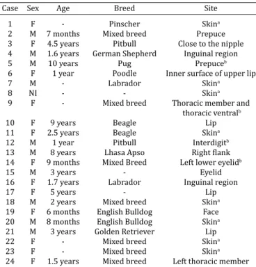
Table 1. Characterization of the dogs affected by papillomatosis
Case Sex Age Breed Site
1 F - Pinscher Skina
2 M 7 months Mixed breed Prepuce 3 F 4.5 years Pitbull Close to the nipple 4 M 1.6 years German Shepherd Inguinal region
5 M 10 years Pug Prepuceb
6 F 1 year Poodle Inner surface of upper lip
7 M - Labrador Skina
8 NI - - Skina
9 F - Mixed breed Thoracic member and thoracic ventralb
10 F 9 years Beagle Lip
11 F 2.5 years Beagle Skina
12 M 1 year Pitbull Interdigitb
13 M 8 years Lhasa Apso Right lank 14 F 9 months Mixed Breed Left lower eyelidb
15 M 3 years - Eyelid
16 F 1.7 years Labrador Inguinal region
17 F 5 years - Lip
18 M 2 years Mixed breed Skina
19 F 6 months English Bulldog Face 20 M 8 months English Bulldog Skina
21 M 3 years Golden Retriever Lip
22 F - Mixed breed Skina
23 F - Mixed breed Skina
hyperplasia and the normal eosinophilic cytoplasm was replaced by pale basophilic cytoplasm in 87.5% of ca-ses (21/24) (Fig.1A). Also, intracytoplasmic eosinophilic material (aggregates of keratin) was observed in 37.5% of cases (9/24). The nuclei of these cells were round or oval with dispersed and condensed chromatin in 95.8% (23/24) and 4.2% of cases (1/24), respectively. Addi-tionally, 1-2 and 1-3 evident nucleolus were observed in 91.7% (22/24) and 8.3% of cases (2/24), respectively. The granular layer was prominent, and the keratohyalin granules were large, round, or irregular in 95.8% of cases (23/24) (Fig.1A). The stratum corneum showed parakera-totic hyperkeratosis and orthokeraparakera-totic hyperkeratosis in 70.8% (17/24) and in 29.2% of cases (7/24), respectively (Fig.1A). Koilocytes (enlarged keratinocytes with eccen-tric pyknotic nuclei surrounded by a clear halo) were ob-served in the spinous and granular layer and their “ghost” cells in the stratum corneum in 70.8% (17/24) and 54.2% of cases (13/24), respectively (Fig.1B). Intranuclear baso-philic inclusions that completely illed the nucleus or had
distinct margination of the nuclear chromatin surrounded by a clear halo in 8.3% of cases (2/24) (Fig.1C) were ob-served in the spinous layer.
The papillomas were supported by small (75.0%; 18/24), moderate (20.8%; 5/24) and severe (4.2%; 1/24) amount of dermal ibrovascular stroma. In addition, 45.8% (11/24) of the papillomas had prominent and dilated ca-pillaries (Fig.1B). Intracytoplasmatic melanin pigment was observed in 45.9% of cases (11/24), with a small amount in rare cells (25%; 6/24); moderate amount in some cells (16.7%; 4/24), and severe amount in all cells (4.2%; 1/24).
Regarding lymphoplasmocytary iniltration, 16.7% (4/24) and 12.5% (3/24) were classiied as focal mild or moderate, respectively. Iniltration of lymphocytes, plasma cells and neutrophils were classiied as focal mild (16.7%; 4/24), focal moderate (8.3%; 2/24), or locally extensive and severe (4.2%; 1/24). Epithelial erosion with lymphocytes, plasma cells and neutrophils was locally extensive in 4.2% of cases (1/24).
Immunohistochemical characterization
The IHC results are reported in Table 2. Immunostai-ning for Papillomavirus in the epithelial cells nuclei was observed in 83.3% of cases (20/24) and with accentua-ted intensity (+++) in 40% (8/20), moderate (++) in 30% (6/20), and discrete (+) in 30% of cases (6/20). Staining occurred in the granular layer and stratum corneum (70%; 14/20), stratum corneum (15%; 3/20), granular layer (10%; 2/20), and in the granular layer, spinous layer, and stratum corneum (5%; 1/20) (Fig.1D).
DISCUSSION
Papillomas are often diagnosed as exophytic (Scott et al. 2001). Cutaneous inverted papilloma is a rare endophytic variant in dogs (Campbell et al. 1988). Exophytic viral pa-pilloma occurs most commonly on the face, ears, and extre-mities, but it may also occur in the mucocutaneous junc-tions (Gross et al. 2005). In this study, lips, inguinal region,
thoracic member, prepuce, and eyelid were the most com-monly affected sites; however, it is not possible to determi-ne the precise site prevalence because this information was not speciied in 33.3% of the clinical records. Endophytic viral papillomas are common in the abdominal and ingui-nal region (Campbell et al. 1988).
There is no known sex predilection and dogs at increased risk are the Great Dane, Irish Setter, and Beagle, while mixed breed dogs are at decreased risk (Goldschmidt & Hendrick, 2002). However, this study suggests that mixed breed dogs were affected more than pure breeds. Exophytic papillomas occur at any age but are most often seen in dogs less than 2 years of age (Gross et al. 2005), and inverted papillomas affect dogs less than 3 years of age (Campbell et al. 1988). This type of tumor is rare in older animals (Narama et al. 1992) and the higher incidence in younger animals supports the idea that older animals have devel-oped immunity as a result of previous exposure to the virus (Chambers et al. 1960).
In this report, typical viral papilloma lesions were ob-served in 87.5% of cases and these lesions comprise mul-tiple inger-like projections of thickened squamous epithe-lium (Gross et al. 2005). The presence of koilocytes, which were seen in 70.8% of these cases, in the spinous layer and their “ghost” cells in the stratum corneum is a sign of viral cytopathic effect, which causes a ballooning degeneration and the nucleus becomes eccentric and picnotic (Gross et al. 1992).
The papillomatosis regression in dogs is spontaneous; therefore, the prognosis is favorable (Goldschmidt & Hen-drick 2002). Usually, disease regression is marked by a moderate, interface iniltrate that primarily consists of T lymphocytes (Gross et al. 2005). In this study, there were inlammatory iniltrates in 62.6% of cases, which suggests that the lesion may be regressing.
Benign lesions usually do not cause clinical problems, except when the site leads to airway obstruction or dyspha-gia. The treatment consists of surgical removal by excision, cryosurgery, or electrosurgery. The treatment with auto-genous vaccines has no proven therapeutic effect in dogs, although some studies suggest that it may prevent disea-se (Sundberg et al. 1994, Scott et al. 2001). A spectrum of proliferative cutaneous lesions occurred in 12 dogs at the injection site of live canine oral papillomavirus vaccine, su-ggesting a viral etiology for the disease. Lesions included epidermal hyperplasia, epidermal cysts, squamous papillo-ma, basal cell epitheliopapillo-ma, and squamous cell carcinoma. Tumor sections revealed papillomavirus antigen in ive of 12 masses (Bregman et al. 1987).
A severe, naturally occurring, nonregressing oral pa-pilloma was observed in a 3.5-year-old neutered female Labrador. The papillomas proved refractory to surgical and medical treatments, including autogenous vaccination and vaccination with capsid (L1) virus-like particles. The papillomas spread to oesophageal mucosa, perioral haired skin, and remote cutaneous sites. Experimental infection of Beagle dogs with this viral isolate resulted in the uncompli-cated development and regression of oral warts within the usual period. These indings support the hypothesis that Table 2. Immunohistochemical characterization of
papillomatosis in dogs
Nº case Diagnostic Immunohistochemical for Papillomavirus
Distribution Intensity
1 Inverted papilloma Granular layer + 2 Inverted papilloma Stratum corneum +
3 Papilloma -
-4 Inverted papilloma Stratum corneum ++
5 Papilloma -
-6 Papilloma Granular layer and ++
stratum corneum
7 Papilloma Granular layer and +G +++C
stratum cor neum
8 Papilloma Granular layer and ++
stratum corneum
9 Papilloma Granular layer and +++
stratum corneum
10 Papilloma -
11 Papilloma Stratum corneum + 12 Papilloma Granular layer and ++
stratum corneum
13 Papilloma Granular layer +++ 14 Papilloma Granular layer and ++
stratum corneum
15 Papilloma Granular layer and +
stratum corneum
16 Papilloma Granular layer and +++
stratum corneum
17 Papilloma Granular layer and +++
stratum corneum
18 Papilloma -
19 Papilloma Granular layer and ++
stratum corneum
20 Papilloma Granular and spinous layer, +++ and stratum corneum
21 Papilloma Granular layer and +
stratum corneum
22 Papilloma Granular layer and +
stratum corneum
23 Papilloma Granular layer and +G +++C
stratum corneum
24 Papilloma Granular layer and +++G +C
stratum corneum
- Negative, + discrete, ++ moderate, +++ accentuated; G granular layer, C
the recurrent lesions are associated with speciic defects in host immunity rather than variations in viral pathogenicity (Nicholls et al. 1999).
The results of the present study conirm that Papilloma-virus was responsible for 83.3% of papillomatous lesions observed in dogs and immunostaining was concentrated in the granular layer and the stratum corneum. The same immunostaining was seen in an outbreak of canine oral pa-pillomavirus in Korea (Yheel et al. 2010). This antibody tar-gets the whole virus, which is formed only in differentiated squamous epithelial cells. Thus, positive reactions tend to be limited to the skin surface and stratum corneum (Gross et al. 2005). Negative IHC cases could be explained by the lack of virus localization in the lesion.
The presence of basophilic intranuclear inclusions in cells from the spinous layer was only noted in two cases. Inclusions are less frequently observed because they are more evident when the biopsy is conducted during the initial development of the papilloma (Goldschmidt et al. 2002).
CONCLUSIONS Our indings indicate that:
1) dogs with papillomatosis were young; 2) mixed breed dogs were affected the most; 3) papilloma lesions occurred mainly in the skin; 4) the exophytic form was predominant;
5) papillomas were caused by Papillomavirus; 6) lesions were caused by viral cytopathic effect; 7) inclusion bodies were rarely observed; and
8) intranuclear immunostaining occurred mainly in the granular layer and in the stratum corneum.
REFERENCES
Bredal W.P., Thoresen S.I., Rimstad E., Aleksandersen M. & Nafstad P.H.J. 1996. Diagnosis and clinical course of canine oral papillomavirus infec-tion. J. Small Anim. Pract. 37:138-142.
Bregman C.L., Hirth R.S., Sundberg J.P. & Christensen E.F. 1987. Cutaneous neoplasms in dogs associated with canine oral papillomavirus vaccine. Vet. Pathol. 24:477-487.
Campbell K.L., Sundberg J.P., Goldschmidt M.H., Knupp C. & Reichmann M. E. 1988. Cutaneous inverted papillomas in dogs. Vet. Pathol. 25:67-71. Chambers V.C. & Evans C.A. 1959. Canine oral papillomatosis. I. Virus assay
and observations on the various stages of the experimental infection. Cancer Res. 19:1188-1195.
Chambers V.C., Evans C.A. & Weiser R.S. 1960. Canine oral papillomatosis. II. Immunologic aspects of the disease. Cancer Res. 20:1083-1093. Ghim S., Newsome J., Bell J., Sundberg J.P., Schlegel R. & Jenson A.B. 2000.
Spontaneously regressing oral papillomas induce systemic antibodies that neutralize canine oral Papillomavirus. Exp. Mol. Pathol. 68:147-151.
Goldschmidt M.H. & Hendrick M.J. 2002. Tumors of the skin and soft tis-sues, p.45-118. In: Meuten D.J. (Ed.), Tumors in Domestic Animals. 4th
ed. Blackwell, Iowa.
Gross T.L., Ihrke P.J. & Walder E.J. 1992. Veterinary Dermatopathology: A macroscopic and microscopic evaluation of canine and feline skin dise-ase. Mosby, St Louis. 520p.
Gross T.L., Ihrke P.J., Walder E.J. & Affolter V.K. 2005. Skin Diseases of the Dog and Cat: Clinical and histopathologic diagnosis. 2nd ed. Blackwell,
Oxford. 932p.
Narama I., Ozaki K., Maeda H. & Ohta A. 1992. Cutaneous papilloma with viral replication in an old dog. J. Vet. Med. Sci. 54:387-389.
Nicholls P.K. & Stanley M.A. 1999. Canine papillomavirus: A centenary re-view. J. Comp. Pathol. 120:219-233.
Nicholls P.K., Klaunberg B.A., Moore R.A., Santos E.B., Parry N.R., Gough G.W. & Stanley M.A. 1999. Naturally occurring, nonregressing canine oral papillomavirus infection: Host immunity, virus characterization, and experimental infection. Virology 265:365-374.
Nicholls P.K., Moore P. F., Anderson D.M., Moore R.A., Parry N.R., Gough G.W. & Stanley M.A. 2001. Regression of canine oral papillomas is associated with iniltration of CD41 and CD81 lymphocytes. Virology 283:31-39. Scott D.W., Miller D.H. & Grifin C.E. 2001. Muller and Kirk’s Small Animal
Dermatology. 6th ed. W.B. Saunders, Philadelphia. 1528p.
Sundberg J.P., Smith E.K., Herron A.J., Jenson A.B., Burk R.D. & Van Ranst M. 1994. Involvement of canine oral papillomavirus in generalized oral and cutaneous verrucosis in a Chinese Shar Pei dog. Vet. Pathol. 31:183-187. Yheel J.Y., Kwon B.J., Kim J.H., Yu C.H., Im K.S., Lee S.S., Lyoo Y.S., Chang