Activated Integrin
a
IIb
b
3
Impairs Proplatelet Formation in
Human Megakaryocytes
Loredana Bury1,2, Alessandro Malara1,2, Paolo Gresele1*, Alessandra Balduini2
1Division of Internal and Cardiovascular Medicine, Department of Internal Medicine, University of Perugia, Perugia, Italy,2Biotechnology Laboratories, Department of Biochemistry, University of Pavia, IRCCS San Matteo Foundation, Pavia, Italy
Abstract
Background:The interaction of megakaryocytes with matrix proteins of the osteoblastic and vascular niche is essential for megakaryocyte maturation and proplatelet formation. Fibrinogen is present in the vascular niche and the fibrinogen receptoraIIbb3is abundantly expressed on megakaryocytes, however the role of the interaction between fibrinogen and
aIIbb3 in proplatelet formation in humans is not yet fully understood. We have recently reported a novel congenital
macrothrombocytopenia associated with a heterozygous mutation of the b3 subunit of aIIbb3. The origin of
thrombocytopenia in this condition remains unclear and this may represent an interesting natural model to get further insight into the role of the megakaryocyte fibrinogen receptor in megakaryopoiesis.
Methodology/Principal Findings: Patients’ peripheral blood CD45+ cells in culture were differentiated into primary megakaryocytes and their maturation, spreading on different extracellular matrix proteins, and proplatelet formation were analyzed. Megakaryocyte maturation was normal but proplatelet formation was severely impaired, with tips decreased in number and larger in size than those of controls. Moreover, megakaryocyte spreading on fibrinogen was abnormal, with 50% of spread cells showing disordered actin distribution and more evident focal adhesion points than stress fibres. Integrin
aIIbb3expression was reduced but the receptor was constitutively activated and a sustained, and substrate-independent,
activation of proteins of the outside-in signalling was observed. In addition, platelet maturation from preplatelets was impaired.
Conclusions/Significance:Our data show that constitutive activation of aIIbb3-mediated outside-in signalling in human
megakaryocytes negatively influences proplatelet formation, leading to macrothombocytopenia.
Citation:Bury L, Malara A, Gresele P, Balduini A (2012) Outside-In Signalling Generated by a Constitutively Activated IntegrinaIIbb3Impairs Proplatelet Formation
in Human Megakaryocytes. PLoS ONE 7(4): e34449. doi:10.1371/journal.pone.0034449
Editor:Neil A. Hotchin, University of Birmingham, United Kingdom
ReceivedOctober 14, 2011;AcceptedMarch 2, 2012;PublishedApril 23, 2012
Copyright:ß2012 Bury et al. This is an open-access article distributed under the terms of the Creative Commons Attribution License, which permits unrestricted use, distribution, and reproduction in any medium, provided the original author and source are credited.
Funding:This work was supported by a Telethon grant (GGP10155) to PG and by a Cariplo Foundation grant (2006.0596/10.8485) to AB. The funders had no role in study design, data collection and analysis, decision to publish, or preparation of the manuscript.
Competing Interests:The authors have declared that no competing interests exist.
* E-mail: grespa@unipg.it
Introduction
Mature megakaryocytes (Mks) migrate to the vascular niche of the bone marrow where they convert the bulk of their cytoplasm into multiple long processes, called proplatelets, that protrude through the vascular endothelium into the sinusoid lumen to release platelets [1,2]. Recently a new intermediate stage in platelet maturation has been described: preplatelets, discoid particles circulating in blood, larger than platelets, that reversibly convert into barbell-shaped proplatelets that in turn generate each two mature platelets after a fission event [3].
Very little is known about the role of specific bone marrow proteins in megakaryocyte differentiation and function. Fibrinogen was shown to be localized in the bone marrow sinusoids of mice and to be essential for proplatelet formation by binding to
megakaryocyteaIIbb3[4]. In fact, mouse megakaryocytes extend
proplatelets when plated on fibrinogen, and treatment withaIIbb3
antagonists strikingly reduces the percentage of megakaryocytes developing proplatelets [4]. However, the interaction between
integrin aIIbb3 and fibrinogen was shown to be essential for
spreading but not for proplatelet formation by human
megakar-yocytes, and in fact while aIIbb3 antagonists almost abolished
adhesion and spreading they did not cause any significant reduction of human proplatelet formation [5]. Indeed, Glanzmann Thrombasthenia (GT), a rare hereditary autosomal recessive bleeding disorder affecting the megakaryocytic lineage and due to quantitative and/or qualitative abnormalities of aIIbb3, is not
associated with thrombocytopenia [6]. Therefore, the role of aIIbb3in proplatelet formation in humans is still controversial.
We have recently described two families with a novel autosomal dominant hereditary mucocutaneous bleeding disorder with macrothrombocytopenia and defective platelet function associated
with a heterozygous mutation (2134+1 G.C) of the ITGB3 gene,
coding for the b3 subunit of aIIbb3 and producing a deletion
(del647-686) of a large part of thebTail Domain (bTD) [7], an
extracellular domain ofb3involved in receptor activation [8]. This
domain, was never described before and it seemed of interest that it was associated with a reduced platelet number and altered platelet morphology.
Purpose of the present study was to analyse megakaryocyte maturation, spreading and proplatelet formation on fibrinogen, and other extracellular matrix proteins, in two patients with the Glanzmann variant macrothrombocytopenia associated with the b3del647-686.
Results
Megakaryocyte differentiation and proplatelet formation
The percentage of CD45+cells differentiated into
megakaryo-cytes was comparable in patients and controls (761.8% vs
9.962.7%, respectively, p = ns). Megakaryocyte maturation
pro-files, classified according to standard criteria [9], were not significantly different between patients and controls, indicating that del647-686 of aˆ3integrin does not affect the differentiation or
maturation of megakaryocytes (Figure 1A).
Proplatelets formed by megakaryocytes in suspension were
instead reduced, with only 1.861.0% of patient megakaryocytes
extending proplatelets vs 7.762.2% of controls after 16 hours
(p,0.05) (Figure 1B), but with a normal morphology. Defective
proplatelet formation was confirmed in experiments with a longer incubation time (24 hours) (data not shown).
On the contrary, when megakaryocytes were plated on fibrinogen proplatelets were numerically comparable to those of
controls (7.763.2% vs 6.760.4% n = 3, p = ns), but presented
important structural alterations, with megakaryocytes showing a spread shape, shorter proplatelet shafts and tips significantly decreased in number and larger in size than those of controls (Figure 1C, 1D and 1E). Interestingly, while pro-platelet formation usually starts from one pole of the megakaryocyte and rapidly leads to the conversion of the entire cytoplasm into proplatelets [2], in our patients pro-platelet formation started at multiple poles of the megakaryocyte cell body (Figure 1E, right). Finally, proplatelet formation from patient’s megakaryocytes incubated with type I collagen or Von Willebrand factor was not different from that of control megakaryocytes [10–12] (on type
I collagen: 0.160.2% vs 0.160.2%, p = ns; on VWF 3.060.6% vs
3.860.2%, p = ns) (Figure 1B).
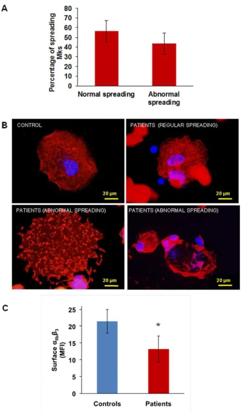
Megakaryocyte spreading
Megakaryocyte spreading on type I collagen was similar in
patients and controls (15.4% vs 18.663.3% of the total adhering
population, p = ns) as well as spreading on Von Willebrand factor (5.2% vs 5.961.3% of the total adhering population, p = ns). On the contrary, spreading on fibrinogen was increased: 31.765.6% of the
total population of adhering megakaryocytes compared to 1062%
of controls (p,0.05). Two different populations were detectable
among patient megakaryocytes: one spreading normally (56.4
611% of the total spread population), and the other abnormally
(43.5611% of the total spread population) (Figure 2A).
Abnormally spread megakaryocytes showed nuclei displaced towards cell periphery, a disordered distribution of actin and focal
adhesion points more evident than stress fibres (Figure 2B lower
panels). Normally spread megakaryocytes, instead, showed central nuclei and an ordered organization of actin in stress fibres and focal
adhesion points, similar to controls (Figure 2B upper panels).
aIIbb3expression and activation
IntegrinaIIbb3was significantly less expressed on the surface of
patients’ megakaryocytes than on that of control megakaryocytes (mean fluorescence intensity: 13.262.1 vs 21.562.2%,
respective-ly, p,0.05) (Figure 2C), in accordance with what we previously
observed with the patients’ platelets [7].
Asb3integrin is also a subunit of theaVb3receptor (CD51/61),
we measured aVb3 by flow cytometry and we found that its
expression was comparable between patients and controls, both in platelets (patients 4.860.6% vs controls 4.860.4%, p = ns) and in megakaryocytes (patients 1361.1% vs controls 10.761.6%, p = ns) (data not shown).
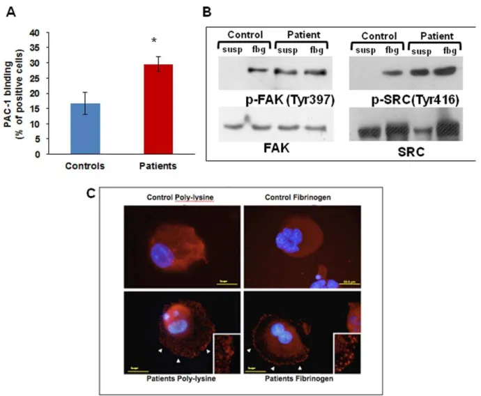
To studyaIIbb3receptor activation we measured the binding of
PAC-1, a monoclonal antibody that binds only to activatedaIIbb3,
to un-stimulated megakaryocytes: 29.560.9% of patients’
mega-karyocytes bound PAC1 vs 16.763.6% of control megakaryocytes
(Figure 3A), showing constitutive activation ofaIIbb3integrin in
patients’ megakaryocytes.
We therefore assessed aIIbb3-triggered outside-in signalling by
measuring the phosphorylation of FAK and Src after adhesion to fibrinogen by western blotting [13].
Src and FAK were phosphorylated in patients’ megakaryocytes also in suspension while in controls phosphorylation was observed
only upon adhesion to fibrinogen (Figure 3B).
We also assessed FAK clustering at immunofluorescence: clustering was clearly evident in patients’ megakaryocytes already
one hour after plating on fibrinogen (Figure 3C right panel),
while with control megakaryocytes it was evident only after 3 hours. Moreover, FAK clusters were observed in patients’ megakaryocytes in suspension, differently from controls were they
were evident only upon contact with fibrinogen (Figure 3C left
panel), consistently with constitutive activation ofaIIbb3.
Conversion of preplatelets into platelets
Very recently a new intermediate form between proplatelets and platelets, the preplatelet, was described [3]. A preplatelet can reversibly convert into a barbell-shaped proplatelet and generate two platelets passing through a ‘‘figure 8’’ structure. We therefore counted ‘‘figure 8’’ structures in platelet rich plasma (PRP) and we observed a significantly lower percentage of them in patients’ PRP
as compared to controls (0.560.7% vs 2.161.2% respectively,
p,0.05) (Figure 4).
Discussion
Extracellular proteins play an important role in megakaryopoi-esis and platelet formation by interacting with their receptors on megakaryocytes [5]. In particular, the vascular niche is enriched in fibrinogen and von Willebrand factor which drive the late phases of megakaryopoiesis and allow proplatelet formation and platelet release [4,5].
Here we show that two patients with a variant form of Glanzmann Thrombasthenia (GT) associated with macrothrom-bocytopenia, which is not normally present in GT, due to a partial
deletion of integrin b3 [7] have megakaryocytes that, despite
normal differentiation, fail to extend proplatelets in suspension, form abnormal proplatelets on fibrinogen, and show reduced preplatelet maturation.
Patient megakaryocytes expressed significantly less aIIbb3 on
their surface, as already seen with platelets [7], but this was constitutively activated, as shown by PAC-1 binding under resting conditions, by faster spreading upon contact with fibrinogen and by FAK clustering and Src and FAK phosphorylation in
suspension. A constitutively activated aIIbb3 in our patient
megakaryocytes is consistent with a deletion involving thebTD,
a portion ofaIIbb3with a key role in maintaining the receptor in its
low affinity conformation, [8] and with our previous observation
that CHO cells expressing the b3 del647-686 mutation bind
fibrinogen without the need of activation [Bury L, Cecchetti L, Giannini S, Corazzi T, Appolloni V, et al. (2010) Impact of a
novel integrin b3 mutation (del647-686), associated with a
Glanzmann’s variant hereditary platelet defect, on GPIIb/IIIa expression and signalling. Blood Transfus 8: OC067].
The expression ofaVb3, the receptor for vitronectin, was instead
normal probably due to the structural differences betweenaIIband
aVin their calf2 domains [8,14], the domain interacting withbTD.
Spreading on fibrinogen showed a peculiar pattern, with half of the population spreading normally, and half showing abnormal spreading, similar to patients’ platelets [7] suggesting that a preferential segregation of the mutantb3subunits in clusters occurs
in some cells but not in others upon ligand binding toaIIbb3[15].
Megakaryocyte spreading and proplatelet formation on Von Willebrand Factor (VWF) were instead normal, which may be
unexpected because VWF is a ligand for aIIbb3. Given that in
platelets [16] and inaIIbb3expressing CHO cells [17] theaIIbb3
-VWF interaction occurs only after integrin activation and that
signalling through GPIb-IX-V activates aIIbb3 [18], it is
conceivable that in megakaryocytes contact with VWF GPIb-IX-V activatesaIIbb3that, in turn, promotes spreading. If this were
the case, in our patients a constitutively activatedaIIbb3would not
perturb VWF-mediated megakaryocyte spreading while it would affect fibrinogen-mediated spreading, where this activation is not required.
Also proplatelet formation on fibrinogen was abnormal in our patients, with a reduced number of proplatelets with enlarged tips,
Figure 1. Megakaryocyte differentiation and proplatelet formation.(A) Megakaryocyte maturation stages of patients did not differ from those of controls. (B) Proplatelet formation (PPF) from patient megakaryocytes in suspension was drastically reduced. When megakaryocytes were plated on type I collagen proplatelet formation was absent, similar to controls, while on fibrinogen and von Willebrand factor the number of megakaryocytes extending proplatelets was normal. *p,0.05 vs control. (C) Representative pictures of proplatelet formation in suspension, in a control subject and a patient (206magnification). Arrows indicate pro-platelets, only one developing proplatelet is evident in the patient sample. (D,
E) Patient megakaryocytes extended a reduced number of proplatelets with abnormal characteristics: a spread shape with shorter than normal proplatelet shafts and tips significantly decreased in number and larger in size than those of controls. *p,0.05 vs control. (F) Representative images of megakaryocytes from patients and controls releasing proplatelets upon adhesion to fibrinogen.
in agreement with two recent reports describing patients with gain-of-function mutations ofaIIbb3associated with thrombocytopenia,
one at the cytoplasmic tail of b3 [19] and the second at the
cytoplasmic domain of aIIb [20]. Differently from these reports,
that demonstrated constitutive aIIbb3 activation only in cells
transfected with the mutant integrin [19,20], our study shows for
the first time a constitutively activated aIIbb3 in patients’
megakaryocytes. Altogether these observations show that an absentaIIbb3 is less disruptive to thrombopoiesis than a
hyper-active receptor, suggesting that outside-in signalling must be ‘‘switched off’’ during platelet production.
Figure 2. Megakaryocyte spreading on fibrinogen andaIIbb3 expression. (A) and (B) When plated on fibrinogen two populations of
megakaryocytes are visible: half of the population spread regularly, while half showed abnormal spreading, with nuclei displayed towards cell periphery, a disordered distribution of actin and focal adhesion points more evident than stress fibres. (C) Flow cytometry showed decreased expression ofaIIbb3on the surface of patient’s megakaryocytes as compared with control megakaryocytes. *p,0.05 vs control.
doi:10.1371/journal.pone.0034449.g002
Our data also suggest that actin remodelling is critical in the late phases of fibrinogen-induced proplatelet formation. In fact,
ligand-binding to aIIbb3 induces the activation of c-Src, normally
associated with the cytoplasmic tail of b3in resting
megakaryo-cytes, and then FAK activation that in turn stimulates actin remodelling leading to cell spreading [21]. A constitutively activated FAK-Src signalling, as observed in our patients’ megakaryocytes, leads to permanent actin polymerization and
Figure 3. IntegrinaIIbb3activation and outside-in signalling.(A) Flow cytometry analysis of PAC-1 binding to resting megakaryocytes is
significantly increased in patients as compared with controls. *p,0.05 vs control. (B) Western blotting showed Src and FAK phosphorylation in patient megakaryocytes in suspension as well as after adhesion onto fibrinogen. (C) Differently from control cells (upper panels), patient megakaryocytes showed FAK clustering already after 1 hour of adhesion onto fibrinogen, and also in suspension (lower panels).
doi:10.1371/journal.pone.0034449.g003
Figure 4. Conversion of preplatelets into mature platelets.In patient’s peripheral blood less ‘‘figure 8’’ shapes are present. Two ‘‘figure 8’’ shapes are circled in white in control blood (left), while no ‘‘figure 8’’ shapes are visible in this picture of patient peripheral blood (right). Arrows show examples of preplatelets.
this may cause abnormal proplatelet formation, as shown by treatment of megakaryocytes with an inhibitor of actin assembly, cytochalasin [22], or by macrothrombocytopenia in mice genetically deficient of ADF or cofilin, two proteins involved in actin depolymerization [23].
Recently it has been shown that in the late maturation steps leading to the formation of platelets, a malleable cytoplasm is essential for the passage from preplatelets, large, oval-shaped circulating platelet precursors, into barbell-shapes, by the twisting of their microtubule cytoskeleton around their centre to yield ‘‘figure 8’’ structures, and finally into two individual platelets [3]. It is therefore conceivable that a rigid, constitutively activated actin network may hinder proplatelet formation and lead to the formation of platelets of an abnormal size, compatible with the reduced maturation of preplatelets into platelets observed in our patients’ blood.
In conclusion, impaired proplatelet formation from megakar-yocytes, together with a normal number of reticulated platelets excluding enhanced platelet destruction, lead us to conclude that macrothrombocytopenia in our patients is due to defective platelet formation. Our results show that constitutive activation ofaIIbb3
-mediated outside-in signalling in human megakaryocytes nega-tively influences proplatelet formation and open new perspectives in the study of the role of the aIIbb3–fibrinogen axis in platelet
formation and related diseases.
Materials and Methods
Cell culture and immunofluorescence
CD45+ cells were separated from peripheral blood of the
patients and healthy controls and cultured as previously described [5,11,24].
All subjects gave written informed consent to the study, which was approved by the Committee on Bioethics of the University of Perugia.
Megakaryocyte differentiation was evaluated at day 14 of culture on cells (16105) cytospun on poly-L-lysine-coated glass
coverslips (Sigma-Aldrich, Milan, Italy) and stained with an anti CD41 antibody or with May Grunwald Giemsa, as previously described [10,25]. CD41 positive cells were classified according to dimensions and nuclear configuration.
To evaluate proplatelet formation and megakaryocyte spreading onto adhesive substrates megakaryocytes at day 14 of culture were separated on a BSA gradient (3–4%), plated onto glass coverslips
coated with 100mg/ml human fibrinogen (Sigma-Aldrich, St.
Louis, MO, USA), 10mg/ml VWF-rich concentrate (Haemate P;
Aventis-Behring, Milan, Italy) or 25mg/ml type I collagen from
bovine tendon (kind gift of prof M.E. Tira, University of Pavia) in 24-well plates (16105cellsperwell), and allowed to adhere for 16 h
at 37uC and 5% CO2. To evaluate proplatelet formation in
suspension, megakaryocytes were seeded in 24 well plates and
incubated for 16 h at 37uC and 5% CO2.
Samples were then analyzed by immunofluorescence as
previously described [5].Analysis was performed on 20 different
fields for each sample. For tips diameter and number measure-ments, at least one hundred tips per sample were measured.
Proplatelet formation and megakaryocyte spreading onto differ-ent substrates were calculated as the percdiffer-entage of proplatelets or spread megakaryocytes over the total megakaryocyte population adhering to the substrate. Normal and abnormal spreading on fibrinogen are expressed as the percentage of normal or abnormal shapes out of the total population of spreading megakaryocytes.
FAK phosphorylation by immunofluorescence was assessed on megakaryocytes plated for 1 hour onto fibrinogen or poly-L-lysine
using an anti phospho-FAK (Tyr 397) antibody (Cell Signalling, Danvers, MA, USA).
Flow cytometry
CD41 expression was analyzed by flow cytometry after incubation of Mks for 30 minutes with the FITC-conjugated mAb anti-CD41 clone P2 (Beckman Coulter, Miami, FL, USA). CD51 expression was analyzed incubating Mks and platelets for 30 minutes with a FITC-conjugated anti-CD51 mAb
(Immuno-tech, Marseille, France). To measureaIIbb3 activation Mks were
incubated for 30 minutes with the PAC-1 FITC mAb (BD Biosciences, Milan, Italy), that recognizes activatedaIIbb3.
PAC-1 binding is expressed as the percentage of megakaryocytes that bind PAC-1 out of the total number of CD41-expressing cells.
For every mAb an isotypic antibody was used as a negative control. Samples were analysed in an EPICS XL-MCL flow cytometer (Beckman Coulter, Miami, FL, USA), equipped with an argon laser operating at 488 nm [26].
SDS page and Western Blotting
Patient and control megakaryocytes were plated for 3 h at 37uC
on 12-well plates pre-coated with 100mg/ml of purified human
fibrinogen or 1% BSA. Cells were then washed twice with PBS and lysed with lysis buffer (40 mM Tris- HCl, 0.3 M NaCl, 1 mM
EDTA, 1 mM NaF, 1 mM Na3VO4, 10ml NP-40, 10mg/ml
leupetin/pepstatin).
An equal amount of proteins were resolved by 8% SDS-polyacrylamide gel electrophoresis (PAGE) and transferred onto nitrocellulose. Membranes were probed with a rabbit anti-phospho–FAK (Tyr 397) or an anti-FAK MoAb, with a rabbit anti-phospho-Src (Tyr416) or an anti-Src (Cell Signalling Technology, Danvers, MA) MoAb and immunoreactive bands were detected using peroxidase-conjugated secondary antibodies and chemiluminescence detection.
Preplatelet ‘‘figure 8’’ counting
Patient and control blood was centrifuged at 100 g for 20 minutes
to obtain PRP; 106platelets were then cytospun on
poly-L-lysine-coated glass coverslips (Sigma-Aldrich, St. Louis, MO, USA), fixed with 4% PFA for 20 minutes, permeabilized with 0.1% Triton-X for 5 minutes, blocked with 3% BSA for 2 hours, stained with an anti-b1 tubulin antibody (a kind gift of professor Joseph Italiano, Boston, USA) and then with a secondary antibody conjugated with Alexa Fluor 488 (Invitrogen, Life Technologies, Grand Island, NY, USA). Specimens were mounted in Mowiol (Calbiochem, Merck, Darmstadt, Germany) and analyzed through a Carl Zeiss Axio Observer. A1 fluorescence microscope, using a 100X/1,4 Plan-Apochromat oil-immersion objective.
‘‘Figure 8’’ preplatelets were counted as the percentage of
‘‘figure 8’’ shapes over the total number of b1 tubulin-positive
elements plated on the slide; the analysis was performed on 20 different fields for each sample.
Statistic analysis
Data are presented as means6SD. T test for unpaired data or
two way ANOVA were used to analyze data, with a significant difference set at p,0.05.
Acknowledgments
The authors thank the patients for their kind collaboration and professor Joseph Italiano for helpful suggestions and for the protocols for the study of preplatelets. Editorial handling from Dr. Sara Orsini is gratefully acknowledged.
Author Contributions
Conceived and designed the experiments: PG AB. Performed the experiments: LB AM. Analyzed the data: LB AM AB. Wrote the paper: LB PG.
References
1. Avecilla ST, Hattori K, Heissig B, Tejada R, Liao F, et al. (2004) Chemokine-mediated interaction of hematopoietic progenitors with the bone marrow vascular niche is required for thrombopoiesis. Nat Med 10: 64–71.
2. Junt T, Schulze H, Chen Z, Massberg S, Goerge T, et al. (2007) Dynamic visualization of thrombopoiesis within bone marrow. Science 317: 1767–70. 3. Thon JN, Montalvo A, Patel-Hett S, Devine MT, Richardson JL, et al. (2010)
Cytoskeletal mechanics of proplatelet maturation and platelet release. J Cell Biol. 191: 861–74.
4. Larson MK, Watson SP (2006) Regulation of proplatelet formation and platelet release by integrinaIIbb3. Blood 108: 1509–14.
5. Balduini A, Pallotta I, Malara A, Lova P, Pecci A, et al. (2008) Adhesive receptors, extracellular proteins and myosin IIA orchestrate proplatelet formation by human megakaryocytes. J Thromb Haemost 6: 1900–7. 6. Nurden AT (2006) Glanzmann Thrombasthenia. Orphanet J Rare Dis 6: 1–10. 7. Gresele P, Falcinelli E, Giannini S, D’Adamo P, D’Eustacchio A, et al. (2009) Dominant inheritance of a novel integrin b3 mutation associated with a hereditary macrothrombocytopenia and platelet dysfunction in two Italian families. Haematologica 94: 663–9.
8. Zhu J, Luo BH, Xiao T, Zhang C, Nishida N, et al. (2008) Structure of a complete integrin ectodomain in a physiologic resting state and activation and deactivation by applied forces. Mol Cell 32: 849–61.
9. Williams N, Levine RF (1982) The origin, development and regulation of megakaryocytes. Br J Haematol 52: 173–80.
10. Balduini A, Malara A, Pecci A, Badalucco S, Bozzi V, et al. (2009) Proplatelet formation in heterozygous Bernard-Soulier syndrome type Bolzano. J Thromb Haemost 7: 478–84.
11. Pecci A, Malara A, Badalucco S, Bozzi V, Torti M, et al. (2009) Megakaryocytes of patients with MYH9-related thrombocytopenia present an altered proplatelet formation. Thromb Haemost 102: 90–6.
12. Chen Z, Naveiras O, Balduini A, Mammoto A, Conti MA, et al. (2007) The May-Hegglin anomaly gene MYH9 is a negative regulator of platelet biogenesis modulated by the Rho-ROCK pathway. Blood 100: 171–9.
13. Parsons JT, Martin KH, Slack JK, Taylor JM, Weed SA (2000) Focal Adhesion Kinase: a regulator of focal adhesion dynamics and cell movement. Oncogene 19: 5606–13.
14. Xiong JP, Mahalingham B, Alonso JL, Borrelli LA, Rui X, et al. (2009) Crystal structure of the complete integrinaVb3ectodomain plus ana/btransmembrane fragment. J Cell Biol. 24; 186: 589–600.
15. Coller BS, Shattil SJ (2008) The GPIIb/IIIa (integrin a´IIbaˆ3) odyssey: a technology-driven saga of a receptor with twists, turns, and even a bend. Blood 112: 3011–25.
16. Kieffer N, Fitzgerald LA, Wolf D, Cheresh DA, Phillips DR (1991) Adhesive Properties of theb3Integrins: comparison of GP IIb-IIIa and the Vitronectin Receptor Individually Expressed in Human Melanoma Cells. The Journal of Cell Biology 113: 451–461.
17. Mekrache M, Legendre P, Kieffer N, Baruch D (2009) Activation of integrin a´IIbaˆ3expressed in Chinese hamster ovary cells is required for interaction with solid-phase von Willebrand factor. British Journal of Haematology 119: 1024–1032.
18. Kasirer-Friede A, Cozzi MR, Mazzucato M, De Marco L, Ruggeri ZM, j etal (2004) Signalling through GP Ib-IX-V activatesaIIbb3independently of other receptors. Blood 103: 3403–3411.
19. Ghevaert C, Salsmann A, Watkins NA, Schaffner-Reckinger E, Rankin A, et al. (2008) A non-synonymous SNP in the ITGB3 gene disrupts the conserved membrane-proximal cytoplasmic salt bridge in theaIIbb3 integrin and co-segregates dominantly with abnormal proplatelet formation and macrothrom-bocytopenia. Blood 111: 3407–14.
20. Kunishima S, Kashiwagi H, Otsu M, Takayama N, Eto K, et al. (2011) Heterozygous ITGA2B R995W mutation inducing constitutive activation of the aIIbb3 receptor affects proplatelet formation and causes congenital macro-thrombocytopenia. Blood 117: 5479–84.
21. Obergfell A, Eto K, Mocsai A, Buensuceso C, Moores SL, et al. (2002) Coordinate interactions of Csk, Src, and Syk kinases withaIIbb3initiate integrin signalling to the cytoskeleton. J Cell Biol 157: 265–75.
22. Italiano JE Jr., Lecine P, Shivdasani RA, Hartwig JH (1999) Blood platelets are assembled principally at the ends of proplatelet processes produced by differentiated megakaryocytes. J Cell Biol. 147: 1299–312.
23. Bender M, Eckly A, Hartwig JH, Elvers M, Pleines I, et al. (2010) ADF/n-cofilin-dependent actin turnover determines platelet formation and sizing. Blood 116: 1767–75.
24. Nurden P, Gobbi G, Nurden A, Enouf J, Youlyouz-Marfak I, et al. (2010) Abnormal VWF modifies megakaryopoiesis: studies of platelets and megakar-yocyte cultures from patients with von Willebrand disease type 2B. Blood 115: 2649–56.
25. Balduini A, D’Apolito M, Arcelli D, Conti V, Pecci A, et al. (2006) Cord blood in vitro expanded CD41 cells: identification of novel components of megakaryocytopoiesis. J Thromb Haemost 4: 848–60.