Factors Bring Open Questions about the HLA Influence
and Gene-Dosage Effects
Luz Marı´a Medrano1, Ba´rbara Dema1, Arturo Lo´pez-Larios1, Carlos Maluenda2, Andre´s Bodas2, Natalia Lo´pez-Palacios3, M. A´ ngeles Figueredo1, Miguel Ferna´ndez-Arquero1, Concepcio´n Nu´n˜ez1* 1UGC de Inmunologı´a, Hospital Clı´nico San Carlos, Instituto de Investigacio´n Sanitaria del Hospital Clı´nico San Carlos (IdISSC), Madrid, Spain,2Servicio de Pediatrı´a, Hospital Clı´nico San Carlos, Instituto de Investigacio´n Sanitaria del Hospital Clı´nico San Carlos (IdISSC), Madrid, Spain,3Servicio de Aparato Digestivo, Hospital Clı´nico San Carlos, Instituto de Investigacio´n Sanitaria del Hospital Clı´nico San Carlos (IdISSC), Madrid, Spain
Abstract
Celiac disease (CD) is a chronic inflammatory disorder triggered after gluten ingestion in genetically susceptible individuals. The major genetic determinants areHLA-DQA1*05andHLA-DQB1*02, which encode the DQ2 heterodimer. These alleles are commonly inherited in cis withDRB1*03:01, which is associated with numerous immune-related disorders, in some cases contributing with a different amount of risk depending on the haplotype context. We aimed at investigating those possible differences involvingDRB1*03:01-carrying haplotypes in CD susceptibility. A family (274 trios) and a case-control sample (369 CD cases/461 controls) were analyzed.DRB1*03:01-carrying individuals were classified according to the haplotype present (ancestral haplotype (AH) 8.1, AH 18.2 or non-conserved haplotype) after genotyping ofHLA-DRB1,-DQA1, -DQB1, -B8, TNF-308, TNF-376 and theTNFaand TNFbmicrosatellites. We observe that the AH 8.1 confers higher risk than the remainingDRB1*03:01-carrying haplotypes, and this effect only involves individuals possessing a single copy ofDQB1*02. CD risk for these individuals is similar to the one conferred by inheritDQA1*05andDQB1*02in trans. It seems that an additional CD susceptibility factor is present in the AH 8.1 but not in otherDRB1*03:01-carrying haplotypes. This factor could be shared with individuals possessing DQ2.5 trans, according to the similar risk observed in those two groups of individuals.
Citation:Medrano LM, Dema B, Lo´pez-Larios A, Maluenda C, Bodas A, et al. (2012) HLA and Celiac Disease Susceptibility: New Genetic Factors Bring Open Questions about the HLA Influence and Gene-Dosage Effects. PLoS ONE 7(10): e48403. doi:10.1371/journal.pone.0048403
Editor:Massimo Pietropaolo, University of Michigan Medical School, United States of America
ReceivedJuly 30, 2012;AcceptedOctober 1, 2012;PublishedOctober 31, 2012
Copyright:ß2012 Medrano et al. This is an open-access article distributed under the terms of the Creative Commons Attribution License, which permits unrestricted use, distribution, and reproduction in any medium, provided the original author and source are credited.
Funding:This work was supported by grants from Fondo de Investigaciones Sanitarias (PI11/00614 and PI10/02876). The funders had no role in study design, data collection and analysis, decision to publish, or preparation of the manuscript.
Competing Interests:The authors have declared that no competing interests exist.
* E-mail: [email protected]
Introduction
Human leukocyte antigen (HLA) is a master piece in the pathogenesis of celiac disease (CD), as first evidenced by the strong genetic association existent between CD susceptibility and certain HLA alleles. This region, located on 6p21, contains hundreds of genes with immunological function and it is characterized by a high gene density and variability and an extensive linkage disequilibrium, which make difficult to pinpoint the causal variant/s. Despite this, CD can be considered a particular disease since the specific HLA alleles involved and their functional implication are well-established. The presence of the DQA1*05
and DQB1*02 susceptibility alleles implies the formation of the aandbchains of the HLA-DQ2 heterodimer, present in around 90–95% of CD individuals. This molecule shows high affinity for peptides resultant from incomplete gluten digestion, which bind and present to antigen specific T cells, triggering the intestinal inflammation prototypical of CD.
In most cases,DQA1*05andDQB1*02are encoded in the same chromosome (DQ2.5 cis) and appear in very strong linkage disequilibrium with DRB1*03:01. In fact, this allele was first associated with CD risk [1]. The DQA1*05, DQB1*02 and
DRB1*03:01alleles can be present in two different haplo-specific
contexts and constitute the so-called ancestral haplotypes (AH) 8.1 and AH 18.2; they can also be found within non-specific allelic combinations and constitute other less frequent haplotypes (hereafter called non-conserved haplotypes). The DRB1*03:01
allele, and consequently, DRB1*03:01 haplotypes, have been associated to numerous immune-mediated disorders, as type 1 diabetes, multiple sclerosis or selective IgA deficiency, among many others; in some cases with the different DRB1*03:01
haplotypes showing a differential contribution to disease risk [2,3]. A differential behaviour between AH 18.2 and AH 8.1 has also been described in CD [4,5], although no relevance has been given to this observation. TheDQA1*05andDQB1*02alleles can also be inherited in trans, each encoded in one chromosome from each parent (DQ2.5 trans).
HLA influence on CD susceptibility shows a dose effect. Individuals can be classified in high or intermediate CD risk according to the number of DQA1*05- and DQB1*02-carrying alleles. Homozigosity for DQ2.5 cis and heterozigosity for DQ2.5 cis with a chromosome possessing a second DQB1*02
allele (DQ2.2) confer the highest risk to develop CD. Hetero-zigosity for DQ2.5 cis in individuals with a single copy of
Additionally to the molecule DQ2.5, the influence of HLA-DQ8 (geneticallyDQA1*03,DQB1*03:02) on the disease is already known. This molecule is present in almost all the CD patients without DQ2.5. However, the genetic influence of the HLA region in CD is not limited to the factors coding DQ2 or DQ8, and several works have attempted to discover new susceptibility factors without much success (see [6] for review). Some variants in the
TNFgene have been suggested as DQ2 independent factors for CD susceptibility, even as the responsible factors for the additional risk present on the AH 8.1. [7]. Last years have witnessed a spectacular increase in the knowledge of the genetic basis of CD, favoured by development of genome wide association studies (GWAS), but these works have not added new information about the HLA contribution because they have been mainly focused on the influence of genes outside this region.
We aimed at investigating the additional genetic contribution to CD susceptibility lying on the HLA region, by focusing in the possible differential contribution of the different DRB1*03:01 -carrying haplotypes.
Materials and Methods
Ethics Statement
This study was approved by the ethical committee (CEIC) of Hospital Clı´nico San Carlos. Samples were obtained after obtaining written informed consent. For children, the informed consent was signed by their parents or legal guardian.
Subjects
A total of 274 trios composed for both parents and the affected child and a case-control series consisting of 369 independent CD patients and 461 ethnically matched healthy controls were studied. CD patients were diagnosed following the European Society for Pediatric Gastroenterology, Hepatology, and Nutrition (ESP-GHAN) [8], 97% are positive for DQ2 and/or HLA-DQ8. Controls correspond mainly to blood donors and laboratory staff. CD samples were consecutively collected in two centres of the same region (Hospital La Paz and Hospital Clı´nico San Carlos, Madrid, Spain) and controls were collected at the Hospital Clı´nico San Carlos. All samples correspond to unrelated Spanish white individuals.
Genotyping
DNA was extracted from fresh peripheral blood leukocytes by a ‘‘salting out’’ procedure. All samples were genotyped for HLA-DRB1, -DQA1 and -DQB1 by PCR-SSOP (Polymerase Chain Reaction-Sequence Specific Oligonucleotide Probe). The different
DRB1*03:01haplotypes were assessed by additional genotyping of theTNFsingle nucleotide polymorphisms (SNPs) -308 (rs1800629) and -376 (rs1800750) and the microsatellites TNFa and TNFb; those polymorphisms were typed as previously described [9,10]. The presence of the HLA-B8 allele was tested by TaqMan technology using the tag SNPs rs6457374 and rs2844535 (Applied Biosystems Inc., Foster City, CA, USA).
We defined the AH 8.1 according to the simultaneously presence of DRB1*03:01, DQA1*05:01, DQB1*02:01, TNF -308A, TNFa2b3 and B*8. AH 18.2 was defined according to carriage of DRB1*03:01, DQA1*05:01, DQB1*02:01, TNF -376A
andTNFa1b5. Haplotypes with all the remaining allelic combina-tions in those loci or markers were designed as non-conserved haplotypes. The TNFmarkers studied were selected because, in
DRB1*03:01subjects, they are haplo-specific for AH 18.2 or AH 8.1.
Statistical Analysis
HLA haplotypes were deduced directly from the pedigree for patients used in the family study. In cases and controls, the EM (Expectation-Maximization) algorithm implemented in the Arle-quin software was used to estimate haplotype frequencies.
The transmission disequilibrium test (TDT) was used to analyse the preferential transmission of one haplotype over the others when analysing family data. This test uses only information provided by heterozygous parents.
Comparisons between groups were performed with the chi-square test using the statistical package EpiInfo v5.00 (CDC, Atlanta, USA). Heterogeneity between haplotype groups was evaluated with Review Manager (RevMan) 5.0 software (Copen-hagen: The Nordic Cochrane Centre, The Cochrane Collabora-tion, 2008).
Results
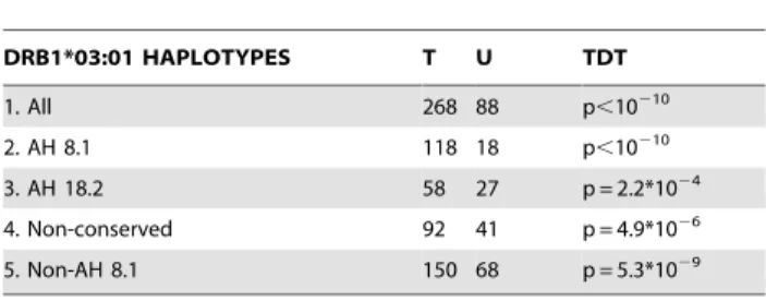
We studied 274 trios to investigate the possibility of a differential transmission of the different DRB1*03:01 haplotypes to the affected child (Table 1). DRB1*03:01 is always preferentially transmitted, independently of its haplotype context (see TDT results, in Table 1). However, the distortion in the transmission of this allele is significantly higher when it is present in the AH 8.1, compared with its presence in the remaining DRB1*03:01 -containing haplotypes (p = 8.7*1024vs. AH 18.2; p = 2.4*1024vs. conserved haplotypes). AH 18.2 and non-conserved haplotypes show a similar preferential transmission to offspring (p = 0.99). These differences are also observed when considering the haplotype transmission from the DRB1*03:01
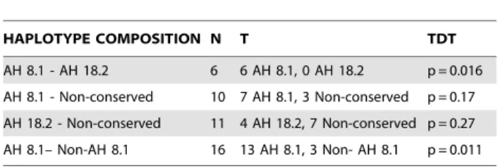
homozygous parents (composed by differentDRB1*03:01 haplo-types) included in the 274 families (Table 2).
We wanted to validate this observation in a case-control sample (Table 3). Since no differences were observed between AH 18.2 and non-conserved haplotypes in the family data, they were combined in subsequent analysis (and called non-AH 8.1). As already known,DRB1*03:01overall appears at significantly higher frequency in CD patients than in controls: 45% vs. 14%, respectively (OR = 4.97 95% CI 3.90–6.34, p,1027); but we additionally show that this case-control difference is higher when considering only the AH 8.1 (OR = 6.53 95% CI 4.47–9.56, p,1027).
Table 1.Transmission data of the diverseDRB1*03:01 -containing haplotypes in the 274 families studied and TDT results.
DRB1*03:01 HAPLOTYPES T U TDT
1. All 268 88 p,10210
2. AH 8.1 118 18 p,10210
3. AH 18.2 58 27 p = 2.2*1024
4. Non-conserved 92 41 p = 4.9*1026
5. Non-AH 8.1 150 68 p = 5.3*1029
T = transmitted; U = untransmitted; TDT = transmission disequilibrium test. Non-AH 8.1 includes AH18.2 and non-conserved haplotypes.
TDT results are calculated after excluding homozygous parents (5 for AH 8.1, 5 for AH 18.2 and 7 forDRB1*03:01non-conserved haplotypes).
Haplotype comparisons: 2 vs. 3: p = 0.00087 OR = 3.05 95% CI 1.48–6.33; 2 vs. 4: p = 0.00024 OR = 3.06 95% CI 1.59–5.94; 3 vs. 4: p = 0.99 OR = 1.00 95% CI 0.54– 1.88; 2 vs. 5: p = 7.8*1025
In CD, it is well established the existence of a dose effect, what implies the existence of different CD risk categories attending to their HLA constitution. We investigated this differential risk contribution of DRB1*03:01-containing haplotypes in those categories and found that only in those individuals carrying a single copy of theDQB1*02allele (individuals DQ2.5 cis+ non-DQ2.2 in Table 3), the presence of AH 8.1 confers additional risk. When considering the HLA risk categories according to gene dosage effects, carriage of the DQ2 molecule in individuals with a single copy of the DQB1*02 allele is considered as conferring intermediate CD risk. A similar risk is conferred by the presence of DQ2.5 trans, although some groups reported this to be an intermediate higher risk group [11]. We compared CD risk in carriers of DQ2.5 trans (48 individuals out of 369 patients and 21 out of 461 controls) to CD risk in carriers of DQ2.5 cis with and without the AH 8.1 and we observed that DQ2.5 trans confers similar risk than DQ2.5 cis with AH 8.1 (heterogeneity: p = 0.91,
I2= 0%) and significantly higher risk than DQ2.5 cis with non-AH 8.1 haplotypes (heterogeneity: p = 0.09, I2= 64%).
Finally, we investigated the possibility that the similar risk conferred by DQ2.5 trans and DQ2.5 cis with AH 8.1 was due to the presence of a common susceptibility factor. In most cases, carriers of the molecule DQ2.5 trans are genetically characterized by being heterozygous DQB1*03:01-DQA1*05:05/DQB1*02:02-DQA1*02:01(serologically DR5/DR7, terms also used hereafter for simplification purposes). We used genotype data corresponding to 6,769 SNPs located in the HLA (29.96–33.19 Mb interval), which were previously obtained in a subset of our Spanish samples (more than 500 CD patients and 300 controls) in the context of the Immunochip Project (http://www.immunobase.org). We selected all the homozygous individuals for AH 8.1, AH 18.2, DR5 (DQB1*03:01-DQA1*05:05) and DR7 (DQB1*02:02-DQA1*02:01) and looked for a chromosomal region shared between AH 8.1 and DR5 (DQB1*03:01-DQA1*05:05) or AH 8.1 and DR7 (DQB1*02:02-DQA1*02:01) but not common to AH 18.2. However, no regions emerged after that search. Note that this result could be affected by the low number of available homozygous individuals, mainly DR5 ( DQB1*03:01-DQA1*05:05) and DR7 (DQB1*02:02-DQA1*02:01) (3 and 4, respectively).
Discussion
Our results evidence the presence of an additional susceptibility factor to CD in the HLA region, which is linked to AH 8.1. This factor only increases susceptibility when appearing in individuals carrying a single copy of the DQB1*02 allele (DQ2.5 cis non-DQ2.2) and split up the HLA intermediate risk group into two groups: one with higher intermediate risk, which is composed of
Table 2.Transmission data of the diverseDRB1*03:01 -containing haplotypes fromDRB1*03:01homozygous parents (N = 27).
HAPLOTYPE COMPOSITION N T TDT
AH 8.1 - AH 18.2 6 6 AH 8.1, 0 AH 18.2 p = 0.016
AH 8.1 - Non-conserved 10 7 AH 8.1, 3 Non-conserved p = 0.17
AH 18.2 - Non-conserved 11 4 AH 18.2, 7 Non-conserved p = 0.27
AH 8.1– Non-AH 8.1 16 13 AH 8.1, 3 Non- AH 8.1 p = 0.011
T = transmitted; TDT = transmission disequilibrium test. doi:10.1371/journal.pone.0048403.t002
Table 3.Frequency and comparison of the diverseDRB1*03:01haplotypes in celiac disease (CD) patients (2N = 738) and controls (2N = 922), overall and classified according to the different HLA CD risk categories.
DRB1*03:01 HAPLOTYPES CD CONTROLS CD vs. CONTROLS
HETEROGENEITY AH 8.1 VS. NON-AH 8.1
2N % 2N % p OR (95% CI) p I2
ALL DRB1*03:01
All 330 44.7 129 14.0 ,1027 4.97 (3.90–6.34)
AH 8.1 141 19.1 42 4.6 ,1027 6.53 (4.47–9.56) 0.06 71%
Non-AH 8.1 189 25.6 87 9.4 ,1027 4.22 (3.16–5.65)
DQ2.5 cis+DQ2.5 cis
All 74 10.0 16 1.7 ,1027 8.99 (5.03–16.28)
AH 8.1 30 4.1 6 0.65 ,1027 9.72 (3.93–28.74) 0.82 0%
Non- AH 8.1 44 6.0 10 1.1 ,1027 8.55 (4.10–18.31)
DQ2.5 cis+DQ2.2
All 104 14.1 13 1.4 ,1027 15.55 (8.39–29.37)
AH 8.1 40 5.4 6 0.7 ,1027 12.96 (5.39–37.64) 0.60 0%
Non- AH 8.1 64 8.7 7 0.8 ,1027 17.77 (8.03–46.30)
DQ2.5 cis+non-DQ2.2
All 152 20.6 100 10.8 ,1027 2.95 (2.21–3.94)
AH 8.1 72 9.8 30 3.3 ,1027
4.66 (2.94–7.44) 0.009 85%
Non-AH 8.1 80 10.8 70 7.6 3.2*1026 2.22 (1.56–3.17)
carriers of the molecule DQ2.5 cis encoded by AH 8.1 and carriers of the molecule DQ2.5 trans; and one with lower intermediate risk, which is composed of carriers of the molecule DQ2.5 cis encoded by either AH 18.2 or non-conserved
DRB1*03:01haplotypes.
The well-known contribution of the DQ2 molecule to CD pathogenesis is genetically based on carriage of DQA1*05 and
DQB1*02. These two alleles are commonly present in the small segment identical by descent among DRB1*03:01-containing haplotypes. However, outside that shared segment, the divergence between DRB1*03:01 haplotypes do not differ from that found between disparate haplotypes [12]; therefore, a susceptibility factor located there is not expected to be present in all DRB1*03:01
haplotypes. On the other hand, previous studies suggested a close evolutionary relationship among DRB1*03:01-containing haplo-types, DR5 and DR7 (DQB1*03:01-DQA1*05:05 and
DQB1*02:02-DQA1*02:01) [13], which could explain the existence of a common susceptibility factor between AH 8.1 and one of those haplotypes. No definitive conclusion can de drawn from our data due to the low number of chromosomes compared. Further analysis including higher sample size is mandatory.
The analysis of the HLA region in GWAS showed, besides the expected peak corresponding to DQ2.5 cis, two SNPs associated to CD risk, both located within or adjacent to HLA-DQA1and
HLA-DQB1 [14]. This is in accordance with our results, which suggest a risk factor common toDRB1*03:01-containing haplo-types and DR5 or DR7 (DQB1*03:01-DQA1*05:05 or
DQB1*02:02-DQA1*02:01), because those are the regions that they shared.
One intriguing issue emerging from our study is why the additional risk factor present in the AH 8.1 does not seem to influence on CD susceptibility when it appears in individuals carrying a second copy of the DQB1*02 allele. In CD, T cell stimulation due to gluten-derived peptides depends on the number and type of HLA-DQ2 molecules expressed. DQ2.5 molecules can bind a high repertoire of gluten peptides, but only a restricted subset is bound to DQ2.2 molecules, which reduce the immuno-genicity of DQ2.2. Additionally, the number of these DQ molecules is also a relevant factor in T cell stimulation and this depends on the number of specific alleles inDQA1andDQB1loci, which determines the possibleab-chain combinations constituting the DQ heterodimers [15]. As a matter of fact, all HLA-DQ molecules are identical in HLA-DQ2.5 homozygous individuals,
which can bind a very high repertoire of gluten peptides and confer the highest CD risk. It could be speculated that in such scenario an additional susceptibility factor has null or limited possibility to increase risk. By the contrary, in HLA-DQ2.5 cis individuals (without a second copy ofDQB1*02, i.e., non-DQ2.2), only one of the four possibleabcombinations constitutes an HLA-DQ2.5 molecule and therefore the presence of a genetic factor which increases immunogenicity against gluten derived peptides could have a relevant impact in increasing CD risk.
The HLA dose effect is also influenced by differences in the kinetic stability of the interaction between HLA molecules and gluten derived peptides, key factor for development of T cell responses against gluten [16,17]. For most peptide ligands, DQ2.5 shows higher binding stability than DQ2.2. The risk variant present in the AH 8.1, and presumably in individuals possessing DQ2.5 trans molecules, could affect the kinetic stability of the interaction HLA-gluten either increasing the number of gluten derived peptides which can bind with high affinity or increasing the binding stability of peptides previously recognised. Boddet al
[17] claimed that T-cell epitopes must be assessed and character-ized in the context of the HLA molecules expressed by the T-cell donor and underlined the relevance that this could have for future peptide-based vaccines. According to that, it would be interesting to establish a comparison of the HLA-DQ molecules present in individuals DQ2.5 cis with AH 8.1, DQ2.5 cis with non-AH 8.1 haplotypes and DQ2.5 trans. Differences in their binding gluten peptides would imply that peptide-based vaccines should look at those individuals differentially.
Much more work deserves this field, with several open questions as which is the causal variant lying on the AH 8.1 and which are their specific functional implications.
Acknowledgments
We are most grateful to Carmen Martı´nez Cuervo and M. A´ ngel Garcı´a Martı´nez for their expert technical assistance.
Author Contributions
Conceived and designed the experiments: CN. Performed the experiments: LMM BD ALL. Analyzed the data: LMM BD ALL MFA CN. Contributed reagents/materials/analysis tools: CM AD AB NLP MAF. Wrote the paper: LMM CN. All authors critically revised the manuscript and approved the final version of the manuscript.
References
1. Keuning JJ, Pena AS, van Leeuwen A, van Hooff JP, va Rood JJ (1976) HLA-DW3 associated with coeliac disease. Lancet 1: 506–508.
2. Baschal EE, Aly TA, Jasinski JM, Steck AK, Noble JA, et al. (2009) Defining multiple common ‘‘completely’’ conserved major histocompatibility complex SNP haplotypes. Clin Immunol 132: 203–214.
3. de la Concha EG, Cavanillas ML, Cenit MC, Urcelay E, Arroyo R, et al. (2012) DRB1*03:01 Haplotypes: Differential Contribution to Multiple Sclerosis Risk and Specific Association with the Presence of Intrathecal IgM Bands. PLoS One 7: e31018.
4. Bolognesi E, Karell K, Percopo S, Coto I, Greco L, et al. (2003) Additional factor in some HLA DR3/DQ2 haplotypes confers a fourfold increased genetic risk of celiac disease. Tissue Antigens 61: 308–316.
5. Bilbao JR, Calvo B, Aransay AM, Martin-Pagola A, Perez de Nanclares G, et al. (2006) Conserved extended haplotypes discriminate HLA-DR3-homozygous Basque patients with type 1 diabetes mellitus and celiac disease. Genes Immun 7: 550–554.
6. Louka AS, Lie BA, Talseth B, Ascher H, Ek J, et al. (2003) Coeliac disease patients carry conserved HLA-DR3-DQ2 haplotypes revealed by association of TNF alleles. Immunogenetics 55: 339–343.
7. de la Concha EG, Fernandez-Arquero M, Vigil P, Rubio A, Maluenda C, et al. (2000) Celiac disease and TNF promoter polymorphisms. Hum Immunol 61: 513–517.
8. Husby S, Koletzko S, Korponay-Szabo IR, Mearin ML, Phillips A, et al. (2012) European Society for Pediatric Gastroenterology, Hepatology, and Nutrition
guidelines for the diagnosis of coeliac disease. J Pediatr Gastroenterol Nutr 54: 136–160.
9. Nedospasov SA, Udalova IA, Kuprash DV, Turetskaya RL (1991) DNA sequence polymorphism at the human tumor necrosis factor (TNF) locus. Numerous TNF/lymphotoxin alleles tagged by two closely linked microsatellites in the upstream region of the lymphotoxin (TNF-beta) gene. J Immunol 147: 1053–1059.
10. Fernandez-Arquero M, Arroyo R, Rubio A, Martin C, Vigil P, et al. (1999) Primary association of a TNF gene polymorphism with susceptibility to multiple sclerosis. Neurology 53: 1361–1363.
11. van Heel DA, Hunt K, Greco L, Wijmenga C (2005) Genetics in coeliac disease. Best Pract Res Clin Gastroenterol 19: 323–339.
12. Traherne JA, Horton R, Roberts AN, Miretti MM, Hurles ME, et al. (2006) Genetic analysis of completely sequenced disease-associated MHC haplotypes identifies shuffling of segments in recent human history. PLoS Genet 2: e9. 13. Sollid LM (2000) Molecular basis of celiac disease. Annu Rev Immunol 18: 53–
81.
14. van Heel DA, Franke L, Hunt KA, Gwilliam R, Zhernakova A, et al. (2007) A genome-wide association study for celiac disease identifies risk variants in the region harboring IL2 and IL21. Nat Genet 39: 827–829.
16. Fallang LE, Bergseng E, Hotta K, Berg-Larsen A, Kim CY, et al. (2009) Differences in the risk of celiac disease associated with DQ2.5 or HLA-DQ2.2 are related to sustained gluten antigen presentation. Nat Immunol 10: 1096–1101.