From the Field to the Lab: Best Practices for
Field Preservation of Bat Specimens for
Molecular Analyses
Angelique Corthals1,2*, Alynn Martin3, Omar M. Warsi4, Megan Woller-Skar3, Winston Lancaster5¤, Amy Russell3, Liliana M. Dávalos4,6
1Department of Sciences, John Jay College of Criminal Justice (CUNY), New York, New York, United States of America,2Department of Pathology, Stony Brook University School of Medicine, Stony Brook, New York, United States of America,3Department of Biology, Grand Valley State University, Allendale, Michigan, United States of America,4Department of Ecology and Evolution, Stony Brook University, Stony Brook, New York, United States of America,5Department of Biological Sciences, California State University, Sacramento, California, United States of America,6Consortium for Inter-Disciplinary Environmental Research, School of Marine and Atmospheric Sciences, Stony Brook, New York, United States of America
¤ Current address: Department of Biology, University of Alabama at Birmingham, Birmingham, Alabama, United States of America
Abstract
Studies in molecular ecology depend on field-collected samples for genetic information, and the tissue sampled and preservation conditions strongly affect the quality of the DNA obtained. DNA yields from different tissue types have seldom been compared, and the rela-tive performance of storage media has never been directly tested, even though these media may influence DNA degradation under field conditions. We analyzed DNA yield from buccal swabs and wing punches harvested from live bats using nucleic acid quantification as well as quantitative PCR for a single-copy nuclear locus. We also compared DNA yields from wing tissue preserved in three media: ethanol, NaCl-saturated dimethyl sulfoxide (DMSO), and silica desiccant. Wing punches yielded more total DNA than did buccal swabs, and wing tissues preserved in silica beads yielded significantly more total and nuclear DNA than those preserved in DMSO or ethanol. These results show that tissue type and preservation media strongly influence the quantity of DNA obtained from non-lethal genetic samples, and based on these effects we provide recommendations for field collection of tissues for genetic analyses.
INTRODUCTION
Collecting samples in the field for molecular studies can be stressful or even lethal for the or-ganism of interest. Ecologists must often sample tissues in a way that both maximizes nucleic acid yields and minimizes stress on the sampled individual. The choices of tissue type and tis-sue preservation protocols are often based on convenience and historical practices instead of optimizing for the highest yield. In this paper, we aim to evaluate alternative, affordable, and
OPEN ACCESS
Citation:Corthals A, Martin A, Warsi OM, Woller-Skar M, Lancaster W, Russell A, et al. (2015) From the Field to the Lab: Best Practices for Field Preservation of Bat Specimens for Molecular Analyses. PLoS ONE 10(3): e0118994. doi:10.1371/ journal.pone.0118994
Academic Editor:Alfred L. Roca, University of Illinois at Urbana-Champaign, UNITED STATES
Received:April 16, 2014
Accepted:December 18, 2014
Published:March 23, 2015
Copyright:© 2015 Corthals et al. This is an open access article distributed under the terms of the Creative Commons Attribution License, which permits unrestricted use, distribution, and reproduction in any medium, provided the original author and source are credited.
Data Availability Statement:All data files are available from the Dryad database (http://dx.doi.org/ 10.5061/dryad.3d727).
Funding:The authors have no support or funding to report.
efficient field collection protocols that do not require euthanizing the animal for downstream molecular analyses. We focus on bats (Mammalia: Chiroptera) based on both our expertise and the burgeoning literature on chiropteran population genetics and phylogenetics [1–5].
Traditionally, the most common mammalian tissue collected in the field has been the liver, because it is both easy to identify and very large [6–15]. If the goal is to retrieve genomic DNA for sequencing, however, the liver is ill suited for preservation as it has several distinct disad-vantages for molecular analysis. First, it requires the euthanasia of the animal being sampled. Second, because of its high enzymatic activity, the liver is also the first organ to go through au-tolysis after death [16]. Third, livers express high levels of stress- and inflammation-related proteins at the moment of euthanasia, as transient hypoxia increases hypoxanthine and xan-thine in liver cells [17]. This may bias transcriptomic or proteomic studies, and contaminates genomic DNA extractions with plentiful RNA transcripts that may interfere with sequence capture methods [18]. The second most common tissue type, muscle, is more resistant to autol-ysis, but also requires the euthanasia of the animal sampled and thus may contribute to popula-tion declines [8,14,19].
Nonlethal sampling of bats usually involves obtaining patagial biopsy samples [20–22] or feces [23]. While Worthington et al. [21] advised storing patagial samples in NaCl-saturated di-methyl sulfoxide (DMSO), ethanol and silica beads are also commonly used as storage media [15,24,25]. These three media are affordable and relatively easy to transport, and have been the preferred method of nonlethal bat tissue preservation in the field. Each medium, however, gen-erates challenges in preserving even robust molecules such as genomic DNA [12,13]. Other agents, such as lysis buffer, or Qiagen’s RNAlaterand AllProtect are used less often. The Qia-gen products are relatively expensive (e.g., RNAlatercosts $315 US/250 ml), and lysis buffer over-lyses tissues when left for a long period of time at room temperature or in field conditions [26,27]. Ideally, field-collected samples should be preserved in liquid nitrogen to preserve the best molecular profile, including fragile molecules [28,29]. This preservation method is rarely used in the field, though, as liquid nitrogen is difficult to transport and attracts negative atten-tion at customs and airports, and refilling tanks is often difficult or impossible in remote loca-tions [8,30–32].
In this paper, we assess several affordable methods of sampling and preserving mammalian tissues without euthanizing individuals. We sampled bat wing punches and mouth swabs aim-ing to uncover the best type of tissue for downstream genomic DNA analyses. The collection of buccal cells using a mouth swab has been extensively used for rabies surveillance [33], and is being introduced here to collect bat epithelial cells. Three common media (ethanol, DMSO, and silica beads) were used to assess tissue preservation methods for wing punches. Genomic DNA was extracted from each sample type and for each medium used. A comparison of DNA yields, using both spectrophotometric nucleic acid quantification and quantitative PCR (qPCR) of a single-copy nuclear gene, was conducted between sample types (wing punch vs. buccal swabs) and between each preservation medium for wing punches (ethanol, DMSO and silica beads). The purpose of both experiments was to evaluate which sample type and which medium produced the best results.
RESULTS
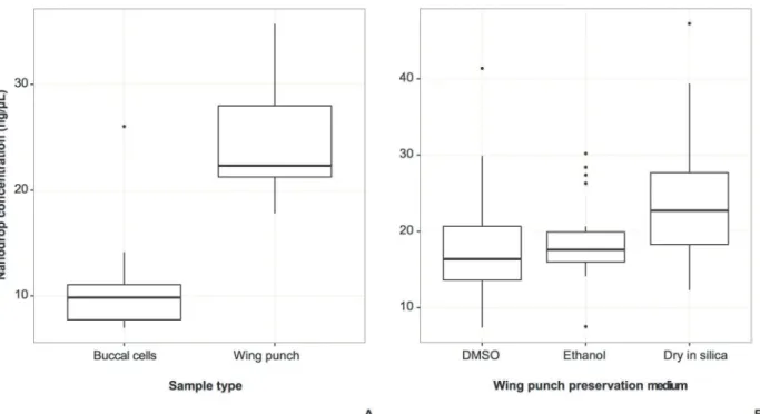
chemicals. Comparisons of total nucleic acid concentration from the sample type experiment showed that wing punches yielded more nucleic acids than buccal cells (Fig. 1A). Comparisons from the preservation medium experiment showed that more nucleic acids were recovered from wing punches preserved dry in silica than from DMSO- or ethanol-preserved wing tissue (Fig. 1B).
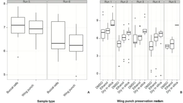
In contrast to the total nucleic acids recovered, target DNA quantity, as assessed by the number of copies of the nuclear gene amplified from buccal cells and wing punches, did not differ significantly between tissue types (Fig. 2A), and there was great variability between runs and individuals sampled (Table 1,S1 Fig.). The best-fit model included group-level coefficients for both qPCR runs and individual bats, but the effect of the sample type was not significant (Table 1). In the preservation medium experiment, wing punches preserved dry in silica in the field consistently yielded more copies of the nuclear gene than punches preserved in ethanol, which in turn produced more copies than punches preserved in DMSO (Table 2,Fig. 2B,S1B Fig.). As with the sample type experiment, the best-fit model accounted for both qPCR runs and individual bats and, in sharp contrast, preservation medium had a significant effect on DNA copies detected (Table 1).Table 2provides an estimate of the effects of individual preser-vation methods, showing that silica beads preserved more copies than ethanol, and both pre-served more copies than DMSO (for every copy prepre-served in DMSO, ~2.4 copies were preserved in ethanol, and ~5.7 in silica).
The number of nuclear gene copies and concentration of total cellular nucleic acids in ex-tracts were significantly associated in wing punches in the preservation medium experiment (Fig. 3). In contrast, nucleic acids and nuclear gene copies from matched buccal cells and silica-preserved wing punches in the sample type experiment were not significantly associated (S2 Fig.).
Fig 1. DNA concentration measured using nanodrop spectrophotometer.A. Comparison between sample types obtained from the same individuals (t(22)= 6.55,P= 1.254e-06). B. Comparison between preservation treatments for wing punch samples obtained from the same individuals (individual effect F(20, 36)= 5.250,MSE= 121.8,P= 8.33e-06; treatment preservation effectF(2, 36)= 6.978,MSE= 161.9,P= 0.00275).
DISCUSSION
Buccal swabs acquired in the sample type experiment required more collector skill and were less bat-friendly than initially anticipated. Though larger bats (e.g.,Brachyphylla, ~35 g) could handle the swabbing with brushes unhurt, smaller bats (e.g.,Pteronotus quadridens, ~5 g) did not fare well. The epidermis of the palate of smaller bats would quickly tear, causing discomfort and sometimes bleeding unless extreme care was applied. The samples themselves were also more prone to inconsistency during collection compared with the wing punches. While a wing punch is easily replicable by using a biopsy punch tool, collecting buccal cells depends on the time the collector is able to handle the bat, as well as the sensitivity of the bat to the swab brush. Though a time of 60 seconds was assigned to the collection of each buccal sample, not all bats handled were able to sustain 60 seconds of brushing without bleeding.
The buccal cells obtained by swabbing, though easy to transport, produced low nucleic acid yields (Fig. 1A), even as they generated similar numbers of copies of target nuclear DNA as wing punches (Fig. 2A). The low yield may also be due to the preservation of the swab in silica beads and/or the lower density of buccal cells in the oral cavity of most bats. It is important to point out that despite the low comparative performance of buccal swabs, they still yielded enough DNA for PCR amplification. While buccal swabs may prove useful for larger bats, we recommend that further studies focus on testing a number of alternatives: 1) using cotton swabs rather than brushes, 2) using Whatman paper‘brushes’to see if buccal cells can be ab-sorbed and preserved on paper better than with nylon bristles, and 3) combining the buccal swabs with preservation in a weakly acid lysis buffer that would lyse cells but preserve nuclei [35].
When comparing preservation media for wing punches, our results conclusively show that wing punches desiccated in silica beads in the field yield significantly greater quantities of DNA compared to samples preserved in ethanol or DMSO. Wing punches are an ideal tissue
Fig 2. Number of copies of single-copy nuclear gene obtained through quantitative PCR (qPCR) amplifications from different sample types and methods of preservation.Results are presented sorted by qPCR run. The models that best fit both datasets accounted for group-level effects of runs and individuals sampled (Table 1). A. Comparison between sample types obtained from the same individuals (no significant effect of sample type was found, see
Table 1). B. Comparison between preservation treatments for wing punch samples obtained from the same individuals (ethanol and dry preservation were significantly better than DMSO preservation, see Tables1and2).
type for nonlethal bat sampling because they allow researchers to release the bat after capture. Punches are usually collected with a sterile biopsy puncher that standardizes the size of each sample independently of the collector’s skill, and can be easily stored in vials without further handling. Previous studies have shown that wounds from similarly-sized wing punches will heal completely in an average of 2–4 weeks [36].
Silica beads are affordable, easy to store, easy to acquire, and lack travel restrictions because they are not liquid or combustible. Compared to silica beads, ethanol and DMSO are inferior storage agents if the goal is to maximize DNA yield. These chemicals are also inherently dam-aging to DNA. Ethanol becomes increasingly acidic over time, breaking the sugar-phosphate backbone of DNA, and forming Maillard products—broken down sugars that may inhibit PCR [12,13,37]. We suggest that future studies test alternative storage media such as ethanol mixed with acid gelling agents to prevent acidification and DNA degradation over time. DMSO is both cytotoxic and genotoxic, and is known to lower base-pair composition dependence [38]. This is a useful feature to facilitate PCR amplification of, for example, a GC-rich sample
Table 1. Models of the natural logarithm of nuclear gene copies detected through qPCR as a function of the sample type (Nobservations= 90,
Nindividual bats= 12, buccal swab brush or wing punch.
Model Group-level intercepts DF logLik AIC LRT P-value
No predictor qPCR run 3 −77.76 161.53 — —
Sample type qPCR run 4 −76.55 161.10 2.426 0.12
No predictor Individual 3 −82.01 170.03 — —
Sample type Individual 4 −81.40 170.79 1.230 0.27
No predictor qPCR run and individual 4 −69.94 147.87 — —
Sample type qPCR run and individual 5 −68.98 147.95 1.916 0.17
No predictor qPCR run 3 −438.88 883.76 — —
Preservation medium qPCR run 5 −417.07 844.13 43.624 3.4e-10
No predictor Individual 3 −359.94 725.87 — —
Preservation medium Individual 5 −298.09 606.19 123.68 0.00
No predictor qPCR run and individual 4 −354.09 716.86 — —
Preservation medium qPCR run and individual 6 −282.32 576.64 145.53 0.00
This includes individual WCL025, not shown inS1A Fig.because it only had a wing punch), or the preservation medium for wing punches (Nobservations= 228,Nindividual bats= 21, silica beads, ethanol, or DMSO. This includes individual ALR074, not shown inS1B Fig.because it only had a wing punch preserved in ethanol). No-predictor models serve as null hypotheses of no effect of the sample type or preservation medium on the number ofrag2copies detected using qPCR. Group-level intercepts of qPCR run and/or individual bat sampled serve to capture effects of unmeasured variability from subtle differences in qPCR starting conditions and bat behavior. Significance was measured using a likelihood ratio test of the model with predictors to its null.
Models with different sample types or preservation media as predictors can be compared to one another using the Akaike Information Criterion (AIC). The model with the lowest AIC is the bestfit to the data. DF = model degrees of freedom, logLik = log-likelihood of modelfit to the data, and LRT = likelihood
ratio test statistic.
doi:10.1371/journal.pone.0118994.t001
Table 2. Estimated coefficients of the effect of wing punch preservation methods on the natural loga-rithm of gene copies detected after accounting for differences between qPCR runs and among indi-vidual bats.
Medium Coefficient Std. error
Dry in silica 6.94 0.39
Ethanol 6.08 0.39
DMSO 5.20 0.39
[39,40]. At high concentration and for a period of years, DMSO buffer becomes deleterious and may cause PCR amplification to fail.
CONCLUSION
We introduced mouth swabbing for collecting bat DNA, and tested techniques of preservation of tissues commonly used in the field. We found that brush-style mouth swabs did not yield as much nucleic acid concentration and distressed the bats more than collecting wing punches, and that storing wing punches in silica beads in the field yielded more DNA of higher quality compared with liquid buffers (ethanol and DMSO). We therefore recommend the use of silica beads as a field preservation medium for wing punches.
MATERIALS AND METHODS
Samples
Tissues were collected during field trips to Puerto Rico and Hispaniola (Dominican Republic) and represent nine species from two families: Mormoopidae (Mormoops blainvillei,Pteronotus parnellii, andP.quadridens) and Phyllostomidae (Brachyphylla cavernarum,B.nana, Ero-phylla sezekorni,Macrotus waterhousii, andMonophyllus redmani, seeS1 Table). The sample collection protocols reported here were approved by Stony Brook University IACUC 20091741, Grand Valley State University IACUC 09-07-A, and California State University at
Fig 3. Relationship between the median number of copies of the single-copy nuclear gene obtained from repeated quantitative PCR reactions and concentration of whole DNA (wing punches in three preservation treatments).The correlation was significant whether including (r= 0.32,p= 0.018), or
excluding the outlier measurements from sample individual ALR096 (r= 0.47,p= 0.00034).
Sacramento IACUC S10-001. Field work, sample collection and export from the Dominican Republic was approved by Ministry of the Environment permit no. 0000986.
For the sample type experiment, individuals caught on Mona Island and Puerto Rico in June 2010 were sampled by both brushing their cheeks and palates using buccal swab brushes and taking 2-mm diameter wing punches. Both brushes and wing punches from this trip were preserved dry in individual tubes and stored dry in a cabinet with permeable boxes of indicator silica beads to reduce humidity and prevent condensation. These samples were extracted short-ly after arrival in the lab in 2010.
For the preservation medium experiment we sampled three 2-mm diameter wing punches from each captured individual during our first field collection to Puerto Rico and the Domini-can Republic in May-June 2009. One punch per individual was preserved in the field dry in 3-mm indicating silica beads, in NaCl-saturated 20% DMSO, and in ethanol. Upon arrival at the lab the punches were frozen at−80°C and preserved ultra-cold until genomic DNA
extrac-tion in January 2011.
Wing tissues for both sample type and preservation medium experiments were obtained using Acu-Punch sterile, disposable 2-mm skin biopsy punches (Acuderm, Inc.). One biopsy punch was used for each individual, and skin samples were taken from regions of the plagiopa-tagium that were neither likely to tear into the edge of the wing, nor close to major blood ves-sels. Bats regularly make tears like these while flying into branches, and similar wounds have been shown to heal completely in less than 30 days [36]. Punches were preserved in ~0.7 g indi-cator silica, in ~1 mL of 20% DMSO solution saturated with NaCl, or in ~1 mL molecular grade 100% ethanol.
For the sample type experiment, Epicentre (Illumina, Inc.) brushes were used to swab the mouth of the bat for 60 s, minimizing bleeding or gagging. A timer was used to ensure that brushes were equally applied to all individuals. This procedure is standard for collecting sam-ples from humans and other large mammals. The mouths of bats, however, do not open in a way that generates large cheek regions. For this reason, the brushes mostly sampled cells from the palate and the tongue.
Extraction protocols
All DNA extractions were conducted in a laboratory that undergoes regular decontamination with hypochloride treatment. Each sample extraction was conducted separately to prevent cross contamination. All extractions were performed in a Bio-Safety Laboratory (BSL)-II cabinet, which was UV-irradiated for 1 hour prior to each sample extraction. All consum-ables, including pipettor tips, micro-centrifuge tubes, and collection tubes as well as the small equipment, such as pipettors, were UV-irradiated in a UV crosslinker for 20 minutes at 1200 x100μJ/cm2. Gloves were also changed between every step of the extraction to prevent
contamination. Mock DNA extractions and control blank PCRs are performed continuously in the laboratory and screened for contamination.
Wing punches from both preservation medium and sample type experiments were placed in individual 1.5μl micro-centrifuge tubes, and DNA was extracted using the QIAmp (Qiagen)
tissue extraction protocol. First, all wing punches were lysed in 15μl of ATL buffer with 10μl
of proteinase K and incubated at 56°C for 3 hours. Second, we added 50μl of AL buffer with
1μl of carrier RNA and 50μl of molecular-grade 99% ethanol, and incubated the solution at
room temperature for 5 min. Third, the solution was purified using the QIAmp micro-col-umns. Finally, the samples were eluted using 55μl of PCR-grade water and stored at 4°C prior
Swab brushes from the sample type experiment were placed in 1.5μl micro-centrifuge
tubes, and DNA was extracted using a modified QIAmp extraction protocol. Cells from the swab brushes were lysed at 56°C for 60 min in 600μl of QIAmp micro kit ATL buffer and 20μl
of proteinase K. Six hundredμl of AL buffer were added, with 1μl of carrier RNA, and
incubat-ed the solution at 70°C for 10 min in a thermomixer shaking at 900 rpm. Three hundrincubat-edμl of
molecular-grade ethanol were added, and pulse-vortexed for each sample for 15 sec. All swab brushes were removed from the micro-centrifuge tubes, and the solution was purified using the QIAmp micro-columns. Finally, each sample was eluted using 55μl of PCR-grade water and
stored at 4°C prior to quantification and amplification.
Measurement of nucleic acids and DNA yield
We expected that both the wing punches and buccal swabs would yield some DNA from non-chiropteran sources such as bacteria, fungi, and insects. Therefore, we used two measures of DNA yield: 1) total nucleic acid concentration using a spectrophotometer (Nanodrop 1000, Thermo Scientific), and 2) number of copies of a single-copy nuclear gene using quantitative PCR (qPCR). One and a halfμL of DNA extract was used to obtain measures of absorbance at
the 260-nm wavelength to determine optical density using the spectrophotometer with a sensi-tivity range from 2.0 to 3700 ng. Optical density measures can comprise RNA, protein, and chemical contamination in addition to DNA. We used the minimum threshold of 1.80 for the 260/280 optical density ratio to check for protein and chemical contamination [34]. Higher val-ues indicate pure nucleic acids. For this reason we refer to optical density measures as nucleic acid quantifications, as they may include some RNA.
To specifically quantify target nuclear DNA rather than total nucleic acids or possible con-taminants, we used a 124-bp fragment of the single-copy nuclear recombination-activating gene 2 (rag2) as the target locus for qPCR. The primers used for the protocol were designed based on chiropteran sequences ofrag2obtained in our laboratory as well as those taken from Genbank: rag2-q2-f1 (5’-ACACCAAACAATGAGCTTTC-3’) and rag2-q2-r1 (5’ -CCA-TATCTGGCTTCAGG-3’). Following primer design, a small subset of samples (N= 5) was used to verify the length of the target product and absence of mispriming using standard PCR. The PCR yielded a single low-molecular weight bright band of the correct length (S3 Fig.).
The Qiagen Quantifast SYBR green PCR kit was used to perform these reactions. The qPCR reactions followed the Qiagen SYBR Green PCR kit handbook, as follows: 12.5μl of qPCR mix,
2.5μl of nuclease free H2O, 2.5μl of each primer, and 5μl of the template for a total reaction
volume of 25μl. The conditions were a two-step cycling protocol with a 5-min PCR activation
step at 95°C, denaturation for 10 min at 95°C and combined annealing and extension steps at 60°C for 30 s for a total number of 35 cycles. Quantitative PCR for each sample was replicated within runs (technical replicate), and in separate runs, resulting in ~4 estimates of gene copies per sample. The standard quantification curve used to estimate the number of copies in the sample was plotted using lambda bacteriophage DNA diluted from 108to 101concentration from a stock solution of 100μg/ml or 1010copies/ml. This curve was used to calculate the total
number of DNA copies in the sample.
Statistical analyses
Quantitative PCR depends strongly and nonlinearly on initial conditions, and requires rep-lication of each DNA sample tested [41,42]. This introduces a multi-level structure to the data: observations of gene copy count are nested within qPCR runs as well as within the samples an-alyzed. Additionally, individual bats may vary in their behavior (when swabbing with the brush) or skin thickness (when taking a wing punch), introducing another source of variation. A hierarchical modeling approach was therefore used to measure the effects of preservation medium or sample type on single-copy gene quantity. The factors of interest for each experi-ment were either the sample type or preservation medium, and their effects were estimated using experiment-wide coefficients [43]. The variation arising from other factors, such as the qPCR run or identity of each bat were modeled as group-specific intercepts. The intercepts for individuals, in particular, were expected to capture variability in number of copies arising from bat species, condition, and time elapsed between collecting the punch in the field and extracting the DNA (as some bats sampled at the beginning of the field trip spent more time in field con-ditions than those captured closer to the date of return to the lab).
Multi-level models were fitted using maximum likelihood in thelmerfunction of the R package lme4 v. 0.999999-0 [44]. The fits of nested models to the data were compared using likelihood ratio tests, and significance of the log-likelihood ratio statistic was approximated by theχ2distribution with degrees of freedom equal to the difference in number of parameters of
the models. For non-nested models, fits were compared using the Akaike Information Criteri-on (AIC), with the best-fit model correspCriteri-onding to the lowest AIC value [45].
Analyses of correlation between DNA concentration and the number of copies detected were conducted by estimating Pearson’srand its significance. To reduce the impact of extreme values in estimating correlations, up to four repeated measurements of gene copies per extract were summarized using the median. All analyses were carried out in R statistical language v. 2.14.2 [46].
Supporting Information
S1 Fig. Number of copies of single-copy nuclear gene obtained through quantitative PCR (qPCR) amplifications from different sample types and methods of preservation.Results are presented sorted by individual sampled. The models that best fit both datasets accounted for group-level effects of runs and individuals sampled (Table 1). A. Comparison between sam-ple types obtained from the same individuals (no significant effect of samsam-ple type was found, seeTable 1, note that individual WCL025 is not shown here). B. Comparison between preser-vation treatments for wing punch samples obtained from the same individuals (ethanol and dry preservation were significantly better than DMSO preservation, seeTable 1, note that indi-vidual ALR074 is not shown here).
(TIFF)
S2 Fig. Relationship between the median number of copies of the single-copy nuclear gene obtained from repeated quantitative PCR reactions and concentration of whole DNA (buc-cal cells and wing punches).The correlation was not significant (r= 0.07,p= 0.766).
(TIFF)
performed to the image. (TIFF)
S1 Table. List of samples, sample types, and preservation media used in the two experi-ments.DMSO is NaCl-saturated dimethyl sulfoxide, ETOH is ethanol, and SG is silica desic-cant.
(DOCX)
Acknowledgments
For assistance in collecting samples we thank Armando Rodriguez-Duran, Ashley Rolfe, and Jean Sandoval in Puerto Rico. In the Dominican Republic we thank the logistical support of Grupo Jaragua in Santo Domingo and Oviedo, and especially Gerson Feliz, Esteban Garrido, Sixto Incháustegui, Yolanda M. Leon, and Miguel Santiago Nuñez-Novas.
Author Contributions
Conceived and designed the experiments: AC LMD. Performed the experiments: AC OW. An-alyzed the data: AM MW-S AR LMD. Contributed reagents/materials/analysis tools: AC AM MW-S WL AR LMD. Wrote the paper: AC AR LMD.
REFERENCES
1. Phillips CD, Phelan G, Dowd SE, McDonough MM, Ferguson AW, Delton Hanson J, et al. Microbiome analysis among bats describes influences of host phylogeny, life history, physiology and geography. Mol Ecol. 2012; 21: 2617–2627. doi:10.1111/j.1365-294X.2012.05568.xPMID:22519571
2. Russell A, Cox M, Brown V, McCracken G. Population growth of Mexican free-tailed bats (Tadarida bra-siliensis mexicana) predates human agricultural activity. BMC Evol Biol. 2011; 11: 88. doi:10.1186/ 1471-2148-11-88PMID:21457563
3. Muscarella RA, Murray KL, Ortt D, Russell AL, Fleming TH. Exploring Demographic, Physical, and His-torical Explanations for the Genetic Structure of Two Lineages of Greater Antillean Bats. PLoS ONE. 2011; 6: e17704. doi:10.1371/journal.pone.0017704PMID:21445291
4. Dumont ER, Dávalos LM, Goldberg A, Voigt CC, Rex K, Santana SE. Morphological innovation, diversi-fication and the invasion of a new adaptive zone. Proc R Soc B. 2012; 279: 1797–1805. doi:10.1098/
rspb.2011.2005PMID:22113035
5. Rojas D, Vale Á, Ferrero V, Navarro L. When did plants become important to leaf-nosed bats? Diversifi-cation of feeding habits in the family Phyllostomidae. Mol Ecol. 2011; 20: 2217–2228. doi:10.1111/j.
1365-294X.2011.05082.xPMID:21481051
6. Atmadja DS, Tatsuno Y, Ueno Y, Nishimura A. The effect of extraction methods. The kind of organ sam-ples and the examination delay on the DNA yields and typing. Kobe Journal of Medical Science. 1995; 41: 197–211. PMID:8869006
7. Baker RJ, Hafner MS. Curation of collections of frozen tissues. Curatorial problems unique to frozen tis-sue collections. In: Dessauer HC, Hafner MS, editors. Collections of Frozen Tistis-sues: Value, Manage-ment, Field and Laboratory Procedures, and Directory of Existing Collections. Lawrence, Kansas: Association of Systematics Collections; 1984. pp. 35–40.
8. Corthals A, Desalle R. An application of tissue and DNA banking for genomics and conservation: The Ambrose Monell Cryo-Collection (AMCC). Syst Biol. 2005; 54.
9. Dessauer HC, Hafner MS, Goodman M. Value of frozen tissue collections for studies in evolutionary bi-ology. In: Dessauer HC, Hafner MS, editors. Collections of Frozen Tissues: Value, Management, Field and Laboratory Procedures, and Directory of Existing Collections. Lawrence, Kansas: Association of Systematics Collections; 1984. pp. 3–5.
10. Florian M-L. The effects of freezing and freeze-drying on natural history specimens. Collection Forum. 1990; 6: 1–7.
12. Hajibabaei M, deWaard JR, Ivanova NV, Ratnasingham S, Dooh RT, Kirk SL, et al. Critical factors for assembling a high volume of DNA barcodes. Philosophical Transactions of the Royal Society B: Biolog-ical Sciences. 2005; 360: 1959–1967. PMID:16214753
13. Zimmermann J, Hajibabaei M, Blackburn DC, Hanken J, Cantin E, Posfai J, et al. DNA damage in pre-served specimens and tissue samples: a molecular assessment. Frontiers in Zoology. 2008; 5: e18. 14. Engstrom MD, Murphy RW, Haddrath O. Sampling vertebrate collections for molecular research:
prac-tice and policies. In: Metsger D, Byers S, editors. Managing the Modern Herbarium: An Interdisciplinary Approach. Vancouver: Elton-Wolf; 1999. pp. 315–330.
15. Velazco PM, Patterson BD. Diversification of the Yellow-shouldered bats, GenusSturnira(Chiroptera, Phyllostomidae), in the New World tropics. Mol Phylogenet Evol. 2013; 68: 683–698. doi:10.1016/j. ympev.2013.04.016PMID:23632030
16. Hanner R, Corthals A, Dessauer HC. Salvage of genetically valuable tissues following a freezer failure. Mol Phylogenet Evol. 2005; 34: 452–455. PMID:15619456
17. Kataoka T, Yoshimoto M, Nakagawa S, Mizuguchi Y, Taguchi T, Yamaoka K. Basic study on active changes in biological function of mouse liver graft in cold storage after low-dose X-irradiation. Journal of Clinical Biochemistry and Nutrition. 2009; 45: 219–226. doi:10.3164/jcbn.09-06PMID:19794932
18. Lemmon AR, Emme SA, Moriarty Lemmon E. Anchored Hybrid Enrichment for Massively High-Throughput Phylogenomics. Syst Biol. 2012; 61: 727–744. doi:10.1093/sysbio/sys049PMID:
22605266
19. Benford G. Saving the "library of life". Proceedings of the National Academy of Sciences, USA. 1992; 89: 11098–11101. PMID:1438320
20. Flanders J, Jones G, Benda P, Dietz C, Zhang S, Li G, et al. Phylogeography of the greater horseshoe bat,Rhinolophus ferrumequinum: contrasting results from mitochondrial and microsatellite data.
Molec-ular Ecology. 2009; 18: 306–318. doi:10.1111/j.1365-294X.2008.04021.xPMID:19192181
21. Worthington Wilmer J, Barratt E. A non-lethal method of tissue sampling for genetic studies of chiropter-ans. Bat Research News. 1996; 37: 1–3.
22. Miller-Butterworth CM, Vonhof MJ, Rosenstern J, Turner GG, Russell AL. Genetic Structure of Little Brown Bats (Myotis lucifugus) Corresponds With Spread of White-Nose Syndrome Among Hibernacu-la. Journal of Heredity. 2014; 105: 354–364. doi:10.1093/jhered/esu012PMID:24591103
23. Boston ESM, Puechmaille SJ, Scott DD, Buckley DJ, Lundy MG, Montgomery IW, et al. Empirical As-sessment of Non-Invasive Population Genetics in Bats: Comparison of DNA Quality from Faecal and Tissue Samples. Acta Chiropt. 2012; 14: 45–52.
24. Russell AL, Medellín RA, McCracken GF. Genetic variation and migration in the Mexican free-tailed bat (Tadarida brasiliensis mexicana). Molecular Ecology. 2005; 14: 2207–2222. PMID:15910338 25. Vázquez-Domínguez E, Mendoza-Martínez A, Orozco-Lugo L, Cuarón AD. High dispersal and
general-ist habits of the batArtibeus jamaicensison Cozumel Island, Mexico: an assessment using molecular genetics. Acta Chiropterologica. 2013; 15: 411–421.
26. Longmire JL, Maltbie M, Baker RJ. Use of "lysis buffer" in DNA isolation and its implication for museum collections. Occasional Papers of the Museum of Texas Tech University. 1997; 163: 1–3.
27. Kilpatrick CW. Noncryogenic Preservation of Mammalian Tissues for DNA Extraction: An Assessment of Storage Methods. Biochem Genet. 2002; 40: 53–62. PMID:11989787
28. Michaud CL, Foran DR. Simplified field preservation of tissues for subsequent DNA analyses. Journal of Forensic Sciences. 2011; 56: 846–852. doi:10.1111/j.1556-4029.2011.01771.xPMID:21480896 29. Borisenko AV, Lim BK, Ivanova NV, Hanner RH, Hebert PDN. DNA barcoding in surveys of small
mam-mal communities: a field study in Suriname. Molecular Ecology Resources. 2008; 8: 471–479. doi:10.
1111/j.1471-8286.2007.01998.xPMID:21585824
30. Seutin G, White BN, Boag PT. Preservation of avian blood and tissue samples for DNA analysis. Can J Zool. 1991; 69: 82–90.
31. Karlsson JOM, Toner M. Long-term storage of tissues by cryopreservation: critical issues. Biomaterials. 1996; 17: 243–256. PMID:8745321
32. Savolainen V, Powell MP, Davis K, Reeves G. DNA and Tissue Banking for Biodiversity and Conserva-tion. ed . Kew: Kew Royal Botanic Gardens. 2006.
33. Racey PA, Hutson AM, Lina PHC. Bat Rabies, Public Health and European Bat Conservation. Zoono-ses and Public Health. 2013; 60: 58–68. doi:10.1111/j.1863-2378.2012.01533.xPMID:22909028 34. Thermo Fisher Scientific. NanoDrop 1000 Spectrophotometer V3. 7 User’s Manual. 2008. pp. 1–105. 35. Loureiro J, Rodriguez E, Dolezel J, Santos C. Comparison of four nuclear isolation buffers for plant
36. Faure PA, Re DE, Clare EL. Wound Healing in the Flight Membranes of Big Brown Bats. J Mammal. 2009; 90: 1148–1156.
37. Gebel T, Koenig A. Impact of dimethyl sulfoxide and examples of combined genotoxicity in the SOS chromotest. Mutation Research/Genetic Toxicology and Environmental Mutagenesis. 1999; 444: 405– 411.
38. Henke W, Herdel K, Jung K, Schnorr D, Loening SA. Betaine Improves the PCR Amplification of GC-Rich DNA Sequences. Nucleic Acids Res. 1997; 25: 3957–3958. PMID:9380524
39. Sahdev S, Saini S, Tiwari P, Saxena S, Singh Saini K. Amplification of GC-rich genes by following a combination strategy of primer design, enhancers and modified PCR cycle conditions. Molecular and Cellular Probes. 2007; 21: 303–307. PMID:17490855
40. Frey UH, Bachmann HS, Peters J, Siffert W. PCR-amplification of GC-rich regions: 'slowdown PCR'. Nature Protocols. 2008; 3: 1312–1317. doi:10.1038/nprot.2008.112PMID:18714299
41. Kitchen RR, Kubista M, Tichopad A. Statistical aspects of quantitative real-time PCR experiment de-sign. Methods. 2010; 50: 231–236. doi:10.1016/j.ymeth.2010.01.025PMID:20109551
42. Derveaux S, Vandesompele J, Hellemans J. How to do successful gene expression analysis using real-time PCR. Methods. 2010; 50: 227–230. doi:10.1016/j.ymeth.2009.11.001PMID:19969088 43. Gelman A. Analysis of variance—Why it is more important than ever. Annals Of Statistics. 2005; 33: 1–
31.
44. Bates D, Maechler M. lme4: Linear mixed-effects models using S4 classes. v. 0.999375-33. ed. 2010. 45. Burnham KP, Anderson DR. Model Selection and Multimodel Inference. ed. New York:
Springer-Ver-lag. 2002.