ISSN 1414-431X
www.bjournal.com.br
www.bjournal.com.br
Volume 45 (12) 1102-1340 December 2012
Braz J Med Biol Res, December 2012, Volume 45(12) 1234-1239
10.1590/S0100-879X2012007500158
doi:
Depressed nNOS expression during spine transition in the
developing hippocampus of FMR1 KO mice
Qin Xu, Zhiwei Zhu, Jialu Xu, Weizhong Gu and Zhengyan Zhao
Institutional Sponsors
The Brazilian Journal of Medical and Biological Research is partially financed by
Faculdade de Medicina de Ribeirão Preto Campus
Ribeirão Preto
Explore High - Performance MS Orbitrap Technology In Proteomics & Metabolomics
analiticaweb.com.br S C I E N T I F I C
BIOMEDICAL SCIENCES
AND
Depressed nNOS expression during spine
transition in the developing hippocampus
of FMR1 KO mice
Qin Xu
1, Zhiwei Zhu
1, Jialu Xu
1, Weizhong Gu
2and Zhengyan Zhao
11Department of Children’s Health Care, Children’s Hospital, Zhejiang University, Hangzhou, Zhejiang, China 2Department of Pathology, Children’s Hospital, Zhejiang University, Hangzhou, Zhejiang, China
Abstract
Nitric oxide (NO), synthesized as needed by NO synthase (NOS), is involved in spinogenesis and synaptogenesis. Immature spine morphology is characteristic of fragile X syndrome (FXS). The objective of this research was to investigate and compare changes of postnatal neuronal NOS (nNOS) expression in the hippocampus of male fragile X mental retardation 1 gene knock-out mice (FMR1 KO mice, the animal model of FXS) and male wild-type mice (WT) at postnatal day 7 (P7), P14, P21, and P28. nNOS mRNA levels were analyzed by real-time quantitative PCR (N = 4-7) and nNOS protein was estimated by Western blot (N = 3) and immunohistochemistry (N = 1). In the PCR assessment, primers 5’-GTGGCCATCGTGTCCTACCATAC-3’ and 5’-GTTTCGAGGCAGGTGGAAGCTA-3’ were used for the detection of nNOS and primers 5’-CCGTTTCTCCTGGCTCAGTTTA-3’
and 5’-CCCCAATACCACATCATCCAT-3’ were used for the detection of β-actin. Compared to the WT group, nNOS mRNA expres
-sion was significantly decreased in FMR1 KO mice at P21 (KO: 0.2857 ± 0.0150, WT: 0.5646 ± 0.0657; P < 0.05). Consistently,
nNOS immunoreactivity also revealed reduced staining intensity at P21 in the FMR1 KO group. Western blot analysis validated
the immunostaining results by demonstrating a significant reduction in nNOS protein levels in the FMR1 KO group compared to the WT group at P21 (KO: 0.3015 ± 0.0897, WT: 1.7542 ± 0.5455; P < 0.05). These results suggest that nNOS was involved in
the postnatal development of the hippocampus in FXS and impaired NO production may retard spine maturation in FXS.
Key words: FXS; nNOS; NO; Dendritic spine; Hippocampus
Introduction
Correspondence: Zhengyan Zhao, Department of Children’s Health Care, Children’s Hospital, Zhejiang University, 57 Zhugan Xiang,
Hangzhou 310003, Zhejiang, China. Fax: +86-571-8703-3296. E-mail: [email protected]
Received November 19, 2011. Accepted April 3, 2012. Available online October 5, 2012. Published December 17, 2012. Fragile X syndrome (FXS) is one of the most prevalent
inherited mental disorders. It is caused by dynamic expansion of CGG repeats in the fragile X mental retardation 1 (FMR1) gene, leading to transcriptional silencing of fragile mental retardation protein (FMRP) (1). FMRP interferes with gene
expression at the RNA level (2).Subjects with FXS and FMR1
knockout (KO) mice have been shown to exhibit immature spine characteristic including elevated density and length of dendritic spines in the brain, which may underlie the cognitive and behavioral abnormalities in FXS (3,4).
During spine morphogenesis, failure to establish normal connections with presynaptic axon terminals may, to some
extent, contribute to an immature appearing profile of spine morphologies (5,6). Changes in synaptic connections are
considered essential for learning and memory formation (7). Nitric oxide (NO), as a neurotransmitter, is synthesized as needed by NO synthase (NOS) from its precursor L-arginine.
Prior studies have shown that NO is involved in spinogenesis and synaptogenesis (8-10). It has also been shown that NO can stimulate axons located around the spines to differentiate into varicosities to form multi-innervated spines (11). Based on the reported role for NO in spinogenesis and synaptogenesis and the observed immature spine morphology characteristic of FXS, we hypothesized that altered NO formation may be involved in spine malformation in FXS. Using an animal model of FXS, FMR1 KO mice, we compared the expression of neuronal NOS (nNOS) mRNA and protein levels from the hippocampi of wild-type (WT) and FMR1 KO mice at four time points after birth.
Material and Methods
Animals
(FVB.129P2-nNOS depression in developing hippocampus of FXS mice 1235
Fmr1tm1Cgr/J) and WT mice (FVB.129P2-Pde6b+ Tyrc-ch/
AntJ), were purchased from the Jackson Laboratory (USA). Animals were housed in a room with a 12-h light/dark cycle
with access to food and water ad libitum. Each male was
housed with a female in a plastic cage. F1 pups were weaned
and separated according to gender at 28 days. Each male F1
mouse was mated with one female when they were 3 months old. At postnatal day 7 (P7), P14, P21, and P28 (day of birth was
P0), 4 to 7 (only 1 to 2 from each litter) male F2 mice from each
group were decapitated. The genotypes of the experimental animals were mapped using PCR. FMR1 KO mice and WT mice were used to assess mRNA and protein levels at each time point. The left hemispheres were used for real-time PCR (4-7 mice per genotype, per age group). The right hemispheres were divided so that 3 of the right hemispheres were used for Western blot analysis and the remaining ones were used for immunohistochemistry. All experimental mice were killed by cervical dislocation and the brains were immediately removed. The hippocampi from each hemisphere were dissected out and stored at -80°C. All procedures used in this study adhered to
the guidelines established by the Animal Ethics Committee of
Zhejiang University, School of Medicine, China.
Genotyping
In the KO mice, the FMR1 gene had been disrupted by targeting a transgene to exon 5 with homologous recombina-tion. DNA extraction was performed on tail snip tissue stored at -80°C. All DNA extraction procedures were performed accord-ing to the protocol of the AxyPrep Multisource Genomic DNA Miniprep Kit (Axygen Biosciences, USA). The FMR1 KO and WT mice were genotyped by PCR from DNA samples using primers provided by the Jackson Laboratory: S1, 5’-CACGAG ACTAGTGAGACGTG-3’ and S2, 5’-CTTCTGGCACCTCC AGCTT-3’ were used to detect the knockout allele of 400 bp, and M1, 5’-TGTGATAGAATATGCAGCATGTGA-3’ and M2, 5’-CTTCTGGCACCTCCAGCTT-3’ were used for the wild-type allele of 131 bp.
Western blot assays
Western blot analysis was performed on right hippocam-pus samples collected from FMR1 KO and WT mice (N = 3 per genotype, per age group). The tissue was lysed in RIPA buffer and protein concentrations were measured using the BCA Protein Assay Kit (Beyotime Institute of Biotechnology, China). FMR1 KO and WT samples containing 50 µg protein
were separated by 6% SDS-PAGE and electrophoretically transferred onto polyvinylidene difluoride membranes
(Bio-Rad, USA). After blocking, blots were incubated with primary antibodies recognizing nNOS (polyclonal antibodies against
nNOS; 1:800; Santa Cruz Biotechnology, USA) and β-actin (monoclonal antibodies against β-actin; 1:3000; Beyotime
Institute of Biotechnology). Blots were then incubated with secondary antibodies (goat-anti-rabbit and goat-anti-mouse,
respectively; Zhongshan Institute of Biotechnology, China) in ECL reagent and exposed to autoradiographic film (Kodak).
After immunoblotting, digitized images of immunoreactive
bands for target (nNOS) and control (β-actin) proteins were
imported into the Image-Pro Plus software (Media Cybernetics, USA). The ratio of nNOS to β-actin was then determined and these values were compared across development for statistical
significance. Background levels were subtracted and values of nNOS/β-actin at the four time points were determined to give
a relative mean density for each age. The results are reported
as means ± SEM.
Immunohistochemistry
The tissue was obtained as described above.Samples
were fixed in 10% neutral formalin, pH 7.4, for 24 h, dehy
-drated with distilled waterfor 30 min, rinsed in 80% ethanol
for 1 h, rinsed twice in 95% ethanol for 1 h each, rinsed twice in 100% ethanol for 1 h each, rinsed twice in absolute xylene for 20 min each, and then embedded in paraffin at 58-60°C for 30 min. After embedding, 4-μm coronal serial sections were cut (Microm HM340 Emicrotome, Microm, Germany). Two
to four hippocampal sections per animal were obtained. The sections were treated with the Vision two-step strategy and
high-temperature antigen retrieval. Four-micron thick paraffin-embedded sections were deparaffinized twice, 10 min each in 100% xylene, and then hydrated with two 5-min rinses in 100% ethanol, two 3-min rinses in 95% ethanol, two 3-min rinses in 80% ethanol, and two 3-min rinses in 70% ethanol. The sec -tions were then rinsed in distilled water and the slides were
placed in a pressure cooker filled with boiling sodium citrate buffer, pH 6.0, and heated under pressure. One minute and
forty seconds after steaming, the pressure cooker was removed from the heating source and cooled down to room temperature with tap water. The container was opened, the slides were rinsed 3 times for 5 min each with phosphate-buffered saline
(PBS), incubated with 3% hydrogen peroxide for 10 min, and
then rinsed three times in PBS for 5 min each. The slides were then incubated with the same primary antibody as used for the
Western blot (1:60) for 1 h in a humid chamber at 37°C. After
three 5-min rinses in PBS, the slides were incubated with anti-rabbit HRP-conjugated secondary antibody (1:30, Zhongshan Institute of Biotechnology) for 30 min at 37°C. The sections were rinsed again in PBS 3 times, 5-min each and staining was then visualized using diaminobenzidine as the chromogen. The slides were observed and examined for color change under
a light microscope (Olympus BX60, Japan) in order to deter -mine when to terminate chromogen development. Antibody staining was followed by a counterstain with hematoxylin for
1 min. For antibody specificity controls, the primary antibody
was omitted from the sections and substituted with PBS as a negative control. Two single-blind pathologists independently assessed and reported the immunohistochemical staining. Representative images were taken of one of the hippocampi stained per genotype, per age.
cDNA preparation
isolated using the AxyPrep Multisource Total RNA Miniprep Kit according to manufacturer instructions (Axygen Biosciences).
The yield of total RNA was quantified spectrophotometrically by measuring the absorbance at 260 and 280 nm and purity was confirmed by the A260/A280 ratio (range: 1.8 ± 0.2). Two
micrograms of total RNA from each sample was reverse tran-scribed into cDNA using the SYBR PrimeScript™ RT-PCR
kit (TaKaRa Code: DRR063A) according to manufacturer
instructions (Takara Bio Inc., Japan).
Real-time PCR
Semi-quantitative real-time PCR was used to examine the expression of nNOS messenger RNA in the left hippocampi using total RNA. cDNAs from reversed transcribed RNA were
amplified and analyzed on a Thermal Cycler 7500 Real-Time
PCR System (Applied Biosystems, USA) using an SYBR PrimeScript™ PCR Kit (Takara Bio Inc.). Two steps of
real-time PCR amplification were run: hot start for 3 min at 94°C, denaturation for 15 s at 94°C, and annealing for 1 min at 62°C for 40 cycles. Melt curve analyses verified the formation of a single-desired PCR product. Mouse β-actin was used as an in -ternal control, and self-designed primer sequences were used, 5’-GTGGCCATCGTGTCCTACCATAC-3’ and 5’-GTTTCG AGGCAGGTGGAAGCTA-3’ for the detection of nNOS, 5’-CCGTTTCTCCTGGCTCAGTTTA-3’ and 5’-CCCCAATACC
ACATCATCCAT-3’ for the detection of β-actin. The amount of
target transcript, normalized by an endogenous reference, was
calculated as 2-ΔCT, where ΔCT are the differences in threshold
cycles for the target (nNOS) and reference (β-actin) measured
in the samples. The level of the endogenous reference gene
is constantly expressed, thus 2-ΔCT as relative fold-change
over the values of the endogenous reference represents the
target gene mRNA levels. The specificity of each reaction was
controlled by melting curve analysis. A negative PCR control containing water instead of cDNA was performed. Forty-seven
FMR1 KO and 47WT mice were analyzed at each time point.
The results are reported as means ± SEM.
Statistical analysis
Statistical analysis was conducted using the SPSS software
(10.0) and data are reported as means ± SEM. The Student t -test was used for time-matched group comparisons. A two-tailed
P value of less than 0.05 was considered to be significant.
Results
Expression of nNOS protein
Immunohistochemical staining detected a wide distribu-tion of nNOS in developing hippocampi of FMR1 KO and WT mice. Positive staining was observed in all developing FMR1 KO and WT mice, localizing diffusely as well as to the neuronal cytoplasm (Figure 1). Among all the time points,
the mostrobust overall immunoreactivity in both genotypes
was observed in hippocampi of P7 mice (Figure 1). However, in the FMR1 KO group nNOS immunoreactivity intensity
particularly in neuronal cell bodies decreased at P14 and P21 and then increased at P28 (see Figure 1B, D, F, and H). There was no obvious decrease in neuronal perikaryon staining intensity in the WT group at P14 and P21 (see
Fig-Figure 1. Immunocytochemical detection of neuronal nitric ox-ide synthase (nNOS) in hippocampi of WT and FMR1 KO mice at P7, P14, P21, and P28 (N = 1). nNOS immunoreactivity was visualized using diaminobenzidine as brown cytoplasmic stain (arrows), whereas nuclear immunoreactivity was visualized us-ing hematoxylin as a blue stain (arrowheads). Representative images from the Cornu ammonis area 1 region of WT (Panels A, C, E, G) and KO (Panels B, D, F, H) pairs are shown in an
age-matched fashion (magnification = 200; scale bar = 50 µm). WT = wild-type mice; FMR1 KO = fragile X mental retardation 1 gene knockout mice; P7, P14, P21, P28 = postnatal days 7, 14,
nNOS depression in developing hippocampus of FXS mice 1237
ure 1A, C, E, and G). In order to substantiate the qualitative immunostaining findings, protein levels
of hippocampal nNOS were examined at each time point by Western blot analysis. In agreement
with the immunohistochemical findings, the level
of nNOS protein in the FMR1 KO group on P21
was significantly lower than that in the WT group (KO = 0.3015 ± 0.0897, WT = 1.7542 ± 0.5455; P < 0.05; Figure 2). The temporal profile of nNOS
protein levels in the WT group demonstrated a decrease in protein levels at P14, followed by a
significant increase by P21 that was maintained
at P28 (Figure 2). FMR1 KO mice also presented a decrease in nNOS protein levels at P14. How-ever, protein levels remained low at P21 and were
significantly lower than those of WT mice at this
age (Figure 2). The nNOS protein levels in FMR1
KO animals significantly increased to WT values
by P28 (Figure 2). These data support the
immu-nohistochemical findings of temporal differences
in nNOS protein levels in the hippocampi of KO and WT mice. More importantly, they indicate that KO mice may present a temporal delay at P21 in a required increase in nNOS protein levels in the hippocampus.
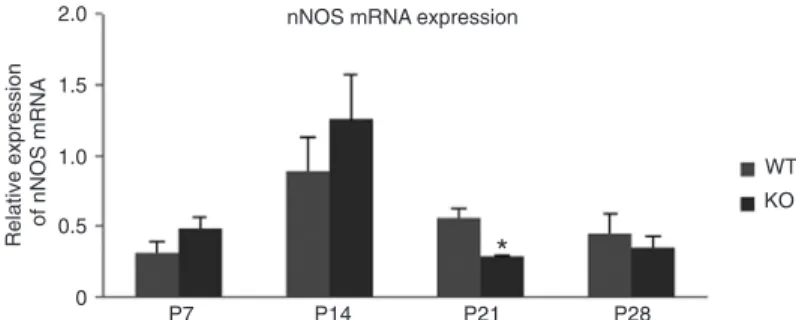
Expression of nNOS mRNA
In order to determine whether the changes in nNOS protein levels might be due to transcriptional regulatory differences, nNOS mRNA levels were compared at each age (P7, P14, P21, and P28)
for each genotype. The profiles of the trends of
nNOS mRNA expression changes in the FMR1 KO and WT groups were similar. The levels of
nNOS mRNA were lower at P7, significantly in
-creased at P14, significantly de-creased at P21,
and remained at those levels on P28(Figure 3).
However, at P21 the FMR1 KO mice showed
sig-nificantly less mRNA compared to WT mice (KO = 0.2857 ± 0.0150, WT = 0.5646 ± 0.0657; P < 0.05; Figure 3). This significant reduction in nNOS
mRNA levels at P21 corresponded precisely with
the significantly lower levels of nNOS protein in
FMR1 KO mice at this age (Figure 2).
Discussion
Current thought regarding spine morphogenesis
as-sumes that spines begin as filopodia, proceed to thin
or stubby spines and ending as mushroom spines (12). However, the neuropathology associated with FXS
exhib-its significantly longer dendritic spines and fewer shorter
spines, as well as more spines with immature appearing morphology and fewer with mature appearing morphology. This has been demonstrated in both FXS brains (3,13,14)
and in brains of FMR1 KO mice, an animal model of FXS (4,15).
In the first to second postnatal week, numerous dendritic filopodia extend to form synaptic contacts with axons (16). At P14, filopodia shrink and more spines are formed (5). At P21, most filopodia complete a functional and morpho -logical transition to spines, suggesting that P21 is a critical period for spinogenesis and synaptogenesis (11,17-19). Subsequent to this, synaptic maturation and stabilization proceed on until P28 (5).
Figure 2. Western blot detection of neuronal nitric oxide synthase (nNOS) in hippocampi of WT and FMR1 KO mice at P7, P14, P21, and P28 (N = 3 per genotype, per age group). A, Representative blots of nNOS protein levels at each age in the hippocampi of the FMR1 KO and WT groups were compared
to β-actin levels (loading control). B, Absorbance of nNOS immunoreactive
bands normalized to their respective β-actin loading control from 3 animals per genotype per age group were averaged and are reported as means ± SEM. *P < 0.05 vs WT on P21 (two-tailed Student t-test). WT = wild-type
mice; FMR1 KO = fragile X mental retardation 1 gene knockout mice; P7,
P14, P21, P28 = postnatal days 7, 14, 21, and 28, respectively.
Figure 3. Comparison of neuronal nitric oxide synthase (nNOS) mRNA in hippocampi of WT and FMR1 KO mice by RT-PCR at P7, P14, P21, and
P28 (N = 4-7 mice per genotype, per age group). ΔCt = Ct (nNOS) - Ct (β-actin), where Ct = cycle threshold, β-actin is the endogenous reference. Relative nNOS mRNA expression: PΔCt = 2-ΔCt, representing nNOS mRNA
levels. The final data in the graph were magnified to 1000 x 2-ΔCt. Data are
reported as means ± SEM. *P < 0.05 vs WT on P21 (two-tailed Student t
-test). mRNA = messenger ribonucleic acid; WT = wild-type mice; FMR1 KO = fragile X mental retardation 1 gene knockout mice; P7, P14, P21, P28 =
The results presented here demonstrate that the pro-tein levels of nNOS in WT mice were relatively high at P7
when dendritic filopodia are abundant, decreased slightly
at P14, dramatically increased to a peak at P21 when
filopodia gradually complete spine transition, and no ad -ditional change occurred at P28 compared to P21 when spines proceed to further stabilization. The protein levels
of FMR1 KO mice were significantly decreased particularly
at P21 compared to WT mice and then increased to levels comparable to those of WT animals by P28. This temporal delay in the expression of nNOS at the critical period of
filopodia to spine transition is likely to cause retardation of
normal spinogenesis and synaptogenesis development in the hippocampus.
The mRNA expression profile of WT mice demonstrated a significant peak of nNOS mRNA expression at P14
compared to all other time points. One possibility for this increase in mRNA at this age may be that nNOS mRNA levels were up-regulated ahead of the increase in protein levels observed at P21 to facilitate a maximal increase in protein levels, thus leading to an increase in NO during this
critical period of filopodia to spine transition. Compared
to the WT group, the nNOS mRNA changes in FMR1 KO mice demonstrated the same trend with the peak of mRNA
levels at P14, but levels of mRNA at P21 were significantly
less than WT controls. Therefore, both protein and mRNA
profiles in the FMR1 KO animals demonstrated a signifi
-cant reduction at P21. This deficiency probably results in
an inadequate production of NO during the critical period for synapse and spine maturation. Although we have not
measured NO levels per se, we suggest that the attenuated
nNOS expression probably results in down-regulation of NO production in FMR1 KO mice. Thus, we speculate that NO
deficiency during spinogenesis and synaptogenesis may
retard dendritic spine maturation in FXS.
There is evidence supporting the notion that NO produc-tion is indeed required for spine maturaproduc-tion. For example, NO as a messenger/modulator in synaptogenesis can stimu-late axons located around the spines to differentiate into varicosities and then form multi-innervated spines (9). This mechanism is mediated by the targeting and mobilization of synaptic vesicles in the presynaptic cell, enhancing trans-mitter release and then activating protein kinase signaling cascades in the postsynaptic cell (20,21). In addition, NO can promote transcription and translation, which are critical for long-term synaptic plasticity and memory formation (22-24). As we know, long-term potentiation (LTP) can increase synaptogenesis and enhance connectivity between neurons to facilitate memory storage. Hippocampal LTP triggered by NO is a process known to be involved in mammalian learning and memory in many studies (25-31). The morphological consequences of hippocampal LTP include enlarged spine heads, shortened spine necks, formation of perforated synapses, bifurcation of spines, and even formation of new spines in the vicinity of the activated spines (32,33). More-over, NOS inhibition during synaptic maturation decreases synapsin I immunoreactivity in rat hippocampi, indicating a reduction in synaptic density (10).
These findings suggest that it is plausible to infer from
our study that impaired NO production may be one of the reasons for spine retardation in FXS.
Acknowledgments
We thank Chaochun Zou (Hangzhou, Zhejiang, China) for technical assistance.
References
1. Brouwer JR, Willemsen R, Oostra BA. The FMR1 gene and fragile X-associated tremor/ataxia syndrome. Am J Med
Genet B Neuropsychiatr Genet 2009; 150B: 782-798.
2. Penagarikano O, Mulle JG, Warren ST. The pathophysiol-ogy of fragile x syndrome. Annu Rev Genomics Hum Genet
2007; 8: 109-129.
3. Grossman AW, Elisseou NM, McKinney BC, Greenough WT.
Hippocampal pyramidal cells in adult Fmr1 knockout mice
exhibit an immature-appearing profile of dendritic spines.
Brain Res 2006; 1084: 158-164.
4. Nimchinsky EA, Oberlander AM, Svoboda K. Abnormal
development of dendritic spines in FMR1 knock-out mice. J
Neurosci 2001; 21: 5139-5146.
5. Matsuno H, Okabe S, Mishina M, Yanagida T, Mori K, Yoshi-hara Y. Telencephalin slows spine maturation. J Neurosci
2006; 26: 1776-1786.
6. Morita A, Yamashita N, Sasaki Y, Uchida Y, Nakajima O,
Nakamura F, et al. Regulation of dendritic branching and spine maturation by semaphorin3A-Fyn signaling. J Neuro-sci 2006; 26: 2971-2980.
7. Centonze D, Siracusano A, Calabresi P, Bernardi G. The
Project for a Scientific Psychology (1895): a Freudian antici -pation of LTP-memory connection theory. Brain Res Brain
Res Rev 2004; 46: 310-314.
8. Nikonenko I, Boda B, Steen S, Knott G, Welker E, Muller
D. PSD-95 promotes synaptogenesis and multiinnervated spine formation through nitric oxide signaling. J Cell Biol
2008; 183: 1115-1127.
9. Nikonenko I, Jourdain P, Muller D. Presynaptic remodeling contributes to activity-dependent synaptogenesis. J Neuro-sci 2003; 23: 8498-8505.
10. Sanchez-Islas E, Leon-Olea M. Nitric oxide synthase inhibi -tion during synaptic matura-tion decreases synapsin I im-munoreactivity in rat brain. Nitric Oxide 2004; 10: 141-149.
11. Grabrucker A, Vaida B, Bockmann J, Boeckers TM. Synapto-genesis of hippocampal neurons in primary cell culture. Cell
Tissue Res 2009; 338: 333-341.
12. Yuste R, Bonhoeffer T. Genesis of dendritic spines: insights from ultrastructural and imaging studies. Nat Rev Neurosci
nNOS depression in developing hippocampus of FXS mice 1239
13. Irwin SA, Patel B, Idupulapati M, Harris JB, Crisostomo RA, Larsen BP, et al. Abnormal dendritic spine characteristics in the temporal and visual cortices of patients with fragile-X syndrome: a quantitative examination. Am J Med Genet
2001; 98: 161-167.
14. Wisniewski KE, Segan SM, Miezejeski CM, Sersen EA,
Rudelli RD. The Fra(X) syndrome: neurological, electro-physiological, and neuropathological abnormalities. Am J
Med Genet 1991; 38: 476-480.
15. Galvez R, Greenough WT. Sequence of abnormal dendritic spine development in primary somatosensory cortex of a mouse model of the fragile X mental retardation syndrome.
Am J Med Genet A 2005; 135: 155-160.
16. Fiala JC, Feinberg M, Popov V, Harris KM. Synaptogenesis via dendritic filopodia in developing hippocampal area CA1.
J Neurosci 1998; 18: 8900-8911.
17. Grutzendler J, Kasthuri N, Gan WB. Long-term dendritic spine stability in the adult cortex. Nature 2002; 420:
812-816.
18. McCroskery S, Bailey A, Lin L, Daniels MP. Transmembrane
agrin regulates dendritic filopodia and synapse formation in
mature hippocampal neuron cultures. Neuroscience 2009;
163: 168-179.
19. Papa M, Bundman MC, Greenberger V, Segal M. Mor-phological analysis of dendritic spine development in pri-mary cultures of hippocampal neurons. J Neurosci 1995; 15:
1-11.
20. Hawkins RD, Kandel ER, Siegelbaum SA. Learning to modu -late transmitter release: themes and variations in synaptic plasticity. Annu Rev Neurosci 1993; 16: 625-665.
21. Hawkins RD, Son H, Arancio O. Nitric oxide as a retrograde messenger during long-term potentiation in hippocampus.
Prog Brain Res 1998; 118: 155-172.
22. Chien WL, Liang KC, Teng CM, Kuo SC, Lee FY, Fu WM.
Enhancement of long-term potentiation by a potent nitric
oxide-guanylyl cyclase activator, 3-(5-hydroxymethyl-2-furyl)-1-benzyl-indazole. Mol Pharmacol 2003; 63:
1322-1328.
23. Ota KT, Monsey MS, Wu MS, Young GJ, Schafe GE.
Synaptic plasticity and NO-cGMP-PKG signaling
coordi-nately regulate ERK-driven gene expression in the lateral
amygdala and in the auditory thalamus following Pavlovian fear conditioning. Learn Mem 2010; 17: 221-235.
24. Taqatqeh F, Mergia E, Neitz A, Eysel UT, Koesling D, Mitt -mann T. More than a retrograde messenger: nitric oxide needs two cGMP pathways to induce hippocampal long-term potentiation. J Neurosci 2009; 29: 9344-9350.
25. Arancio O, Kiebler M, Lee CJ, Lev-Ram V, Tsien RY, Kandel
ER, et al. Nitric oxide acts directly in the presynaptic neuron
to produce long-term potentiation in cultured hippocampal neurons. Cell 1996; 87: 1025-1035.
26. Bon CL, Garthwaite J. On the role of nitric oxide in hip -pocampal long-term potentiation. J Neurosci 2003; 23:
1941-1948.
27. Garthwaite J, Boulton CL. Nitric oxide signaling in the central nervous system. Annu Rev Physiol 1995; 57: 683-706.
28. Mutlu O, Ulak G, Belzung C. Effects of nitric oxide synthase inhibitors 1-(2-trifluoromethylphenyl)-imidazole (TRIM) and
7-nitroindazole (7-NI) on learning and memory in mice.
Fundam Clin Pharmacol 2011; 25: 368-377.
29. Schuman EM, Madison DV. Nitric oxide and synaptic func -tion. Annu Rev Neurosci 1994; 17: 153-183.
30. Schuman EM, Madison DV. A requirement for the intercellu -lar messenger nitric oxide in long-term potentiation. Science
1991; 254: 1503-1506.
31. Schuman EM, Madison DV. Locally distributed synaptic
potentiation in the hippocampus. Science 1994; 263:
532-536.
32. Yuste R, Bonhoeffer T. Morphological changes in dendritic spines associated with long-term synaptic plasticity. Annu
Rev Neurosci 2001; 24: 1071-1089.
33. Harris KM, Fiala JC, Ostroff L. Structural changes at den-dritic spine synapses during long-term potentiation. Philos