Apolipoprotein B-mediated Control of
agr
Type III
Staphylococcus aureus
Quorum-sensing
Pamela R. Hall1,2*, Bradley O. Elmore1, Cynthia H. Spang1,2, Susan M. Alexander2, Brett C. Manifold-Wheeler1,2, Moriah J. Castleman1, Seth M. Daly1,2, M. Michal Peterson2, Erin K. Sully2, Jon K. Femling3, Michael Otto4, Alexander R. Horswill5, Graham S. Timmins1, Hattie D. Gresham2,6
1Department of Pharmaceutical Sciences, University of New Mexico College of Pharmacy, Albuquerque, New Mexico, United States of America,2Research Service, New Mexico Veterans Affairs Health Care Service, Albuquerque, New Mexico, United States of America,3Department of Emergency Medicine, University of New Mexico, Albuquerque, New Mexico, United States of America,4Laboratory of Human Bacterial Pathogenesis, National Institute of Allergy and Infectious Diseases, The National Institutes of Health, Bethesda, Maryland, United States of America,5Department of Microbiology, Roy J. and Lucille A. Carver College of Medicine, University of Iowa, Iowa City, Iowa, United States of America,6Department of Internal Medicine, University of New Mexico School of Medicine, Albuquerque, New Mexico, United States of America
Abstract
Staphylococcus aureuscontains an autoinducing quorum-sensing system encoded within theagroperon that coordinates expression of virulence genes required for invasive infection. Allelic variation withinagrhas generated fouragr specific groups,agr I–IV, each of which secretes a distinct autoinducing peptide pheromone (AIP1-4) that drivesagr signaling. Becauseagr signaling mediates a phenotypic change in this pathogen from an adherent colonizing phenotype to one associated with considerable tissue injury and invasiveness, we postulated that a significant contribution to host defense against tissue damaging and invasive infections could be provided by innate immune mechanisms that antagonizeagr
signaling. We determined whether two host defense factors that inhibit AIP1-induced agrI signaling, Nox2 and apolipoprotein B (apoB), also contribute to innate control of AIP3-inducedagrIII signaling. We hypothesized that apoB and Nox2 would function differently against AIP3, which differs from AIP1 in amino acid sequence and length. Here we show that unlike AIP1, AIP3 is resistant to direct oxidant inactivation by Nox2 characteristic ROS. Rather, the contribution of Nox2 to defense againstagrIII signaling is through oxidation of LDL. ApoB in the context of oxLDL, and not LDL, provides optimal host defense againstS. aureus agrIII infection by binding the secreted signaling peptide, AIP3, and preventing expression of theagr-driven virulence factors which mediate invasive infection. ApoB within the context of oxLDL also binds AIP 1-4 and oxLDL antagonizesagrsignaling by all fouragralleles. Our results suggest that Nox2-mediated oxidation of LDL facilitates a conformational change in apoB to one sufficient for binding and sequestration of all four AIPs, demonstrating the interdependence of apoB and Nox2 in host defense againstagrsignaling. These data reveal a novel role for oxLDL in host defense againstS. aureusquorum-sensing signaling.
Citation:Hall PR, Elmore BO, Spang CH, Alexander SM, Manifold-Wheeler BC, et al. (2013) Nox2 Modification of LDL Is Essential for Optimal Apolipoprotein B-mediated Control ofagrType IIIStaphylococcus aureusQuorum-sensing. PLoS Pathog 9(2): e1003166. doi:10.1371/journal.ppat.1003166
Editor:Alice Prince, Columbia University, United States of America
ReceivedJune 19, 2012;AcceptedDecember 17, 2012;PublishedFebruary 14, 2013
This is an open-access article, free of all copyright, and may be freely reproduced, distributed, transmitted, modified, built upon, or otherwise used by anyone for any lawful purpose. The work is made available under the Creative Commons CC0 public domain dedication.
Funding:This work was supported by NIH grants AI090917 (PRH), AI064926 (HDG), AI078921 (ARH), the Intramural Research Program of the National Institute of Allergy and Infectious Diseases, NIH, ZIA AI000904-10 (MO), and by the Department of Veterans Affairs (PRH, HDG). This project was also supported in part by the Dedicated Health Research Funds of the University of New Mexico School of Medicine allocated to the Signature Program in Cardiovascular and Metabolic Disease (PRH). SMD was supported by the University of New Mexico Biology of Infectious Diseases and Inflammation Training Grant (T32-AI007538) and MJC was supported under the Research Supplements to Promote Diversity in Health-Related Research Program (AI091917-03S1). The funders had no role in study design, data collection and analysis, decision to publish, or preparation of the manuscript.
Competing Interests:The authors have declared that no competing interests exist.
* E-mail: [email protected]
Introduction
Staphylococcus aureus uses global gene regulation to coordinate gene transcription required for survival within distinct host niches [1–10]. One of these global regulators is a four gene operon,agr, that encodes a quorum sensing system that combines secretion of an autoinducing peptide pheromone (AIP) with a sensor regulator. Activation of this system upregulates genes for toxins, hemolysins, lytic enzymes, and metabolic pathways that are required for a phenotypic change in this pathogen from an adherent colonizing phenotype to one associated with significant tissue injury and
to control tissue damage and cell injury have not been fully elucidated [24–29]. In this regard, we previously reported that both the Nox2 NADPH oxidase and the major structural protein of very low and low density serum lipoproteins (VLDL, LDL), apolipoprotein B (apoB), antagonize AIP dependent activation of its cognate receptor within the agrI allele [24,27]. Because three additional agr alleles are represented within the species S. aureus and because all four alleles are associated with significant disease in humans [30,31], we postulated that either or both of these innate immune barriers could be important for antagonism of signaling by otheragrtypes.
Eachagr allele encodes a unique secreted AIP that differs in amino acid content and length but contains a common thiolactone bond that creates a 5-membered ring essential for biologic function [11]. Importantly, secretion of AIP represents an opportunity for host or environmental control ofagrsignaling by either direct modification of key amino acids, cleavage of the thiolactone bond, proteolytic degradation, or sequestration to prevent AIP binding to its receptor, AgrC. For AIP1, reactive oxygen species (ROS) generated by the Nox2 NADPH oxidase expressed in phagocytes and other cells directly modify a key C-terminal member of the thiolactone ring, a methionine, to form methionine sulfoxide [27]. While retaining its cyclic structure, this modification is sufficient to render AIP1 biologically inactive. In addition, the large structural protein of serum LDL, apoB, binds directly to cyclic AIP1, but not its inactive linear form, preventing its activation of its cognate receptor AgrC [24]. Importantly, loss of either Nox2 or apoB in the form of LDL is sufficient to promote agrI-mediated invasive infection beyond epithelial and mucosal barriers. To extend these studies to the otheragr alleles, we first focused on the contribution of these two barriers to host antagonism ofagrIII signaling. AIP3 is shorter and composed of amino acids that are more resistant to oxidant modification as compared to AIP1 and therefore might be less amenable to control by either apoB or ROS. We hypothesized that Nox2 and apoB within the context of its serum lipoproteins would differ in the
molecular mechanism by which they antagonizeagrIII signaling and that this difference would impact the susceptibility to invasive infection in mouse models that lack these innate barriers.
Here, we show that optimal host innate defense againstagr III-mediated signaling requires binding and sequestration of AIP3 by apoB, not in the form of LDL but in LDL oxidized by Nox2. Importantly, these studies revealed an important role for oxLDL in binding to and antagonizing signaling by all fouragralleles. In addition, while ROS directly inactivate AIP1 and 4, they do not affect the biologic function of either AIP 2 or 3. Thus, the contribution of Nox2 in antagonizingagrsignaling for these alleles is primarily through the production of oxLDL. OxLDL-mediated antagonism of agr signaling inhibits agr driven virulence factor expression by all four alleles, providing a mechanistic basis for its importance in preventing invasive S. aureus infection which we demonstrate in a murine agrIII-mediated skin infection model. Therefore, while best studied for its contribution to atherosclerosis, our data reveal a novel role for oxLDL as a host defense effector that controlsS. aureus agr-mediated signaling.
Results
Nox2 oxidation of LDL is required for optimal apolipoprotein B-mediated antagonism ofagr III-signaling
Innate immunity controls agrI-mediated invasive S. aureus infection by early extravasation of activated neutrophils and apoB-containing plasma lipoproteins that act to antagonizeagrI signaling [24,27]. Induction ofagrsignaling requires AIP cyclized through a 5-membered thiolactone ring. AIP binds and activates AgrC which generates phosphorylated AgrA. Phosphorylated AgrA activates the agr P3 promoter generating the effector molecule RNAIII. ApoB recognizes the cyclic, active form of AIP1 and prevents binding to and signaling through its cognate AgrC receptor [24]. Whereas AIP1 and AIP3 differ in amino acid sequence and in length of the amino terminal tail, they both contain the thiolactone ring (Fig. 1A). We postulated that apoB would antagonize AIP3-dependent activation of AgrC, but that conformational changes of apoB might be required for optimal antagonism of the smaller peptide pheromone of theagrIII strains. In addition to binding theagrP3 promoter, phosphorylated AgrA directly enhances transcription of genes encoding phenol soluble modulin virulence factors (PSM alpha, PSM beta) [32–34]. ApoB alone significantly inhibited transcription of psma in clinical isolates USA300 LAC (agrI) and USA400 MW2 (agrIII) [32,35– 37], whereas LDL at equivalent apoB concentration did not inhibitpsmatranscription in theagrIII strain MW2 (Fig. S1A). The
ability of apoB alone to inhibitagrIII-mediated signaling indepen-dent of LDL suggests that the conformation of apoB in LDL is not optimal for binding AIP3. Oxidation of LDL is known to alter the conformation of apoB within the lipoprotein particle [38–40]. We predicted that apoB present in oxLDL would significantly antagonize agrIII-mediated signaling compared to LDL. Using strainagrIII MW2 ([agr::P3-yfp] in which activation of theagr::P3 promoter drives expression of YFP [41], both apoB alone and oxLDL at equivalent apoB concentration significantly antagonized agr::P3 promoter activation as compared to LDL (Fig. 1B). These results were not due to lipoprotein effects on bacterial viability as demonstrated by the number of colony forming units (cfus). Intriguingly, LDL and oxLDL were equally efficacious in antagonism of AIP1 dependent signaling (Fig. S1B), indicating that apoB within either particle is sufficient to neutralize AIP1. To confirm that apoB, and not a lipid component of oxLDL was responsible for antagonizing AIP3-mediated signaling, we
deter-Author Summary
Staphylococcus aureusis a common colonizer of humans but can also cause severe, invasive infection.S. aureususes a secreted peptide-based communication system, agr, to induce production of virulence factors needed for invasive infection. Allelic variation has generated fouragrtypes,agr
I–IV, and each secretes a distinct autoinducing peptide (AIP1-4) that differs in amino acid sequence and length. Understanding host factors that prevent signaling by each of the four agr specific groups (agrI–IV) could provide opportunities for prevention of infection or therapeutic intervention. We previously demonstrated that apolipo-protein B (apoB), the major structural apolipo-protein of very low and low density lipoproteins (VLDL, LDL), binds to the secretedagrI peptide, AIP1, and preventsagrsignaling. In addition, the NADPH oxidase Nox2 produces reactive oxygen species which directly modify and inactive AIP1. Here we examined the role of apoB and Nox2 in defense against agrIII-signaling. We found that apoB in oxidized LDL, but not in native LDL, mediated optimal binding of AIP3. Also, unlike AIP1, Nox2 did not directly inactivate AIP3. Rather Nox2 contributed to defense against agr III-signaling by oxidizing LDL. Furthermore, we found that oxLDL bound all four AIPs and antagonizedagrsignaling by each agr allele in vitro. These results expand our understanding of host defense against S. aureus agr
Figure 1. Optimal apolipoprotein B-mediated antagonism of agrIII-signaling requires oxidation of LDL via Nox2.(A) Schematic representation of AIP1 and AIP3. (B) StrainagrIII MW2 [agr::P3-yfp] was cultured overnight with broth control or 10 nM LDL, apoB or oxLDL, and
agr::P3 activation was measured by flow cytometry. Colony forming units (cfus) were determined by plating on sheep blood agar. (C) Anti-apoB antibody reverses antagonism ofagr::P3 promoter activation in MW2. StrainagrIII MW2 [agr::P3-yfp] was cultured overnight with broth control, 10 nM apoB or oxLDL, along with 30 nM control IgG or 30 nM apoB-specific IgG. (D) LDL, ApoB or oxLDL binding to immobilized AIP3 was measured by SPR following lipoprotein incubation with apoB-specific IgG or control IgG. Data were normalized to the mean6SEM of LDL binding in the absence of antibody. (E) Blood was collected from wild-type orNox22/2mice. After clearing, the serum was heat inactivated and diluted to 10% in TSB for overnight culture with strainagrIII MW2 [agr::P3-yfp].agr::P3 promoter activation was measured by flow cytometry. Data reported are the mean6SEM normalized to broth control. (F) StrainagrIII MW2 [agr::P3-yfp] cultured 4 h with 100 nM AIP plus 10% sera fromNox22/2mice along with 50 nM LDL or 50 nM oxLDL. Data points represent the mean6SEM normalized to broth control. (G) Immunoblot detection of apoB and oxidized LDL. Control LDL and oxLDL plus serum from wild-type andNox22/2mice were vacuum transferred to nitrocellulose and stained for oxLDL using monoclonal antibody E06 or rabbit polyclonal antibody to apoB. A representative blot is shown. Band intensity was quantified using Carestream Molecular Imaging software (New Haven, Connecticut), and data normalized to oxLDL or wild-type sera with E06/apoB ratios equal to 1. (H) StrainagrIII MW2 [agr::P3-yfp] was cultured overnight with exogenous AIP3 (50 nM) and 10 nM of either human LDL, human oxLDL, LDL purified from wild-type mice, LDL purified fromNox22/2mice or broth control. ns, not significant; *, p,0.05; **, p,0.01; ***, p,0.001.
mined the effects of antibodies against apoB on the ability of apoB and oxLDL to inhibitagrIII-dependent signaling. Pre-treatment of apoB and oxLDL with an antibody specific to a linear peptide within apoB, but not with an isotype control antibody, significantly inhibited apoB-mediated antagonism of agr::P3 promoter activa-tion in this agrIII strain (Fig. 1C). Thus apolipoprotein B is sufficient to antagonize agrIII-dependent signaling and optimal antagonism requires apoB presented in the context of oxLDL rather than LDL.
ApoB antagonism ofagrtype I-mediated virulence entails direct binding to AIP1 [24], and based on the results of our signaling assays, we predicted that oxLDL would bind AIP3 at significantly higher levels relative to LDL. We analyzed binding of LDL, apoB and oxLDL, at equivalent apoB concentrations to immobilized AIP1 and AIP3 via Surface Plasmon Resonance (SPR). As expected, oxLDL and apoB bound at significantly higher levels to cyclic AIP3 as compared to LDL (Fig. 1D). In contrast, all 3 bound equally to AIP1 (Fig. S1C). These data support our results on the requirement of oxLDL for antagonism of AIP3-dependent signaling, whereas LDL alone was sufficient to antagonize AIP1 signaling. None of the 3 lipoproteins bound significantly to linear AIP3 (L-AIP3) (data not shown), proving that apoB recognition of AIP3 requires the active, cyclic conformation of AIP3. Further-more, AIP3 binding by apoB and oxLDL was significantly inhibited by antibody specific to a linear peptide within apoB but not control antibody (Fig. 1D), indicating that apoB is responsible for binding of oxLDL to AIP3. The ability of the anti-peptide antibody to reverse functional antagonism in the reporter assay and to block AIP3 binding by both apoB and oxLDL, but not LDL, suggests a conformation-dependent AIP3 binding site within apoB that is also present in oxLDL but absent in LDL. Therefore, the mechanism by which oxLDL antagonizes agrIII signaling includes enhanced binding of AIP3 relative to LDL, and this binding is mediated by apoB and not the lipid components of oxLDL.
Although AIP expressed from each of the fouragralleles differ in amino acid sequence and length, the peptides share a common five-membered thiolactone bond. To determine whether oxLDL universally bindsS. aureusAIPs and antagonizes signaling by each of the fouragralleles, we also measured oxLDL binding to AIP2 and AIP4 (Figure S1D,E) by SPR. OxLDL bound immobilized AIP2 and AIP4, but not the inactive linear peptides, in a dose-dependent manner again illustrating the necessity of the thiolactone bond for apoB-dependent binding. Furthermore, oxLDL antagonizedagr::P3 promoter activation by both anagrII (AH430 – SA502A) andagrIV (AH1872 – MN TG) clinical isolate (Fig. S1F). These data suggest that oxLDL could act as a universal innate inhibitor ofagr-signaling mediated by each of the fourS. aureus agralleles.
ROS production by the NADPH oxidase Nox2 is essential for control of invasive S. aureus infection [42–44], and facilitates oxidation of LDL [45,46]. We postulated that Nox2 would contribute to apoB-mediated control ofagrIIIS. aureusvirulence by oxidation of LDL. If correct, LDL from the serum of Nox2 knockout mice [42] would primarily be in the form of native LDL whereas LDL from the serum of wild-type mice would include oxLDL and would better inhibitagrIII-signaling relative to LDL from Nox22/2 mice. We examined serum from wild-type and Nox22/2mice for antagonism ofagr::P3 promoter activation in the agrIII isolate MW2 and for the presence of LDL and oxLDL. As predicted, serum from wild-type mice, but not from Nox22/2 mice, significantly antagonized agr::P3 promoter (Fig. 1E), and addition of oxLDL, but not LDL, to serum fromNox22/2 mice restored agrIII-antagonism to wild-type levels (Fig. 1F). Relative
serum levels of oxLDL and LDL from wild-type andNox22/2 mice were determined by immunoblot analysis using an antibody against apoB and a monoclonal antibody (E06) which detects epitopes present in oxLDL but absent in LDL [47,48]. Serum from wild-type mice had a significantly higher E06/apoB ratio compared toNox22/2mice (Fig. 1G), indicating that serum from wild-type mice contains more oxLDL than serum fromNox22/2 mice. Although significantly reduced compared to wild-type mice, serum ofNox22/2 mice had some E06 reactivity, indicating the presence of small amounts of oxLDL resulting from oxidation mechanisms distinct from Nox2. Interestingly, the amount of E06 positivity in the LDL control fraction reflects a nominal oxidation level in LDL preparations that may vary by preparation, method and storage. This variable level of nominal oxidation likely resulted in the small but significant LDL antagonism of agr III-signaling observed in some experiments (Fig. 1B). To prove that apoB-containing lipoprotein oxidized by Nox2 is the serum component from wild-type mice responsible for blocking agrIII activation, we measured the ability of LDL purified from the serum of wild-type versus Nox22/2 mice to antagonize agr III-dependent quorum-sensing signaling. At equivalent protein concentrations, LDL purified from the serum of Nox22/2 mice did not inhibit agr::P3 promoter activation in the agrIII isolate MW2 while LDL from wild-type mice significantly inhibitedagrIII signaling (Fig. 1H). There was no significant inhibition ofagr::P3 promoter activation in the presence of eitherNox22/2LDL or our human LDL control with minimal EO6 positivity, further demonstrating the requirement for oxLDL inagrIII-antagonism. In addition, purified LDL from wild-type mice had a 30% greater lipid oxidation level compared to LDL fromNox22/2mice (Fig. S2A). Thus, the ability of serum to controlagrIII-signalingin vitrois dictated by the presence of oxLDL within that serum and Nox2 is a major contributor to oxidation of serum LDL. These data indicate that Nox2 contributes to host control ofagrIII-mediated quorum sensing in part via oxidation of LDL that induces a conformational change in apoB required for optimal AIP3 binding and sequestration.
Reduction of serum apoB levels inNox2knockout mice but not wild-type mice significantly increases
susceptibility toagrIIIS. aureusinvasive infection
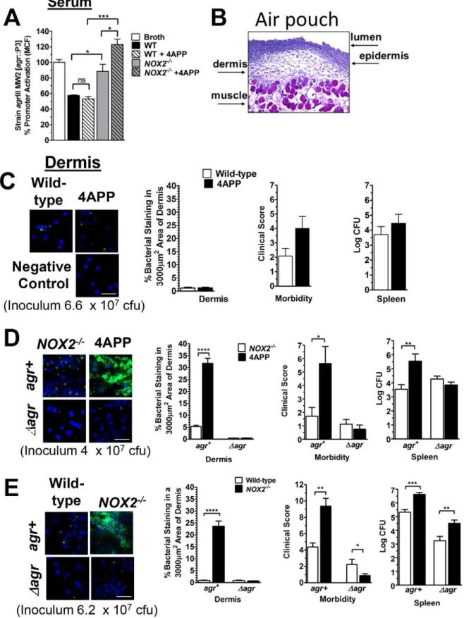
post-equivalent serum antagonism of agrIII-signaling. In contrast, serum from Nox22/2 mice, that contains significantly reduced levels of oxLDL compared to wild-type mice (Fig. 1G), was not optimal for inhibition ofagr::P3 promoter activation in theagrIII isolate MW2 and serum from 4APP-treatedNox22/2 mice was even less effective (Fig. 2A). This suggests that loss of LDL oxidized by both Nox2 and other mechanisms results in the greatest impairment inagrIII-antagonism.
Treatment of wild-type mice with 4APP reduced plasma levels of LDL (Fig. S2B) but did not alter the ability of the mice to oxidize LDL still being secreted from the liver (Fig. S2C). Because serum from vehicle and 4APP treated wild-type mice had similar levels of oxLDL and equally antagonized agrIII-signaling in vitro (Fig. 2A), we predicted that 4APP treatment alone would not increase the susceptibility of wild-type mice to invasive MW2 infection. Using an air-pouch model of infection [24,27,28,51–54] to determine agrIII-dependent invasion of bacteria from the epidermis into the dermis (Fig. 2B), we infected wild-type mice treated with either 4APP or vehicle with a dose of MW2 that wild-type mice are able to maintain at the epithelial barrier. There was no increased susceptibility of the 4APP treated mice to this dose (Fig. 2C). These results confirmed ourin vitrodata that the level of oxLDL in these mice was sufficient to prevent agrIII-quorum sensing required for invasive infection. These data demonstrate that whereas reduction of LDL in the serum of Nox2 competent mice is sufficient to increase susceptibility toagr type I S. aureus infection [24], it does not increase susceptibility toagrIII invasive infection because oxLDL levels are sufficient for protection.
Our serum data indicate that in the absence of Nox2, the resulting reduction of oxLDL significantly impairs apoB-mediated antagonism of agrIII-dependent quorum sensing indicating that apoB within oxLDL is primarily responsible for suppressingagrIII signaling (Fig. 2A). Therefore, we predicted that 4APP treatment of mice lacking Nox2 would significantly increase susceptibility to agrIII-dependent MW2 invasive infection beyond the loss of Nox2 alone. Because Nox22/2 mice are highly susceptible to S. aureus infection due to the critical role of reactive oxidants in host defense against this pathogen [42], and because we predicted that susceptibility would be further increased following 4APP treat-ment, a reduced inoculum of MW2 was used in these experiments (Fig. 2D) relative to the other infection studies (Fig. 2C,E). As predicted, Nox2 knockout mice treated with 4APP prior to infection with MW2 had significantly increased bacterial invasion into the dermis, morbidity and bacterial burden in the spleen compared to control treatedNox22/2mice (Fig. 2D). Moreover, Nox22/2 mice treated with 4APP were not more susceptible to infection with an isogenic agr deletion mutant of MW2 (Dagr)
compared to controls, confirming that the contribution of Nox2 to apoB-mediated host defense is specific to control ofagr-mediated pathogenesis. In contrast, infection ofNox22/2mice with MW2 at an inoculum readily controlled by wild-type mice resulted in significant dermal invasion, morbidity and bacterial burden in the spleen as compared to wild-type mice. In addition, infection with the MW2 agr deletion mutant also resulted in increased dissemination to the spleen (Fig. 2E). Therefore, unlike the agr -specific role of apoB, the protective role of Nox2 is not limited to control ofagr-mediated virulence but has broader implications for
host antibacterial defense such as contributing to direct killing of bacteria in the spleen [46,55–57].
These data confirm that apoB-mediated serum antagonism of agrIII-signalingin vitro is a predictor of in vivosusceptibility to S. aureus agrIII-dependent invasive infection. In addition, the increased susceptibility to agrIII-mediated invasive infection following reduction of serum apoB is dependent upon the presence or absence of Nox2, indicating a novel role for Nox2 in host defense against agrIII-dependent infection by promoting apoB-mediated antagonism of quorum sensing. Whereas reduction of apoB in the context of LDL is not sufficient to increase susceptibility to agrIII-mediated invasive infection, reduction of apoB in the form of oxLDL is sufficient.
oxLDL antagonizesagrIII-mediated virulence factor transcription and expression in MRSA and MSSA clinical isolates
Quorum-sensing through agr regulates expression of over 100 genes, many of which encode virulence factors necessary for invasive infection [11,13,58]. To confirm that the role of apoB extends to agrIII-dependent transcription and translation of virulence factors and that this protection was not limited to isolate MW2, we first determined whether oxLDL inhibited agr III-dependent transcription by randomly selected MRSA and methi-cillin sensitive S. aureus (MSSA) agrIII clinical isolates. OxLDL significantly inhibited transcription of both theagreffector molecule RNAIII and of a keyagrregulated virulence factor, alpha hemolysin (Hla), by both MRSA and MSSAagrIII clinical isolates (Fig. 3A,B). Likewise, oxLDL significantly inhibited production of Hla as determined by functional assay (Fig. 3C). This effect was not due to direct lipoprotein interaction with Hla because neither LDL nor oxLDL reduced the activity of purified Hla (Fig. S3A). Although two of the selected clinical isolates failed to produce the virulence factor staphylococcal lipase, oxLDL again significantly inhibited agr III-dependent lipase secretion as determined by functional assay of the remaining isolates (Fig. 3D). The decrease in lipase activity was not due to direct inhibition by the lipoproteins (Fig. S3B). These results extend our observations of oxLDL-mediated control of agr III-signaling and invasive infection to virulence factor expression by current clinical isolates of both MRSA and MSSA indicating that the contribution of oxLDL as a barrier to agrIII infection is not limited to a single genetic background of the pathogen.
AIP binding and agr::P3 promoter activation analyses suggest that oxLDL would also inhibit agr-dependent transcription and virulence factor translation byagrI, agrII and agrIV MRSA and MSSA clinical isolates. As predicted, oxLDL antagonized agr -dependent transcription of RNAIII and hla along with production of functional Hla in clinical isolates representing agrI, agrII and agrIV alleles (Fig. S3C–E). Therefore, oxLDL can serve as a universal agr antagonist by inhibitingagr-signaling and virulence factor expression byagrI–IV clinical isolates.
ROS-dependent oxidation of LDL but not of AIP3 mediates antagonism ofagrIII-dependent quorum sensing
Based on published literature and ourin vivodata [45,46,59–62], we postulated that in vitrooxidation of LDL by ROS would be sufficient to promote apoB-mediated antagonism ofagr
III-signal-infection, the following parameters were determined and data reported as the mean6SEM: (Left to right) Representative confocal images of dermis from indicated pouches stained with TO-PRO-3 (Invitrogen; blue) and anti-S. aureusantibody (green fluorophore) (Scale bar = 20mm); Quantification of bacterial density in dermis of pouches; Morbidity was scored on a 0–14 point scale and was based on weight loss, appearance, level of dehydration, mobility and responsiveness; Bacterial burden (Log CFU) in spleen. (C) Wild-type mice treated with 4APP or vehicle control. (D)Nox22/2
ing. Previousin vitrostudies have shown that isolated neutrophils induce lipid oxidation of LDL [45,46,63]. Therefore, we first determined the ability of HOCl, a ROS released by activated neutrophils via Nox2 and myeloperoxidase [57,64], to enhance inhibition of agr::P3 promoter activation by LDL in the agrIII isolate MW2. Exposure of LDL to HOCl significantly increased antagonism of AIP3-induced agr::P3 promoter activation com-pared to control LDL (Fig. 4A). This effect was dependent upon oxidative modification of LDL because the ROS scavenger N-acetylmethionine (NAM) blocked the increase in antagonism. To demonstrate that increasedagrIII-antagonism following oxidation of LDL is not limited to a single ROS, we evaluated singlet oxygen
for the ability to enhance agrIII antagonism by LDL. Singlet oxygen (1O2) is a strong oxidizing agent released by activated
neutrophils and contributes significantly to neutrophil extracellular trap (NET) formation, ozone mediated bacterial killing and ozone formation in atherosclerotic plaques as well as conformational changes of apoB within LDL [38,56,65,66]. In addition, 1O2
induces lipid oxidation of LDL [63,67]. As expected, exposure of LDL to1O2(Fig. 4B) significantly increased the ability of LDL, but
not apoB or oxLDL, to antagonizeagrIII-signaling and antagonism increased along with the oxidant dose (Fig. 4C). In addition, increased antagonism ofagrIII-signaling by1O
2or HOCl-treated
LDL corresponded with a significant increase in LDL lipid Figure 3. Effect of oxLDL on antagonism ofagrIII dependent virulence factor production by MRSA and MSSA clinical isolates.(A–D) Bacteria were cultured overnight in the presence of media control or oxLDL (50 nM). (A) RNAIII and (B)hlatranscription relative to 16S rRNA was determined by qRT-PCR (C) Alpha-hemolysin content of culture supernatants was measured by the ability to lyse rRBCs. (D) Supernatants from (C) were also assessed for lipase activity determined by rate of cleavage of the triglyceride substrate tributyrin. Data reported as the mean6SEM. *, p,0.05; **, p,0.01; ***, p,0.001; ****, p,0.0001.
oxidation compared to control (Fig. 4D). Therefore, in vitro oxidation of LDL by ROS representative of activated Nox2 is sufficient to significantly increase apoB-mediated antagonism of agrIII-dependent quorum sensing compared to untreated LDL.
Extravasation of activated neutrophils to sites ofagrIS. aureus infection makes available extracellular ROS that provide defense in part by oxidizing the C-terminal methionine of AIP1 rendering it biologically inactive [27]. Because AIP3 lacks this methionine and the susceptibility of the common AIP thiolactone to oxidative inactivation has not been addressed, we postulated that AIP3 would be resistant to inactivation by ROS relative to AIP1. At concentrations of HOCl that readily inactivated AIP1, AIP3 retained biologic function (Figure 4E). At higher HOCl concen-trations, there was a partial loss in AIP3 function that most likely resulted from fragmentation of the peptide as mass spectrometry failed to identify AIP3 species with charge to mass ratios suggestive
of linearization or addition of oxygen atoms (data not shown). In contrast, mass spectrometry clearly revealed oxidation of the AIP1 methionine residue as was previously reported (data not shown) [27]. To further demonstrate the resistance of AIP3 to ROS-mediated inactivation, we assayed singlet oxygen for the ability to inactivate AIP3. As expected, AIP3 was resistant to inactivation by
1
O2under conditions in which AIP1 signaling decreased rapidly as
a function of time (Fig. 4F). These results were not due to changes in bacterial growth resulting from exposure to residual ROS, as the cfus were consistent between both treated and untreated groups (data not shown). Because both AIP1 and AIP3 contain the 5-membered thiolactone ring, these results suggest that this ring is not a target for inactivation under the conditions examined here. To confirm that the AIP thiolactone is not a primary target for oxidative modification, we evaluated the ability of excess ethylthioacetate, a thiolactone mimetic, to protect AIP1 from Figure 4. ROS-dependent oxidation of LDL but not of AIP3 mediates antagonism ofagrIII-dependent quorum sensing.(A, B) LDL, apoB or oxLDL at equimolar concentrations of apoB were treated as shown with (A) HOCl for 30 min at 37uC, with or without prior addition of the ROS scavenger N-acetylmethionine (NAM), or (B) singlet oxygen (5 min on ice). 10 nM ROS-treated lipoproteins or controls were cultured overnight withagrIII MW2 [agr::P3-yfp] plus 50 nM AIP.agr:P3 promoter activation was measured by flow cytometry. (C) StrainagrIII MW2 [agr::P3-yfp] was cultured overnight with 50 nM AIP3 plus LDL treated with increasing concentrations of singlet oxygen. (D) Measurement of LDL lipid oxidation before and after exposure to singlet oxygen or HOCl. Commercially available oxLDL and bovine serum albumin are given as controls. Data represent the mean6SEM. (E) AIP1 and AIP3 were incubated with buffer or HOCl for 30 min at 37uC, treated with NAM to scavenge remaining ROS, and cultured at 50 nM for 2 h withagrI isolate LAC [agr::P3-yfp] oragrIII MW2 [agr::P3-yfp], respectively. Data was measured by flow cytometry. Data points represent the mean6SEM normalized to 100% activation by untreated AIP and statistics are in reference to this control. (F) AIP1 and AIP3 were exposed to singlet oxygen by incubation with rose bengal in the presence or absence of light, prior to culture withagrI isolate LAC [agr::P3-yfp] oragrIII MW2 [agr::P3-yfp]. Data points represent the mean6SEM normalized to 100% activation by untreated AIP and statistics are in reference to this control. (G) AIP1 was combined with buffer or a 10-fold molar excess of ethylthioacetate prior to exposure to singlet oxygen, followed by culture withagrI isolate LAC [agr::P3-yfp]. Data points represent the mean6SEM of the mean channel fluorescence. ns, not significant; *, p,0.05; **, p,0.01; ***, p,0.001; ****, p,0.0001.
oxidative loss of function by 1O2. At 10-fold molar excess,
ethylthioacetate did not protect AIP1 from inactivation by 1O2
(Fig. 4G), indicating that the thiolactone does not compete with the AIP1 methionine for oxidative modification and therefore the thiolactone is not a primary target of oxidation. These data demonstrate that these ROS do not provide defense againstagr III-mediated signaling by direct oxidative inactivation of AIP3 but rather through enhancement of apoB-mediated control ofagr III-virulence by oxidative modification of LDL.
ROS-mediated inactivation of AIP1 but not AIP3 suggests that methionine is the primary target for oxidative inactivation of AIP. To extend these observations to the role of ROS in oxidative inactivation of other AIPs, we postulated that1O2would inactivate
AIP4, which includes a C-terminal methionine residue, but that AIP2 which lacks methionine would be resistant to inactivation. As predicted, exposure of AIP4 to1O2significantly inhibited
AIP4-inducedagr::P3 promoter activation in theagrIV isolate MN TG, whereas AIP2 function remained unchanged (Fig. S4). These data suggest that in addition to oxidation of LDL, ROS can play an independent role in defense againstagrI andagrIV quorum sensing through direct oxidative inactivation of AIP1 and AIP4.
Exogenous oxLDL restoresin vivoantagonism ofagr III-signaling
Serum proteins extravasate into inflamed tissues and aid in host defense by providing antibody and complement necessary for opsonization [68]. We predicted that extravasated oxLDL present at the site of infection would also contribute to host defense by antagonizingagrIII invasive infectionin vivo. To address this, we first determined relative levels of oxLDL and LDL in lavages of air-pouches of wild-type and Nox22/2 mice 28 hours after
infection with MW2. As expected, oxLDL was present in lavages from wild-type mice who were protected fromagrIII invasion, but was largely absent in lavages from Nox22/2 mice which were highly susceptible to MW2 invasion (Fig. 5A). From these results, we predicted that the addition of oxLDL, but not native LDL, to the air-pouch at the time of infection of Nox22/2 mice would inhibit agrIII-signaling. To test this we infected air-pouches of 4APP-treatedNox22/2mice withagrIII isolate MW2 [agr::P3-yfp] in the presence of oxLDL, LDL or buffer control. Oxidized LDL, but not LDL, introduced into the air-pouch at the time of infection significantly antagonized AIP3-mediatedagr::P3 activation in the resulting air-pouch lavage four hours after infection (Fig. 5B). Weight loss is a primary measure of morbidity in this model and mice treated with oxLDL at the time of infection lost significantly less of their total body weight at four and eight hours post-infection compared to controls (Fig. S5). Thus the presence of oxLDL in the air-pouch at the time of infection significantly inhibited bothagr -signaling and morbidity as measured by weight loss. Both the presence of oxLDL in air-pouch lavages from infected wild-type mice and restoration ofagrIII antagonism by addition of oxLDL, but not LDL, to the air-pouch of 4APP-treatedNox22/2 mice, indicates that Nox2-mediated apoB control of agrIII-dependent signaling works directly at the site of infection to preventagrtype III-dependent signaling and its pathological consequences.
Discussion
Host innate immunity is critical for maintaining a defensive barrier against opportunistic pathogens such asStaphylococcus aureus which colonize skin and mucosal surfaces. In order to breach these defensive barriers,S. aureususes theagrquorum sensing system to
Figure 5. Exogenous oxLDL restores in vivo antagonism ofagrIII-signaling.(A) Control LDL and oxLDL plus air-pouch lavage from MW2 infected wild-type andNox22/2mice was vacuum transferred to nitrocellulose and stained for oxLDL using monoclonal antibody E06 or rabbit polyclonal antibody to apoB. A representative blot is shown. Band intensity was quantified using Carestream Molecular Imaging software (New Haven, Connecticut), and data normalized to oxLDL or wild-type lavage with E06/apoB ratios equal to 1. (B) Air-pouches were generated on the backs of 8 to 12 week oldNox22/2mice treated with 4APP as previously described. At time zero, 4
6107cfu of early exponential phaseagrIII isolate MW2 [agr::P3-yfp] were injected into the air-pouch along with saline control or the following as indicated: 100 nM AIP3 and either buffer control, 100 nM LDL or 100 nM oxLDL. After 4 h pouches were lavaged andagr::P3 promoter activation assessed by flow cytometry. Data reported are the mean6
coordinate expression of virulence genes needed for invasion and to evade host defensive mechanisms. The fact that most humans are able to limitS. aureusinfections to minor ones of skin and skin structures [69] suggests that host factors capable of inhibiting quorum sensing signaling mediated by each of the fouragralleles could contribute to host defense against invasive infection. This antagonism would thus promote host bacterial killing and clearing mechanisms. Here, we extend our knowledge of host inhibition of S. aureus agrI-signaling to host control of quorum-sensing depen-dent virulence mediated by agrIII. Importantly, our studies into host control ofagrIII signaling revealed an important role for the conformation of apoB within lipoprotein particles such that oxLDL was found to be a universal inhibitor of agrsignaling in all fouragralleles. We previously reported that both apolipoprotein B, the structural protein of VLDL and LDL, and ROS generated by the phagocyte NADPH oxidase (Nox2), provide unique barriers against S. aureus agrI-mediated virulence and the loss of either significantly increases susceptibility to invasive infection [24,27]. Specifically, Nox2 derived ROS directly inactivate AIP1 by methionine oxidation whereas the apoB component of LDL binds and sequesters AIP1 to prevent agrI-mediated virulence. This current work demonstrates that the roles of apoB and Nox2 in defense against agrIII-mediated infections are interdependent. Unlike AIP1, AIP3 is resistant to oxidation and direct inactivation by ROS characteristically produced by Nox2. Instead, the contribution of Nox2 to host innate defense against S. aureus agrIII-dependent quorum sensing is through oxidation of LDL. ApoB in the context of oxLDL, and not LDL, provides optimal host defense against S. aureus agrIII infection by binding the secreted signaling peptide, AIP3, and preventing expression of the agr-driven virulence factors which mediate invasive infection. Our results suggest that oxidation of LDL facilitates a conformational change in apoB to one required for optimal binding of AIP3 and apoB-mediated defense against agrIII-dependent virulence. In-triguingly,agrI-,agrII- andagrIV-signaling was also antagonized by oxLDLin vitro, suggesting that apoB within oxLDL is sufficient for antagonism of signaling by each of the fouragralleles. Each of the four S. aureus agr alleles are associated with invasive human infections which may range in severity from mild skin and soft tissue infections to severe disease such as osteomyelitis, endocar-ditis, necrotizing pneumonia and bacteremia [31]. Our data suggest that oxLDL may provide protection against S. aureus infections of any agr type in which agr-signaling contributes to pathogenesis. In contrast, we would predict that oxLDL would play a minimal role in host defense in chronic or device related infections in whichagrsignaling may be less relevant [70–73].
Our data show that LDL and oxLDL are present at the site of infection and prevent quorum-sensing dependent invasion ofagrIII S. aureusinto the underlying dermis. This suggests that other serum proteins which extravasate to the site of infection may also contribute to defense against agr-mediated signaling. Recently, Surewaardet al[74] described a novel role for VLDL, LDL and HDL in binding and inactivation of phenol soluble modulins (PSM), an important group of agr-regulated S. aureus virulence factors. Among these lipoproteins, HDL was attributed with the highest PSM scavenging activity and, using a wide variety ofin vitro assays, demonstrated inhibition of all PSMs investigated. Howev-er, unlike our data demonstrating apoB binding and antagonism of AIP1-4, lipoprotein-mediated inhibition of PSMs was attributed to the lipid and not the protein components of the lipoprotein particles. Another serum component, hemoglobin, also suppresses agrfunction when released from red blood cells [26,75]. Onceagr -signaling is initiated and PSMs as well as hemolysins needed to lyse RBCs for release of hemoglobin have begun to be expressed, both
lipoprotein-mediated scavenging of PSMs and hemoglobin-medi-ated suppression of agr-signaling could clearly contribute to prevention of furtheragr-mediated virulence and invasion of host tissues. Although these additional mechanisms of controlling quorum-sensing dependentS. aureusvirulence may contribute in part to ourin vivoresults, the following factors support the primary role of apoB-mediated control ofagr-dependent virulence in our model: 1) apoB binds immobilized AIP1-4 and apoB and oxLDL antagonizeagr-dependent P3 promoter activation, RNAIII,psma
andhlatranscription and Hla productionin vitroin both laboratory strains and clinical isolates, 2) addition of oxLDL, but not LDL, to MW2 infected air-pouches of LDL-deficientNox22/2 mice was sufficient to suppress quorum-sensing independent of other lipoproteins and 3) compared to untreated mice, 4APP-treated wild-type mice with equivalent oxLDL levels were not more susceptible to MW2 invasive infection, suggesting that oxLDL-mediated control ofagrIII-dependent virulence factor expression predominated and offset the need for lipoprotein scavenging of PSMs. Therefore, apoB plays a unique role in inhibition ofagr signaling upon initiation of infection. All of these findings highlight the host’s multi-tiered innate defense system to combatS. aureus agr-mediated virulence and suggest that conditions which result in decreased lipoprotein levels may contribute on more than one level to host susceptibility toS. aureusinvasive infection.
Our data comparing oxLDL and LDL binding of AIP1 versus AIP3 demonstrate that whereas oxLDL provides optimal binding and antagonism of AIP3, both lipoprotein particles are equally efficient at binding AIP1. Such differences in the ability of apoB-containing lipoprotein particles to bind and antagonize these AIPs may result from multiple factors. One explanation could be that although both peptides contain the conserved thiolactone bond, the length and sequence variation between AIP1 and AIP3 may suggest that each has a unique binding site on apoB. Because oxidation of LDL is known to alter the conformation of apoB within the particle, oxLDL may present apoB in the ideal conformation for binding AIP3 [39,40,76], whereas the binding site for AIP1 may be located in a region of apoB not subject to conformational change. The importance of apoB conformation in antagonizing agrIII-virulence is supported by the ability of an apoB-specific antibody to block AIP3 binding by both apoB and oxLDL, but not LDL. Further investigations using antibody blocking and competition binding studies should help to elucidate differences in binding of AIP 1-4 by the different apoB-containing lipoprotein particles and to determine whether there is a single site or multiple sites for AIP binding to apoB. At present however, the specific binding site(s) for AIP 1-4 within the three-dimensional structure of this 515 kDa protein and the relevant binding affinities require further investigation.
endotoxin coming from the gut, could contribute to oxidation of LDL by constitutive release of Nox2-derived ROS in response to endotoxin released by gut microbiota [82,83]. Epidermal Langer-hans cells might also contribute to constitutive ROS production as they survey the skin for microbes [84–86]. Therefore, Nox2 activity in these other cell types may contribute to a basal level of circulating oxLDL, which in turn may play a sentinel role in innate immune defense against both agrI and agrIII-mediatedS. aureusinvasive infection.
Additional roles for both ROS and apoB in innate defense against S. aureus have recently been reported. First, Sun and colleagues demonstrated that oxidation of amino acid Cys199 of AgrA disrupted DNA binding and inhibited agrI-dependent expression of RNAIII and psmain vitro[87]. This mechanism of oxidation sensing by the agr system is viewed as a bacterial mechanism to counter oxidative stress. However, oxidation of the AgrA disulfide redox switch may also be considered a form of host defense againstS. aureus agr-signaling. This mechanism is distinct from ROS-mediated inactivation of AIP or oxidation of LDL as oxidation of either AgrA, AIP1 or LDL in isolation is sufficient to antagonizeagr-signaling. Second, apoB and LDL were shown to inhibit LTA-induced cytokine release from immune cells through direct interaction with LTA [88]. Thus, apoB could modulate the host response toS. aureusindependent of its role in antagonizing agr-signaling. If this were the primary function of apoB and oxLDL in our current work we would have expected to see a significant role for reduced serum apoB in susceptibility to an agr negative infection in which LTA would be equivalent and we did not. Together, these reports demonstrate multiple distinct and inde-pendent roles for both ROS and apoB in bothagr-dependent and agr-independent host defense againstS. aureusinfection.
Although to our knowledge this is the first description of oxLDL as an innate defense factor controlling quorum-sensing dependent virulence, oxLDL contributes in other direct and indirect ways to host defense. For example, oxLDL inhibits hepatitis C virus cellular entry in vitro [89] and reduces infection by the malaria parasite Plasmodium falciparum by binding the scavenger receptor SR-BI which is also utilized by the parasite [90]. OxLDL may also indirectly contribute to host antimicrobial defense. Autoantibodies generated against lipid components of oxLDL have demonstrated protection against infection by some strains of Streptococcus pneumoniae andHaemophilus influenzae[91]. However, other bacte-ria-oxLDL interactions are harmful to the host. For example, certain strains of Helicobacter pylori enhance atherosclerosis by increasing serum oxLDL [92] and the mitogenic activity of Chlamydia pneumoniain vascular smooth muscle cells is enhanced by oxLDL [93]. Although our understanding of the myriad contri-butions of oxLDL to host antimicrobial defense or microbial pathogenesis is clearly limited, the identification of oxLDL as a barrier againstS. aureus agrdependent quorum-sensing by each of the fouragralleles provides new insight into the role of oxLDL in host defense.
Materials and Methods
Ethics statement
Animal work in this study was carried out in strict accordance with the recommendations in the Guide for the Care and Use of Laboratory Animals of the National Institutes of Health, the Animal Welfare Act and US federal law. The protocol was approved by the Institutional Animal Care and Use Committee (IACUC) of the Research Service of the New Mexico VA Health Care Service.
Reagents
Synthetic AIPs were synthesized by Biopeptide Co., Inc (San Diego, CA) as described [94] and stored in DMSO at280uC. Purified human LDL and oxLDL (Biomedical Technologies Inc., Stoughton, MA and Kalen Biomedical, Montgomery Village, MD) and purified apoB (US Biological, Swampscott, MA) were assayed for apoB content using a commercially available kit (ALerCHEK, Inc., Springvale, Maine). The following reagents were obtained as follows: tributyrin, 4-aminopyrazolo-(3,4-D) pyrimidine (4APP), HOCl, Rose Bengal, N-acetymethionine (NAM), ethylthioacetate, (Sigma-Aldrich, St. Louis, MO); E06 monoclonal Ab (Avanti Polar Lipids, Alabaster, AL); Rabbit anti-apoB (Abcam, Cambridge, MA); Goat anti-anti-apoB (Santa Cruz Biotechnology, Santa Cruz, CA); Goat IgG (R&D Systems, Minneapolis, MN).
Bacterial strains and cultures
The bacterial strains used in this study were as follows: AH1677 (strain agr I LAC [agr:P3-yfp]), AH1747 (strain agr III MW2 [agr:P3-yfp]), AH430 (strainagrII 502a [agr:P3-yfp]) and AH1872 (strain agrIV MN TG [agr:P3-yfp]) were provided by Dr. Alex Horswill (University of Iowa) [41]; USA300 strain LAC and USA400 strain MW2 and theiragrdeletion mutants were provided by Dr. Mike Otto.agrIV clinical isolates (NRS165 and NRS166) were obtained through the Network on Antimicrobial Resistance inStaphylococcus aureus(NARSA) program supported under NIAID, NIH Contract No. HHSN272200700055C. Additional clinical MRSA and MSSA isolates were provided by Dr. Larry Massey, New Mexico VAHCS. To generate synchronized early exponen-tial phase, non-fluorescent bacteria, frozen stocks were cultured in Trypticase soy broth (TSB) (Becton Dickinson, Franklin Lakes, NJ) as described [28]. CFU were determined after washing and sonication to disrupt clumps by plating serial dilutions on blood agar (Becton Dickinson, Franklin Lakes, NJ).
RNAIII promoter activation assays
Early exponential phase, nonfluorescent reporter bacteria (26107/ml) were incubated in TSB in polystyrene tubes with shaking (200 rpm, 37uC) for the indicated times with either broth, synthetic AIP, AIP treated with ROS, or antagonists including lipoprotein particles or apoB. After incubation, bacteria were washed by centrifugation at 3000 rpm for 4 min at 4uC in PBS with 0.1% Triton X-100, sonicated, cultured for CFU where indicated, and then fixed with 1% paraformaldehyde containing 25 mM CaCl2 for analysis by flow cytometry (Accuri C6, BD Accuri Cytometers, Ann Arbor, MI). Promoter activation was demonstrated as fluorescence induction and measured as the mean channel fluorescence (MCF) of YFP-positive bacteria.
Quantitative RT-PCR
experiment was performed in duplicate and samples assayed in triplicate. The primer-probe sequences used were as follows: psma forward primer 59 -TATCAAAAGCTTAATCGAA-CAATTC-39, psma probe 59
-6-FAM-AAAGACCTCCTTTG-TTTGTTATGAAATCTTATTTACCAG-BHQ-2-39, psma
re-verse primer 59- CCCCTTCAAATAAGATGTTCATATC-39, hla forward primer 59 -ACAATTTTAGAGAGCCCAACTGAT-39,hla probe 59 -6-FAM-AAAAAGTAGGCTGGAAAGTGATA-BHQ-2-39,hlareverse primer 59 -TCCCCAATTTTGATTCAC-CAT-39, RNAIII forward primer 59 -AATTAGCAAGTGAG-TAACATTTGCTAGT-39, RNAIII probe 59 -6-FAM-AGT- TAGTTTCCTTGGACTCAGT-GCTATGTATTTTTCTT-BHQ-2-39,RNAIIIreverse primer 59 -GATGTTGTTTACGATAGCT-TACATGC-39,16Sforward primer 59- TGATCCTGGCTCAG-GATGA-39, 16S probe 59 -6-FAM-CGCTGGCGGCGTGCC-TA-BHQ-2-39, 16S reverse primer 59 -TTCGCTCGACTTG-CATGTA-39.
Virulence factor assays
Lipase and alpha hemolysin activity was measured using 0.45mm filtered cultured supernatants from bacterial strains grown overnight as described above in 5 ml TSB with and without 50 nM LDL or oxLDL. Addition of LDL or oxLDL to supernatants from untreated cultures was used as additional controls. Lipase was measured as described using a triglyceride substrate, tributyrin [96]. Alpha hemolysin was measured as described using rabbit erythrocyte lysis [97]. One unit of hemolytic activity was defined as the amount of bacterial supernatant able to liberate half of the total hemoglobin from the erythrocytes.
agr typing
agr typing of clinical isolates was performed using PCR as previously described with minor modifications [98]. In brief, cell pellets from overnight bacterial cultures were suspended in Tris-EDTA buffer (10 mM Tris, 1 mM Tris-EDTA), added to 0.1 mm Zirconia beads (Biospec) and lysed using a bead beater (Biospec). Following centrifugation, the liquid phase was diluted 50-fold and specific DNA quantified by real-time PCR using an ABI7300 Real-Time PCR system with Taqman Gene Expression master mix, along withagr type specific primers and probes [98]. Each assay included positive control DNA fromagrgroup I isolate LAC, agrgroup II isolate 502A andagrgroup III isolate MW2.
Oxidant treatment of AIP and lipoproteins
Oxidation of AIP or LDL was performed using HOCl or singlet oxygen. HOCl (Sigma) was diluted and incubated with AIP or antagonist (lipoprotein or apoprotein) at 37uC for 30 min. The HOCl concentration was determined by its absorbance at 292 nm (pH 12, e292= 350 M21
Ncm21) [27]. Reactions were carried out in sterile microfuge tubes in 100ml volumes and contained 1mM AIP or 2mM lipoprotein based on apoB concentration. Residual ROS were scavenged by the addition of 10 mM NAM following HOCl treatment, before addition to the bacterial reporter assay. Singlet oxygen was generated using 10mM Rose Bengal with or without exposure to 150 W light for 5 min unless otherwise noted [99,100]. To control for heat induction, light was applied through a cold water filled 9.5 cm filter positioned directly above the samples. Sample volumes and concentrations were identical to those used for the HOCl oxidation assays. A tenfold molar excess of ethylthioacetate relative to AIP1 was used for analysis of thiolactone susceptibility to oxidation. LDL oxidation was assayed using either the LPO Assay kit or by determination of 8-isoprostane levels [101,102] using the 8-Isoprostane EIA Kit and 9-Isoprostane Affinity Purification kit (Cayman Chemical Co, Ann
Arbor, MI) as per the manufacturer’s instructions. For determi-nation of 8-isoprostane levels, butylhydroxytoluene was added to LDL samples to 0.005% and samples were flash frozen and stored at280uC until analysis.
Immunoassay
Immunblot analyses were performed to detect apoB and oxLDL in mouse serum and air-pouch lavages. Samples were loaded onto nitrocellulose membranes, presoaked in Tris Buffered Saline (TBS), using a microfiltration apparatus (BioRad, Hercules, CA). Slots were washed with additional TBS following sample application. Membranes were blocked for 30 min using Super-Block (Pierce, Rockford, IL), then probed with SuperSuper-Block containing rabbit anti-apoB or E06 anti-oxLDL for 30 min at room temperature (RT). Unbound primary antibody was removed by washing 36with TBS, 0.1% Tween20 (TBST), followed by incubation with the appropriate alkaline phosphatase (AP) conjugated secondary antibody also in SuperBlock. After 30 min at 25uC. RT, blots were washed 36with TBST and 16with TBS. Blots were developed using nitro-blue tetrazolium and 5-bromo-4-chloro-39-indolyphosphate (NBT/BCIP, Pierce). Band intensity was quantified using Carestream Molecular Imaging software (Rochester, NY, USA).
Surface plasmon resonance
Surface plasmon resonance was performed using a Biacore
6100 (Biacore Life Sciences, GE Healthcare) to analyze the interaction of the analytes (LDL, apoB and oxLDL) with immobilized ligand (AIP) as previously described [24]. N-terminally biotinylated AIP in both native and linear conforma-tions was immobilized on streptavidin sensor chips (GE Health-care, Piscataway, NJ) according to the manufacturer’s protocol. The chips were regenerated with 50 mM NaOH, 1 M NaCl and then washed with immobilization buffer (10 mM Hepes, pH 7.4, 150 mM NaCl, 3 mM EDTA). Biotinylated AIPs were pulsed onto the chip at a concentration of 1mM for 420 s at a flow rate of 10ml/min followed by extensive washing. For binding studies, analytes were applied at 10 nM in running buffer (10 mM Hepes, pH 7.4, 250 mM NaCl, 3 mM EDTA) at a flow rate of 10ml/min with a contact time of 60 s and a dissociation time of 60 s. Chip platforms were regenerated using a 60 s wash with 0.5% SDS followed by 120 s stabilization period. For each experiment specific binding was measured as the RU generated by analyte binding to the test surface minus RU generated by analyte binding to the reference surface (streptavidin without biotin-AIP). The Biacore evaluation software (6100 Version 1.0) was used to analyze the results. All analyses were performed at 25uC.
Mice
Air pouch infection model
Subcutaneous air pouches were created and infected with early exponential phase bacteria at the indicated dose as previously described [27,28]. Twenty-eight hours post infection, the mice were scored for morbidity by the following scale: appearance: 0–4; natural behavior: 0– 3; hydration status (skin pinch test): 0–3; provoked behavior: 0–4. The morbidity score is the sum of the scores in the 4 categories with a maximum of 14 at which point the mice were considered moribund. In addition, weight loss was measured and pouch, lavage and spleen CFU were determined as described [24,27].
Tissue invasion assay
Basolateral pouch tissue was excised, rinsed in PBS and fixed in 3% paraformaldehye containing 0.1 mM CaCl2 and 0.1 mM MgCl in PBS. Pouch tissue was then rinsed with PBS and stored in 25% sucrose until freezing. Pouches were frozen in O.C.T. compound using liquid nitrogen. Cryo blocks were stored at 280uC until sectioned. Cryosections (10mm) were cut onto slides, fixed and permeabilized with acetone at 220uC for 5 minutes, rehydrated with PBS, and blocked overnight with 0.5% BSA, 5% normal rabbit serum, 0.1% Triton-X-100 in PBS. Slides were incubated for 1 h at 4uC with anti-S. aureusantibody (GeneTex, San Antonio, TX) conjugated with Alexafluor 488 (Molecular Probes/Invitrogen). Slides were rinsed and mounted in Prolong Gold Antifade (Molecular Probes/Invitrogen). Images were acquired on a Zeiss LSM 510 confocal microscope. Bacterial density as a measure of tissue invasion into the dermis was quantified from LSM images using Slidebook software (Intelligent Imaging Innovations, Denver, CO). Regions of interest were selected using histiologic criteria for the dermis (3000mm2) and the total area and the portion of that area stained for S. aureus quantified in microns2. Values are displayed as percentage of square microns of staining within the identified area. A minimum of 20 sections were examined for each experimental condition.
FPLC isolation of mouse LDL
LDL was isolated from mouse serum basically as previously described [103,104]. Specifically, protease inhibitors and antioxi-dants were immediately added to freshly prepared serum (Roche Complete Protease Inhibitor Cocktail Tablets, 1 mM EDTA, 1 mM DTT and 20 mM L-ascorbate), and the serum and lipoprotein fraction were stored under an argon atmosphere. LDL was separated using gel filtration chromatography at 4uC with a GE Life Sciences A¨ TKA FPLC system and two Superose 6 10/300 GL columns in series, using 20 mM HEPES, 250 mM NaCl, 1 mM EDTA buffer at a flow rate of 0.3 mL min-1. Up to 600mL serum was loaded, 1 mL fractions collected, and the eluent was monitored by the absorbance at 280 nm. The LDL containing fractions were supplemented with protease inhibitors and antioxidants, pooled and concentrated with Amicon Ultra centrifugal filter (100 kDa MWCO). Immediately before use, LDL was buffer exchanged and concentrated into PBS using centrifugal filters. Total protein concentration was determined using A280= 1 mg/ml.
Statistical analyses
Data are displayed as the mean 6 SEM. In vitro data were analyzed by the Student’s t test and thein vivoresults by the Mann-Whitney U test for nonparametrics.
Supporting Information
Figure S1 OxLDL antagonizes AIP binding andagr::P3 promoter activation mediated byagrI,agrII andagrIV alleles.(A) Quantification ofpsmatranscript relative to 16S RNA
produced by LAC or MW2 cultured 1 h in the presence of 50 nM AIP, plus 50 nM LDL or apoB. Data points represent the mean6 SEM normalized to psma induction by AIP plus broth alone. Statistics are in reference to AIP plus broth. (B)agrI isolate LAC [agr::P3-yfp] oragrIII MW2 [agr::P3-yfp] was cultured for 2 h with 50 nM AIP plus 10 nM LDL or oxLDL. Data points represent the mean 6 SEM normalized to broth control. (C) LDL, apoB or oxLDL binding to immobilized AIP1 was measured by SPR. Data were normalized to the mean 6 SEM of LDL binding. (D) Schematic representation of AIP2 and AIP4. (E) OxLDL binding to immobilized AIP2 and AIP4 was measured by SPR. (F) OxLDL antagonizesagrII andagrIV P3 promoter activation. AH430 (agrII) and AH1874 (agrIV) were cultured overnight with broth control or 10 nM oxLDL and agr::P3 promoter activation measured by flow cytometry. ns, not significant; *, p,0.05; **, p,0.01; ***, p,0.001.
(TIF)
Figure S2 Effect of Nox2 deletion on oxidation of LDL and of 4APP on serum cholesterol associated with apoB-containing lipoprotein particles.(A) Lipid oxidation of LDL purified from the serum ofNox22/2 mice relative to LDL from wild-type mice. (B) Serum was collected from wild-type mice treated with vehicle control or 4APP and the cholesterol content of the LDL/VLDL fraction determined. (C) Lipid oxidation of LDL purified from the serum of 4APP-treated wild-type mice relative to LDL from control treated mice. Data reported as the mean6 SEM. *, p,0.05; **, p,0.01.
(TIF)
Figure S3 OxLDL antagonizes RNAIII transcription and agr-dependent virulence factor transcription and ex-pression by MRSA and MSSA agrI, II and IV clinical isolates.(A)Addition of LDL or oxLDL to (A) purified Hla or (B) supernatant containing lipase has no direct impact on the activity of either virulence factor as measured by functional assay. (C–E) Clinical isolates were cultured 2 h with 50 nM AIP (agrI and II) or overnight without exogenous AIP (agrIV) plus 10 nM oxLDL or media control. Transcription of RNAIII and hla relative to 16S rRNA was measured by qRT-PCR and Hla expression was assessed by functional assay and reported as percent activity relative to media control. *, p,0.05; **, p,0.01; ***, p,0.001; ****, p,0.0001.
(TIF)
Figure S4 Singlet oxygen mediates oxidative inactiva-tion of AIP4 but not AIP2.AIP2 and AIP4 were exposed to singlet oxygen by incubation with rose bengal in the presence or absence of light, prior to culture with (A) AH430 or (B) AH1872, respectively. agr::P3 promoter activation was measured by flow cytometry. Data points represent the mean6SEM of the mean channel fluorescence. ****, p,0.0001.
(TIF)
Figure S5 Exogenous oxLDL reduces morbidity in S.
aureus agrIII infected 4APP-treatedNox22/2 mice at 4
and 8 h post-infection. Air-pouches were generated on the backs of 8 to 12 week old Nox22/2 mice treated with 4APP as previously described. At time zero, 46107cfu of early exponential
Acknowledgments
We thank Dr. Richard S. Larson for use of critical equipment and Genevieve Phillips for expert assistance in confocal microscopy. Images in this paper were generated in the University of New Mexico & Cancer Center Fluorescence Microscopy Shared Resource, funded as detailed at: http://hsc.unm.edu/crtc/microscopy/Facility.html.
Author Contributions
Conceived and designed the experiments: PRH GST HDG. Performed the experiments: PRH BOE CHS SMA BCMW MJC SMD MMP EKS. Analyzed the data: PRH JKF HDG. Contributed reagents/materials/ analysis tools: ARH MO. Wrote the paper: PRH HDG.
References
1. Malachowa N, Whitney AR, Kobayashi SD, Sturdevant DE, Kennedy AD, et al. (2011) Global changes inStaphylococcus aureusgene expression in human blood. PloS One 6: e18617.
2. Palazzolo-Ballance AM, Reniere ML, Braughton KR, Sturdevant DE, Otto M, et al. (2008) Neutrophil microbicides induce a pathogen survival response in community-associated methicillin-resistant Staphylococcus aureus. Journal of Immunology (Baltimore, Md.: 1950) 180: 500–509.
3. McAdow M, Missiakas DM, Schneewind O. (2012)Staphylococcus aureussecretes coagulase and von willebrand factor binding protein to modify the coagulation cascade and establish host infections. Journal of Innate Immunity 4: 141–148. 4. Cheng AG, McAdow M, Kim HK, Bae T, Missiakas DM, et al. (2010) Contribution of coagulases towardsStaphylococcus aureusdisease and protective immunity. PLoS Pathogens 6: e1001036.
5. Torres VJ, Stauff DL, Pishchany G, Bezbradica JS, Gordy LE, et al. (2007) A
Staphylococcus aureusregulatory system that responds to host heme and modulates virulence. Cell Host Microbe 1: 109–119.
6. Smith EJ, Visai L, Kerrigan SW, Speziale P, Foster TJ. (2011) The sbi protein is a multifunctional immune evasion factor ofStaphylococcus aureus. Infection and Immunity 79: 3801–3809.
7. Cheung AL, Bayer AS, Zhang G, Gresham H, Xiong YQ. (2004) Regulation of virulence determinants in vitro and in vivo in Staphylococcus aureus. FEMS Immunology and Medical Microbiology 40: 1–9.
8. Yarwood JM, McCormick JK, Paustian ML, Kapur V, Schlievert PM. (2002) Repression of theStaphylococcus aureusaccessory gene regulator in serum and in vivo. Journal of Bacteriology 184: 1095–1101.
9. Koprivnjak T, Weidenmaier C, Peschel A, Weiss JP. (2008) Wall teichoic acid deficiency inStaphylococcus aureusconfers selective resistance to mammalian group IIA phospholipase A(2) and human beta-defensin 3. Infection and Immunity 76: 2169–2176.
10. Pang YY, Schwartz J, Thoendel M, Ackermann LW, Horswill AR, et al. (2010)
Agr-dependent interactions of Staphylococcus aureus USA300 with human polymorphonuclear neutrophils. Journal of Innate Immunity 2: 546–559. 11. George EA, Muir TW. (2007) Molecular mechanisms ofagrquorum sensing in
virulent staphylococci. Chembiochem 8: 847–855.
12. Diep BA, Otto M. (2008) The role of virulence determinants in community-associated MRSA pathogenesis. Trends in Microbiology 16: 361–369. 10.1016/j.tim.2008.05.002.
13. Cheung GY, Wang R, Khan BA, Sturdevant DE, Otto M. (2011) Role of the accessory gene regulator agr in community-associated methicillin-resistant
Staphylococcus aureuspathogenesis. Infection and Immunity 79: 1927–1935. 14. Heyer G, Saba S, Adamo R, Rush W, Soong G, et al. (2002)Staphylococcus aureus
agrand sarA functions are required for invasive infection but not inflammatory responses in the lung. Infection and Immunity 70: 127–133.
15. Montgomery CP, Boyle-Vavra S, Daum RS. (2010) Importance of the global regulatorsagrand SaeRS in the pathogenesis of CA-MRSA USA300 infection. PloS One 5: e15177.
16. Kobayashi SD, Malachowa N, Whitney AR, Braughton KR, Gardner DJ, et al. (2011) Comparative analysis of USA300 virulence determinants in a rabbit model of skin and soft tissue infection. The Journal of Infectious Diseases 204: 937–941.
17. Bubeck Wardenburg J, Schneewind O. (2008) Vaccine protection against
Staphylococcus aureuspneumonia. The Journal of Experimental Medicine 205: 287–294.
18. Kennedy AD, Bubeck Wardenburg J, Gardner DJ, Long D, Whitney AR, et al. (2010) Targeting of alpha-hemolysin by active or passive immunization decreases severity of USA300 skin infection in a mouse model. The Journal of Infectious Diseases 202: 1050–1058.
19. Giese B, Glowinski F, Paprotka K, Dittmann S, Steiner T, et al. (2011) Expression of delta-toxin byStaphylococcus aureusmediates escape from phago-endosomes of human epithelial and endothelial cells in the presence of beta-toxin. Cellular Microbiology 13: 316–329.
20. Jarry TM, Memmi G, Cheung AL. (2008) The expression of alpha-haemolysin is required forStaphylococcus aureusphagosomal escape after internalization in CFT-1 cells. Cellular Microbiology 10: 1801–1814.
21. Qazi SN, Counil E, Morrissey J, Rees CE, Cockayne A, et al. (2001) agr
expression precedes escape of internalizedStaphylococcus aureusfrom the host endosome. Infection and Immunity 69: 7074–7082.
22. Bayles KW, Wesson CA, Liou LE, Fox LK, Bohach GA, et al. (1998) IntracellularStaphylococcus aureusescapes the endosome and induces apoptosis in epithelial cells. Infection and Immunity 66: 336–342.
23. Wesson CA, Liou LE, Todd KM, Bohach GA, Trumble WR, et al. (1998)
Staphylococcus aureus agrandsarglobal regulators influence internalization and induction of apoptosis. Infection and Immunity 66: 5238–5243.
24. Peterson MM, Mack JL, Hall PR, Alsup AA, Alexander SM, et al. (2008) Apolipoprotein B is an innate barrier against invasive Staphylococcus aureus
infection. Cell Host Microbe 4: 555–566.
25. Horswill AR, Nauseef WM. (2008) Host interception of bacterial communi-cation signals. Cell Host Microbe 4: 507–509.
26. Pynnonen M, Stephenson RE, Schwartz K, Hernandez M, Boles BR. (2011) Hemoglobin promotesStaphylococcus aureusnasal colonization. PLoS Pathogens 7: e1002104.
27. Rothfork JM, Timmins GS, Harris MN, Chen X, Lusis AJ, et al. (2004) Inactivation of a bacterial virulence pheromone by phagocyte-derived oxidants: New role for the NADPH oxidase in host defense. Proceedings of the National Academy of Sciences of the United States of America 101: 13867–13872. 28. Rothfork JM, Dessus-Babus S, Van Wamel WJ, Cheung AL, Gresham HD.
(2003) Fibrinogen depletion attenuates Staphylococcus aureus infection by preventing density-dependent virulence gene up-regulation. Journal of Immunology (Baltimore, Md.: 1950) 171: 5389–5395.
29. Li J, Wang W, Xu SX, Magarvey NA, McCormick JK. (2011)Lactobacillus reuteri-produced cyclic dipeptides quenchagr-mediated expression of toxic shock syndrome toxin-1 in Staphylococci. Proceedings of the National Academy of Sciences of the United States of America 108: 3360–3365.
30. Jacobsson G, Gustafsson E, Andersson R. (2008) Outcome for invasive
Staphylococcus aureusinfections. European Journal of Clinical Microbiology & Infectious Diseases : Official Publication of the European Society of Clinical Microbiology 27: 839–848.
31. Jarraud S, Mougel C, Thioulouse J, Lina G, Meugnier H, et al. (2002) Relationships between Staphylococcus aureus genetic background, virulence factors,agrgroups (alleles), and human disease. Infection and Immunity 70: 631–641.
32. Wang R, Braughton KR, Kretschmer D, Bach TH, Queck SY, et al. (2007) Identification of novel cytolytic peptides as key virulence determinants for community-associated MRSA. Nature Medicine 13: 1510–1514.
33. Queck SY, Khan BA, Wang R, Bach TH, Kretschmer D, et al. (2009) Mobile genetic element-encoded cytolysin connects virulence to methicillin resistance in MRSA. PLoS Pathogens 5: e1000533. 10.1371/journal.ppat.1000533. 34. Queck SY, Jameson-Lee M, Villaruz AE, Bach TH, Khan BA, et al. (2008)
RNAIII-independent target gene control by theagrquorum-sensing system: Insight into the evolution of virulence regulation in Staphylococcus aureus. Molecular Cell 32: 150–158.
35. [Anonymous]. (1999) From the centers for disease control and prevention. Four pediatric deaths from community-acquired methicillin-resistant Staphylococcus aureus–Minnesota and North Dakota, 1997–1999. JAMA : The Journal of the American Medical Association 282: 1123–1125.
36. Pan ES, Diep BA, Carleton HA, Charlebois ED, Sensabaugh GF, et al. (2003) Increasing prevalence of methicillin-resistantStaphylococcus aureusinfection in California jails Clinical Infectious Diseases : An Official Publication of the Infectious Diseases Society of America 37: 1384–1388.
37. Centers for Disease Control and Prevention (CDC). (2003) Methicillin-resistant
Staphylococcus aureusinfections among competitive sports participants–Colorado, Indiana, Pennsylvania, and Los Angeles County, 2000–2003 MMWR. Morbidity and Mortality Weekly Report 52: 793–795.
38. Wentworth P, Jr, Nieva J, Takeuchi C, Galve R, Wentworth AD, et al. (2003) Evidence for ozone formation in human atherosclerotic arteries. Science (New York, N.Y.) 302: 1053–1056.
39. Jayaraman S, Gantz DL, Gursky O. (2007) Effects of oxidation on the structure and stability of human low-density lipoprotein. Biochemistry 46: 5790–5797. 40. Chehin R, Rengel D, Milicua JC, Goni FM, Arrondo JL, et al. (2001) Early
stages of LDL oxidation: Apolipoprotein B structural changes monitored by infrared spectroscopy. Journal of Lipid Research 42: 778–782.
41. Malone CL, Boles BR, Lauderdale KJ, Thoendel M, Kavanaugh JS, et al. (2009) Fluorescent reporters forStaphylococcus aureus. Journal of Microbiological Methods 77: 251–260.
42. Pollock JD, Williams DA, Gifford MA, Li LL, Du X, et al. (1995) Mouse model of X-linked chronic granulomatous disease, an inherited defect in phagocyte superoxide production. Nature Genetics 9: 202–209.
43. Guide SV, Stock F, Gill VJ, Anderson VL, Malech HL, et al. (2003) Reinfection, rather than persistent infection, in patients with chronic granulomatous disease. The Journal of Infectious Diseases 187: 845–853. 44. Jackson SH, Gallin JI, Holland SM. (1995) The p47phox mouse knock-out
model of chronic granulomatous disease. The Journal of Experimental Medicine 182: 751–758.