EVALUATION OF ANTIHYPERTENSIVE DRUGS IN
PATIENTS WITH OBSTRUCTIVE SLEEP APNEA AND IN
RATS SUBMITTED TO CHRONIC INTERMITTENT
HYPOXIA
LUCÍLIA CATARINA DAS NEVES DIOGO
Tese para obtenção do grau de Doutor em Ciências da Vida
na Especialidade de Farmacologia
na NOVA Medical School
EVALUATION OF ANTIHYPERTENSIVE DRUGS
IN PATIENTS WITH OBSTRUCTIVE SLEEP APNEA
AND IN RATS SUBMITTED TO CHRONIC
INTERMITTENT HYPOXIA
Lucília Catarina das Neves Diogo
Orientadora:
Maria Emília Saraiva Monteiro, Professora Catedrática de Farmacologia
Co-orientadoras:
Ana Luísa Trigoso Papoila da Silva, Professora Auxiliar de Bioestatística
Cristina Bárbara Prista Caetano, Professora Associada de Pneumologia com
Agregação
Tese para obtenção do grau de Doutor em Ciências da Vida na Especialidade de
Farmacologia
The experimental work was performed at Chronic Diseases Research Center
(CEDOC), Nova Medical School/ Faculdade de Ciências Médicas
da Universidade Nova de Lisboa and in the
The research described in this thesis was financially supported by
Fundação para a Ciência e Tecnologia (FCT - Portugal)
Grants: SFRH/BD/48335/2008 and PTDC/SAU-TOX/112264/2009
and CEDOC (Chronic Diseases Research Center)
The scientific content of the present thesis has been included in the
publication of the following
international scientific periodicals with
referees:
Diogo LN, Pinto P, Bárbara C, Monteiro EC, Papoila AL. Neck circumference and body mass
index as independent predictors of hypertension misclassification in patients suspected of obstructive sleep apnea. Blood Press Monit. 2015 Feb;20(1):8-15. doi: 10.1097/MBP.0000000000000080.
Diogo LN and Monteiro EC (2014). The efficacy of antihypertensive drugs in chronic
intermittent hypoxia conditions. Front. Physiol. 5:361. doi: 10.3389/fphys2014.00361.
Diogo LN, Pinto P, Bárbara C, Papoila AL, Monteiro EC. The association between
antihypertensive medication and blood pressure control in patients with obstructive sleep apnea (Advances in Experimental Medicine and Biology, in press).
Diogo LN, Faustino IV, Afonso RA, Pereira SA, Monteiro EC, Santos AI. Losartan voluntary
oral administration – a less stressful approach in rats (Journal of the American Association for Laboratory Animal Science,in press).
Diogo LN, Pereira SA, Nunes AR, Faustino IV, Afonso RA, , Santos AI, Monteiro EC. Efficacy
of carvedilol in reversing hypertension induced by chronic intermittent hypoxia (in submission).
Other manuscripts published during the author’s doctoral studies not
explicitly related with the content of the present thesis:
Sílvia V. Conde, Sacramento JF, Guarino MP, Gonzalez C, Obeso A, Diogo LN, Monteiro EC, and Ribeiro MJ. Carotid body, insulin and metabolic diseases: unraveling the links. Front. Physiol. 5:418. doi: 10.3389/fphys.2014.00418.
Inês Martins, Inês Matias, Lucília N Diogo, Emília C. Monteiro, Sérgio Dias. Chronic intermittent hypoxia affects hematopoiesis and modulates the bone marrow microenvironment (in submission).
In equal parts, to Manuel,
to my parents,
i
Table of Contents
List of Tables ...iii
List of Figures ...v
List of Acronyms and Abbreviations ...vii
Acknowledgments ………...xi
Abstract …...xiii
Resumo …...xv
Thesis Outline …...xix
Chapter I- Introduction ...1
Chronic intermittent hypoxia-related disorders ...3
OSA and Hypertension: How relevant is this linkage? ...5
OSA and Hypertension: What is the problem? ...6
What models are available to study Hypertension related to OSA? …………..…………....8
What are the mechanisms involved in the pathogenesis of Hypertension related to OSA?.19 What is already known concerning the efficacy of AHDs? ...22
Chapter II- General and Specific Aims ………...31
Chapter III- General Methods ...35
Clinical Studies ...37
Ethics ………...37
Subjects and Study Design ...37
Predictors of HT misclassification …...37
Association between antihypertensive medication and BP control ………...38
Data collection form and clinical assessment …...40
Sleep Evaluation ………...40
Twenty-four-hour ambulatory blood pressure monitoring ...41
Continuous Positive Airway Pressure therapy ...42
Statistical Analysis ...42
Experimental Studies ...44
Ethics ...44
Animals ...44
Experimental Protocols ...44
Efficacy of Carvedilol in reversing HT induced by CIH ...44
Voluntary oral administration as an alternative method to gavage ...52
ii
Chapter IV- Results ………...57
Section 1 ...59
Predictors of HT misclassification ...61
Association between antihypertensive medication and BP control ...69
Section 2 ………...87
Efficacy of Carvedilol in reversing HT induced by CIH ...89
Voluntary oral administration as an alternative method to gavage …...111 Chapter V- Discussion and Conclusion …...127
Summary of relevant findings ...129
How the present work fits what is already known? ...130
What are the main limitations? ...142
What is still unknown and should be addressed? ...143
What are the added value and the impact of the present work to the field? ………….146
Chapter VI- References …..………...147
iii
List of Tables
INTRODUCTION
Table 1:CPAP effect on blood pressure ………...7
Table 2: Reports on the effects of CIH on blood pressure ……...14
Table 3: Studies of the efficacy of AHDs in OSA patients ...23
Table 4: Studies evaluating the effects of AHDs on BP in animal models of CIH ………...29
RESULTS Section 1 Predictors of HT misclassification Table 1:Baseline characteristics .………...………...64
Table 2: Anthropometric data by sex ………..……...65
Table 3: Multivariable analysis in which BMI and NC were identified as independent predictors of hypertension misclassification ………...66
Association between antihypertensive medication and BP control Table 1:Hypertensive OSA patient characteristics at baseline ………...75
Table 2:ABPM data at baseline …...75
Table 3: Antihypertensive regimens in patients with OSA ...76
Table 4: Association between anti-hypertensive regimens/ number of anti-hypertensive drugs and BP control at baseline and after CPAP adaptation ………...77
Table 5: ABPM data at baseline and after CPAP adaptation of patients with OSA, hypertension and CPAP mean daily use ≥ 4 hours …...78
v
List of Figures
INTRODUCTION
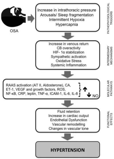
Figure 1: Schematic diagram summarizing the pathways by which intermittent
hypoxia leads to hypertension ...22
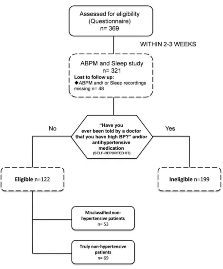
GENERAL METHODS Figure 1: Predictors of HT misclassification: patient eligibility and follow-up...38
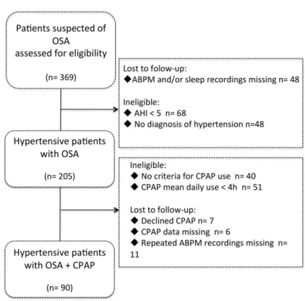
Figure 2: Association between antihypertensive medication and BP control: patient eligibility and follow-up ...39
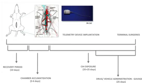
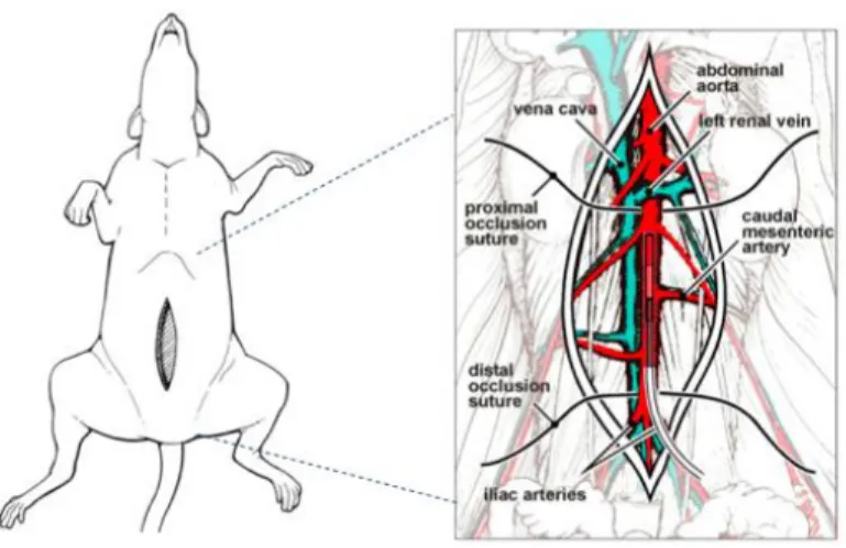
Figure 3: Experimental design of the study performed to evaluate the effects of carvedilol on HT related to CIH ...46
Figure 4: DSI PhysioTel® Receivers ...47
Figure 5: The PA-C40 Transmitter ...47
Figure 6: Surgical procedure for abdominal aorta cannulation with intraperitoneal cavity device placement ...49
Figure 7: Intraperitoneal placement of the transmitter ...49
Figure 8: Typical Oxycycler AT Series System ...50
Figure 9:Graphical representation of oxygen levels inside the CIH chambers …………...51
Figure 10: Method for voluntary ingestion of carvedilol and losartan with three different vehicles ………...54
RESULTS Section 1 Predictors of HT misclassification Figure 1:Flowchart of the study protocol ………...63
Figure 2: Correlation between waist circumference (WC) and BMI ………...65
Figure 3: Influence of BMI and NC on hypertension misclassification resulting from fitting a GAM to the data. Minimum P-value approach: optimal BMI and NC cut-off points for misclassified nonhypertension …………...66
vi
Section 2
Efficacy of Carvedilol in reversing HT induced by CIH
Figure 1: Effect of CIH on body weight in comparison with the growth curve for body weight
gain of male Wistar rats born in the NOVA Medical School animal facility...96
Figure 2: Grouped data showing the daily average recordings of (A) MAP, systolic BP,
diastolic BP and (B) heart rate of rats submitted to CIH for 35 days (n=20) …...97
Figure 3: Effect of CIH on (A) mean arterial blood pressure; (B) systolic blood
pressure; (C) diastolic blood pressure and (D) heart rate, in male Wistar rats (n=20) ...97
Figure 4: Grouped data showing the daily average recordings of (A) MAP and (B)
heart rate of rats administered with MC 0.5% for 25 days and submitted to
60 days of CIH (control-vehicle group; n=5) ...98
Figure 5: Effect of CVD (A) 10 mg/kg (n=5); (B) 30 mg/kg (n=7) and (C)
50 mg/kg (n=8) daily administration (25 days) on mean arterial blood pressure
of rats submitted to 60 days of CIH ...99
Figure 6: Effect of CVD (A) 10 mg/kg (n=5); (B) 30 mg/kg (n=7) and (C) 50 mg/kg (n=8)
daily administration (25 days) on heart rate of rats submitted to 60 days of CIH ...100
Figure 7: The HPLC chromatograms resulting from derivatization with MCF
of blank rat plasma, calibration sample – rat plasma spiked with 100 ng/mL of R-(+)-CVD and S-(-)-CVD, plasma sample of a normoxic rat treated with 50 mg/Kg/day of the racemic CVD and plasma sample of a rat exposed to
chronic intermittent hypoxia treated with 50 mg/Kg/day of racemic CVD ………...101
Figure 8: Ratio S-(-)-CVD / (R-(+)-CVD + S-(-)-CVD) in rats exposed to normoxia
and chronic intermittent hypoxia treated with 50 mg/Kg/day of racemic CVD ……...101
Voluntary oral administration as an alternative method to gavage
Figure 1: Method for voluntary ingestion of losartan with three different vehicles:
nut paste, peanut butter and sugar-dough ……...115
Figure 2: Mean glycaemia values (mg/dl) in the serum of Wistar rats fed with 0.5 g of
nut paste, peanut butter and sugar dough vs. time. Comparison of the three vehicles
effects on glycaemia at 14 and 28-day time points ………...………118
Figure 3: Mean total cholesterol concentrations (mg/dl) in the serum of Wistar rats
fed with 0.5 g of nut paste, peanut butter and sugar dough vs. time ...119
Figure 4: Mean triglycerides concentrations (mg/dl) in the serum of Wistar rats
fed with 0.5 g of nut paste, peanut butter and sugar dough vs. time ...120
Figure 5: Losartan plasma concentrations (μg/ml), from blood collected by cardiac puncture
vii
List of Acronyms and Abbreviations
ABPM ACEi AHDs AHI AP-1 ARB AT II BMI BP CA CB CCB CHI COPD
Ambulatory Blood Pressure Monitoring
Angiotension-Converting Enzyme Inhibitor
Antihypertensive Drugs
Apnea-Hypopnea Index
Activator Protein-1
Angiotensin II Receptor Blocker
Angiotensin II
Body Mass Index
Blood Pressure
Cathecolamines
Carotid Body
Calcium Channel Blocker
Chronic Intermittent Hypoxia
Chronic Obstructive Pulmonary Disease
CPAP CRP CSA CVD DBP EDTA ET-1 FiO2 GAMs GAV HDL-C HIF-1α HPLC HR HT
Continuous Positive Airway Pressure
C- Reactive Protein
Central Sleep Apnea
Carvedilol
Diastolic Blood Pressure
Ethylenediaminetetraacetic Acid
Endothelin-1
Fraction of Inspired Oxygen
Generalized Additive Models
Gavage
High-Density Lipoprotein-Cholesterol
Hypoxia-Inducible Factor α
High-Performance Liquid Chromatography
Heart Rate
Hypertension
ICAM-1 IH
Intercellular Adhesion Molecule
viii
IL INN LDL-C LST MAP MC MCF MNH NADPH NC NH NO eNOS NF-κB NOX2 NTS NUT Nx OSA InterleukinInternational Non-proprietary Name
Low-Density Lipoprotein- Cholesterol
Losartan
Mean Arterial Blood Pressure
Methylcellulose
(-)-menthyl chloroformate
Misclassified Non-Hypertensive Patients
Nicotinamide Adenine Dinucleotide Phosphate
Neck Circumference
Non-Hypertensive Patients
Nitric Oxide
Endothelial Nitric Oxide Synthase
Nuclear Factor-κ-light Chain Enhancer of Activated B Cells
NADPH oxidase 2
Nucleus of the Solitary Tract
Nut Paste
Normoxia
Obstructive Sleep Apnea
PaCO2 PB PD PRA PVN RAAS RCT RERAs ROS SaO2 SBP SD SDB SEM
Partial Pressure of Carbon Dioxide
Peanut Butter
Pulmonary Hypertension
Plasma Renin Activity
Paraventricular Nucleus Neurons
Rennin-Angiotensin-Aldosterone System
Randomized Controlled Trials
Respiratory Effort Related Arousals
Reactive Oxygen Species
Arterial Hemoglobin Oxygen Saturation
Systolic Blood Pressure
Sugar Dough
Sleep-Disordered Breathing
Standard Error of the Mean
ix
SNASOD
Sympathetic Nerve Activity
Superoxide Dismutase Mimetic
SPSS TC TGL TNF-α TSP VEGF WC
Statistical Package for the Social Sciences
Total Cholesterol
Triglycerides
Tumour Necrosis Factor α
Trigger Sleep Blood Pressure monitoring
Vascular Endothelial Growth Factor
xi
Acknowledgements
First of all, I would to express my heartfelt gratitude to my supervisor Professor Emília Monteiro, whose thoughtful consideration and guidance have been truly important. During the last six years you have been a constant source of creativity, strength and enthusiasm, being all that a mentor should be. Thank you for encouraging me and for allowing me to grow as a researcher. To work with you has been a great honour and a real pleasure to me!
I would also like to gratefully thank Professor Ana Luísa Papoila, my co- supervisor, for sharing with me her incredible statistical knowledge and for all those infinite hours that we spent “listening” to the data. Thank you also for your true generosity over the years and for just being unconditionally there for me in all the moments of discouragement and frustration. A simple “Never give up!” really meant a lot to me!
To Professor Cristina Bárbara, also co-supervisor of this thesis, I would like to thank her for her helpful advice during the study design and support during the recruitment. I learned so much, from you and your team, during the almost two years that I spent in Hospital Pulido Valente!
I would also like to extend my sincerest thanks to Professor Ana Isabel Moura Santos for her scientific advice and knowledge within the field of laboratory animal science, but foremost for her friendship and unconditional support throughout these last years. I am also very grateful to Professor Pedro Freire Costa for his many insightful comments and suggestions. “The quick brown fox jumped over the lazy dog’s back” was really a useful tip!
To all my colleagues at NOVA Medical School and CEDOC, especially Teresa Monteiro, Sílvia Conde, Joana Batuca, Maria João Ribeiro, Joana Sacramento, Clara Dias, Nádia Grilo, Aline Marinho, Inês Faustino, Joana Gaspar and Fátima Martins, for all their help and support during this long journey. A special thanks to Rita Nunes for her true friendship, unconditional support and for all the hours we spent chatting and discussing experiments and results; Ricardo Afonso for being my “right hand” during the surgical procedures (our “four-hands” surgeries were a success!!!); Sofia Pereira, my dearest friend and great researcher, for all her support, interest and encouragement (Believe! The best is yet to come!) and also to Daniela Vasconcelos who started this beautiful adventure with me!
xii
I would also like to thank physicians, in particular Professor Paula Pinto, and cardio-respiratory technicians from Centro Hospitalar Lisboa Norte, EPE (CHLN) Sleep Unit (Hospital Pulido Valente) for their valuable support during the patient recruitment period.
I will always be grateful to all my friends for sheltering me over the years. A special thanks to Raquel Salvaterra, my best friend for almost two decades, for being there to listen, party and comfort. You rock girl!
Lastly, I would like to extend my sincerest thanks and appreciation to my loving parents, Diogo and Madalena, who set a shining example from the first day of my life. Words cannot express how grateful I am for your emotional support no matter what path I choose. To my brother Zé, thank you for your infinite support throughout everything. To Paulo for being a true partner and for just being there for me with all his encouragement and support. To my “BIG” Manuel, the rainbow of my life, who is much more than any mother could ask for. Your smile and complicity have been my daily dose of inspiration. I hope that I have not lost too much of your “busy days” and I promise that our Disney trip will happen soon!! Finally, to my grandfather Oswaldo who unfortunately is no longer with me and whom I miss terribly, thank you for all the great moments we shared. This PhD thesis is a tribute to you!
xiii
Abstract
Hypertension (HT) is a highly prevalent condition, although under diagnosed, in patients with obstructive sleep apnea (OSA). These conditions are closely related and 24-hour ambulatory blood pressure monitoring (ABPM) seems to be the most accurate measurement for diagnosing hypertension in OSA. However, this diagnostic tool is expensive and time-consuming and, therefore, not routinely used. On the other hand, although continuous positive airway pressure (CPAP) is considered the gold standard treatment for symptomatic OSA, its lowering effect on blood pressure (BP) seems to be modest and, therefore, concomitant antihypertensive therapy is still required. Data on antihypertensive drug regimens in patients with OSA are scarce and specific therapeutic guidelines for the pharmacological treatment of hypertension in these patients remain absent. The use of animal models of CIH, which mimic the HT observed in patients with OSA, is extremely important since it is imperative to identify preferred compounds for an adequate BP control in this group of patients. However, studies aimed at investigating the antihypertensive effect of antihypertensive drugs in this animal model are insufficient, and most reports on CIH animal models in which drugs have been tested were not designed to respond to pharmacological issues. Moreover, when testing antihypertensive drugs (AHDs) it becomes crucial to ensure the selection of a non-invasive and stress-free method for drug delivery. Although gavage is effective and a widely performed technique for daily dosing in laboratory rodents, it comprises a sequence of potentially stressful procedures for laboratory animals that may constitute bias for the experimental results.
The overall goal of the present translational research was to contribute to identify more effective AHDs for the treatment of hypertension in patients with OSA and investigate underlying mechanisms of systemic effects associated with OSA, as well as its modulation by AHDs. The specific aims were: first, to find new predictors based on anthropometric measures to identify patients that misclassify themselves as non-hypertensive, and thereby promote the selective use of ABPM; second, to investigate a hypothetical association between ongoing antihypertensive regimens and BP control rates in patients with OSA, before and after CPAP adaptation; third, to determine, in a rat model of CIH-induced hypertension, the efficacy of carvedilol (CVD), a nonselective beta-blocker with intrinsic anti-α1-adrenergic activity and antioxidant properties; fourth, to explore the effects of CIH on the pharmacokinetics profile of CVD and fifth, to investigate an alternative method to gavage, for chronic administration of AHDs to laboratory rats.
xiv
ongoing antihypertensive medication profile registration. In the second phase, a rat experimental model of HT induced by a paradigm of CIH that simulates OSA was used.
The main findings of this work were: first, body mass index (BMI) and neck circumference (NC) were identified as independent predictors of hypertension misclassification in patients suspected of OSA; second, in patients with OSA, BP control is independent of both the antihypertensive regimen and the number of antihypertensive drugs, either before or after CPAP adaptation; third, although the doses of 10, 30 and 50 mg/Kg of CVD promoted a significant reduction in heart rate, no decrease in mean arterial pressure was observed; fourth, the S/(R+S) ratios of CVD enantiomers, between rats exposed to CIH and normoxic conditions, were different and fifth, voluntary ingestion proved to be an effective method for a controlled daily dose administration, with a define timetable, that is independent of handling and restraint procedures.
In conclusion, the clinical study showed that BP control in OSA patients is independent of both the antihypertensive regimen and the number of antihypertensive drugs. Additionally, our results highlight the lack of validity of self-reported hypertension and suggest that all patients suspected of OSA with undiagnosed hypertension and with a BMI and NC above 27 Kg/m2 and 39 cm should be screened for hypertension, through ABPM. The results attained in the rat model of HT related to CIH suggest that the blockade of the sympathetic nervous system together with the putative pleiotropic effects of carvedilol is not able to revert hypertension induced by CIH and point out that the pharmacokinetic changes induced by CIH on S/(R+S) ratio are not apparently responsible for the lack of efficacy of carvedilol in reversing this particular type of hypertension. Finally, the results here presented support the use of voluntary oral administration as a viable alternative to gavage for chronic administration of a fixed dose of AHDs.
Key words: ambulatory blood pressure monitoring, antihypertensive drugs, carvedilol, chronic
xv
Resumo
A hipertensão arterial (HA) é uma patologia altamente prevalente, embora claramente subdiagnosticada, em doentes com síndrome de apneia obstrutiva do sono (SAOS). Estas duas patologias apresentam uma estreita relação e a monitorização ambulatória da pressão arterial (MAPA), por um período de 24 horas, parece ser o método mais preciso para o diagnóstico de hipertensão em doentes com SAOS. No entanto, esta ferramenta de diagnóstico para além de ser dispendiosa e envolver um número acrescido de meios técnicos e humanos, é mais morosa e, por conseguinte, não é utilizada por rotina no contexto do diagnóstico da SAOS. Por outro lado, apesar da aplicação de pressão positiva contínua nas vias aéreas (CPAP – Continous Positive Airway Pressure) ser considerada a terapêutica de eleição para os doentes com SAOS, o seu efeito no abaixamento da pressão arterial (PA) parece ser modesto, exigindo, por conseguinte, a implementação concomitante de terapêutica anti-hipertensora. Acontece que são escassos os dados relativos aos regimes de fármacos anti-hipertensores utilizados em doentes com SAOS e, acresce ainda que, as guidelines terapêuticas para o tratamento farmacológico da HA, neste grupo particular de doentes, permanecem, até ao momento, inexistentes. A utilização de modelos animais de hipóxia crónica intermitente (CIH), que mimetizam a HA observada em doentes com SAOS, revela-se extremamente importante, uma vez que se torna imperativo identificar fármacos que promovam um controle adequado da PA neste grupo de doentes. No entanto, estudos concebidos com o intuito de investigar o efeito anti-hipertensor dos fármacos neste modelo animal revelam-se insuficientes e, por outro lado, os escassos estudos que testaram fármacos anti-hipertensores neste modelo não foram desenhados para responder a questões de natureza farmacológica. Acresce ainda que se torna imprescindível garantir a escolha de um método para administração destes fármacos que seja não invasivo e que minimize o stress do animal. Embora a gavagem seja uma técnica indiscutivelmente eficaz e amplamente utilizada para a administração diária de fármacos a animais de laboratório, ela compreende uma sequência de procedimentos geradores de stress para os animais e, que podem por conseguinte, constituir um viés na interpretação dos resultados obtidos.
xvi
em terceiro lugar, avaliar a eficácia do carvedilol (CVD), um fármaco bloqueador β-adrenérgico não selectivo com actividade antagonista α1 intrínseca e propriedades anti-oxidantes num modelo animal de hipertensão induzida pela CIH; em quarto lugar, explorar os efeitos da CIH sobre o perfil farmacocinético do CVD; e, em quinto lugar, investigar um método alternativo à gavagem para a administração crónica de fármacos anti-hipertensores a animais de laboratório. Com este intuito, na primeira fase deste projecto, fizemos uso de uma amostra com um número apreciável de doentes com SAOS (n=369), que acorreram, pela primeira vez, à consulta de Patologia do Sono do CHLN e que foram submetidos a um estudo polissonográfico do sono, à MAPA e que preencheram um questionário que contemplava a obtenção de informação relativa ao perfil da medicação anti-hipertensora em curso. Numa segunda fase, utilizámos um modelo experimental de HT no rato induzida por um paradigma de CIH.
Do nosso trabalho resultaram os seguintes resultados principais: em primeiro lugar, o índice de massa corporal (IMC) e o perímetro do pescoço (PP) foram identificados como preditores independentes de “auto-classificação errónea” da HA em doentes com suspeita de SAOS; em segundo lugar, não encontramos qualquer associação com significado estatístico entre os vários esquemas de fármacos anti-hipertensores bem como o número de fármacos incluídos nesse esquemas, e o controle da PA (antes e depois da adaptação do CPAP); em terceiro lugar, apesar das doses de 10, 30 e 50 mg/kg de carvedilol terem promovido uma redução significativa da frequência cardíaca, não foi observado qualquer decréscimo na PA no nosso modelo animal; em quarto lugar, as razões S/(R+S) dos enantiómeros do CVD nos animais expostos à CIH e a condições de normóxia revelaram-se diferentes; e, em quinto lugar, a administração oral voluntária mostrou ser um método eficaz para a administração diária controlada de fármacos anti-hipertensores e que é independente da manipulação e contenção do animal.
xvii
viabilidade da utilização da administração oral voluntária, em alternativa à gavagem, para a administração crónica de uma dose fixa de fármacos anti-hipertensores.Palavras-chave: Administração oral voluntária, apneia obstructiva do sono, carvedilol,
xix
Thesis Outline
The present thesis is divided into five main chapters, enumerated with Roman numbers, whose content is summarized below.
Chapter I is a general introduction to the thesis, summarizing the most relevant topics of literature in chronic intermittent hypoxia-related disorders, namely obstructive sleep apnea, and its close linkage with hypertension. Special focus is given to the efficacy of pharmacological interventions to revert hypertension related with chronic intermittent hypoxia conditions in both animals and humans. The content of this chapter has been included in the publication of a review article.
In Chapter II, we summarize the starting points for the establishment of the main goals of our
research line. The general and specific research aims that will be addressed in this thesis are also listed in this chapter.
Chapter III contains the description of the general methods used during both clinical and
experimental studies.
In Chapter IV, we present the results of this thesis, published or submitted to international peer-reviewed journals. Section 1 comprises two original articles regarding the results of the study performed in humans. Section 2 includes one original article concerning the results attained in the animal model of hypertension induced by chronic intermittent hypoxia and the other relating to an alternative method to gavage, for chronic administration of antihypertensive drugs to laboratory animals.
Chapter IV summarizes the main conclusions and limitations of our work and presents the
I
I
NTRO
DU
CTION
3
Chronic intermittent hypoxia-related disorders
Is is well established that intermittent hypoxia (IH) affects control of breathing, the autonomic nervous system and the cardiovascular system (Foster et al., 2007). Chronic intermittent hypoxia (CIH) is a feature that is present in interstitial lung disease (Fletcher et al., 1992a) and sleep-disordered breathing (SDB), and it has also been shown to occur in patients with hepatopulmonary syndrome (Tanné et al., 2005; Ogata et al., 2006; Palma et al., 2008). Since several years ago, there has been growing interest concerning CIH due to the high relevance of the part assumed to be played by sleep-related breathing disorders in chronic diseases.
4
apneas are observed in polysomnograms of patients with OSA, they are normally ignored because we do not presently understand their potencial clinical relevance.
Nowadays, it is well known that the outcomes of these sleep-related breathing disorders can lead to vascular diseases, contributing to a considerable increase in overall cardiovascular risk. The desaturation-reoxygenation sequence, a typical pattern coupled with the majority of respiratory events, is thought to be responsible for most of the associated cardiovascular morbidity (Lévy et al., 2012). Although OSA has been associated with several cardiovascular conditions, it has been more closely etiologically connected to systemic HT (Kapa et al., 2008), and the link between HT and OSA is now widely accepted and supported by different findings. Most episodes of OSA are coupled with sleep disruption, which per se increases sympathetic nerve activity and blood pressure (Morgan et al., 1996). In addition, the occurrence of arousals appears to enhance the pressor effects of asphyxia during OSA (Morgan et al., 1998), contributing synergistically to blood pressure increase. In any case, studies in both animals and humans underline the major role of hypoxia itself in promoting an increase in blood pressure (Brooks et al., 1997b; Tamisier et al., 2011).
Regarding CSA, this SDB, like OSA, is strongly linked to cardiac disease and cardiovascular outcomes (Brenner et al., 2008). Indeed, the majority of patients with CSA have underlying cardiovascular disease, primarily heart failure, which is considered the most common risk factor for CSA, followed by atrial fibrillation (Bradley et al., 1992). Moreover, like OSA, CSA has been implicated in heart failure pathophysiology (Mehra, 2014) and occurs in 30–50% of patients with left ventricular dysfunction and heart failure caused by HT, cardiomyopathy and ischemic heart disease (Bradley and Floras, 2003). Thus, CSA has significant co-morbidity with many cardiac conditions, which clearly contributes to an increase in the associated mortality and morbidity.
I
NTRO
DU
CTION
5
erythropoietin subsequent to hypoxemia), is the occurrence of right ventricle hypertrophy leading to cor pulmonale (Levitzky, 2008). The prevalence of this chronic cardiopulmonary condition among patients with SDB is estimated to range from 17% to 52% (Minic et al., 2014), and 20–30% of untreated OSA patients suffer from PH (Dumitrascu et al., 2013). Even if PH in this group of patients is typically not severe (Badesh et al., 2010), OSA patients with PH have a higher mortality rate than OSA patients without PH (Minai et al., 2009). A recent meta-analysis shows that CPAP is associated with a mild but statistically significant reduction in pulmonary artery pressure in OSA patients (Sun et al., 2014). This decrease might translate into a better outcome in patients with PH secondary to OSA. However, more studies are needed to confirm this assumption.OSA and Hypertension: How relevant is this linkage?
Since 2003, OSA has formally been recognized as a frequent and important secondary cause of HT and is one of the first causes to be screened mainly in patients with a suggestive phenotype, refractory HT and a non-dipping profile (Chobanian et al., 2003; Mancia et al., 2007). More recently, OSA has been identified as an independent risk factor for HT (Lavie et al., 2000; Peppard et al., 2000; Marin et al., 2012), as one of the major clinical conditions that favors poorly controlled HT (Oliveras and Schmieder, 2013), and as the most common condition associated with resistant HT (Pedrosa et al., 2011). OSA and HT are two prevailing risk factors for several cardiovascular events (Wang and Vasan, 2005; Baguet et al., 2009). Due to their high prevalence and cardiovascular morbidity (Wolf et al., 2007; Malhotra and Loscalzo, 2009), OSA and HT are now acknowledged as public health problems. Epidemiological data show that the estimated overall prevalence of HT among patients with OSA is approximately 50% and an estimated 30–40% of hypertensive patients are diagnosed with OSA (Calhoun, 2010), confirming the bidirectional relationship between OSA and HT. Moreover, OSA and HT are chronic diseases mostly diagnosed in active adults and because of the associations between OSA and obesity and advancing age, the public health burden of OSA related to cardiovascular disease is expected to rise in the coming years (Dempsey et al., 2010). The use of both antihypertensive drugs (AHDs) and CPAP in these patients is for life and consequently
treatment is associated with a high impact both in terms of costs and in patients’ quality of life.
6
OSA and Hypertension: What is the problem?
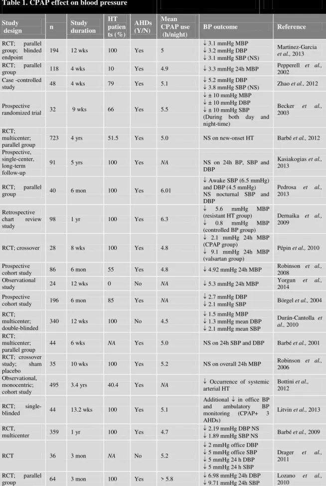
CPAP is considered the gold standard treatment for mild, moderate and severe OSA due to its remarkable ability in providing pneumatic splitting of the upper airway and effectiveness in reducing the apnea-hypopnea index (AHI), symptoms, and cardiovascular morbidity and mortality (Hla et al., 2002; Pepperell et al., 2002; Wolf et al., 2007; Epstein et al., 2009; Mannarino et al., 2012). Besides preventing hypoxemia, sleep disturbance and apnea episodes, CPAP reduces sympathetic activity, systemic inflammation and oxidative stress (Yorgun et al., 2014). However, the results found for the effectiveness of CPAP on blood pressure (BP) control are still controversial. Table 1 summarizes the results of original studies in which the effect of CPAP on BP has been analysed.
Whereas some studies and meta-analyses (Bakker et al., 2014; Varounis et al., 2014) have reported modest effects for CPAP in lowering BP, others tend to support the beneficial effect of CPAP treatment on BP reduction and attenuating the risk of developing HT. In any case, although the lowering effect of CPAP on BP is relevant in terms of overall cardiovascular risk reduction, this effect is very limited when compared to the performance of AHDs in patients with essential HT (Pépin et al., 2010). Thus, treating HT in patients with sleep apnea is proving to be a dificult task and there is consensus that the use of AHDs is mandatory. In spite of this, data on AHDs regimens in patients with OSA are scarce and there is a lack of specific therapeutic guidelines for the pharmacological treatment of HT in these patients. Furthermore, the effects of antihypertensive agents on OSA patients are not consistent (Parati et al., 2012) and there are no data on the efficacy of specific AHDs regimens when associated with CPAP.
A new treatment for OSA patients is the oral appliance/mandibular advancement device (Guralnick and Bakris, 2012). Oral appliance therapy is an important alternative to CPAP for some patients with mild to moderate OSA (Iftikhar et al., 2013). Despite a recent study (Andrén et al., 2013) and a recent meta-analysis (Iftikhar et al., 2013) which have shown some beneficial effects of this device in reducing blood pressure measurements, larger and longer randomized control trials are needed to confirm the effects of oral appliance therapy on BP control.
I NTRO DU CTION
7
Table 1. CPAP effect on blood pressure
Study
design n Study duration HT patien ts (%) AHDs (Y/N) Mean CPAP use (h/night)
BP outcome Reference
RCT; parallel group; blinded endpoint
194 12 wks 100 Yes 5
3.1 mmHg MBP 3.2 mmHg DBP 3.1 mmHg SBP (NS)
Martinez-Garcia et al., 2013 RCT; parallel
group 118 4 wks 10 Yes 4.9 3.3 mmHg 24h MBP
Pepperell et al., 2002
Case -controlled
study 48 4 wks 79 Yes 5.1
5.2 mmHg DBP
3.8 mmHg SBP (NS) Zhao et al., 2012
Prospective
randomized trial 32 9 wks 66 Yes 5.5
± 10 mmHg MBP ± 10 mmHg DBP ± 10 mmHg SBP (During both day and night-time)
Becker et al., 2003
RCT; multicenter; parallel group
723 4 yrs 51.5 Yes 5.0 NS on new-onset HT Barbé et al., 2012
Prospective, single-center, long-term follow-up
91 5 yrs 100 Yes NA NS on 24h BP, SBP and DBP
Kasiakogias et al., 2013
RCT; parallel
group 40 6 mon 100 Yes 6.01
Awake SBP (6.5 mmHg) and DBP (4.5 mmHg) NS nocturnal SBP and DBP
Pedrosa et al., 2013
Retrospective chart review study
98 1 yr 100 Yes 6.3
5.6 mmHg MBP (resistant HT group)
0.8 mmHg MBP (controlled BP group)
Dernaika et al., 2009
RCT; crossover 28 8 wks 100 Yes 4.8
2.1 mmHg 24h MBP (CPAP group)
9.1 mmHg 24h MBP (valsartan group)
Pépin et al., 2010
Prospective
cohort study 86 6 mon 55 Yes 4.8 4.92 mmHg 24h MBP
Robinson et al., 2008
Observational
study 24 12 wks 0 No NA 5.3 mmHg 24h MBP
Yorgun et al., 2014
Prospective
cohort study 196 6 mon 85 Yes NA
2.7 mmHg DBP
2.1 mmHg SBP Börgel et al., 2004 RCT;
multicenter; double-blinded
340 12 wks 100 No 4.5
1.5 mmHg MBP 1.3 mmHg mean DBP 2.1 mmHg mean SBP
Durán-Cantolla et al., 2010 RCT;
multicenter; parallel group
44 6 wks NA Yes 5.0 NS on 24h SBP and DBP Barbé et al., 2001
RCT; crossover study; sham placebo
35 10 wks 100 Yes 5.2 NS on overall 24h MBP Robinson et al., 2006
Observational, monocentric; cohort study
495 3.4 yrs 40.4 Yes NA Occurrence of systemic arterial HT
Bottini et al., 2012
RCT;
single-blinded 44 13.2 wks 100 Yes 5.1
Additional in office BP and ambulatory BP monitoring (CPAP+ 3 AHDs)
Litvin et al., 2013
RCT,
multicenter 359 1 yr 100 Yes 4.7
2.19 mmHg DBP NS
1.89 mmHg SBP NS Barbé et al., 2009
RCT 36 3 mon NA No 5.2
2 mmHg office DBP 5 mmHg office SBP 5 mmHg 24 h DBP 5 mmHg 24 h SBP
Drager et al., 2011
RCT; parallel
group 64 3 mon 100 Yes > 5.8
6.98 mmHg 24h DBP 9.71 mmHg 24h SBP
Lozano et al., 2010
8
What models are available to study Hypertension related to OSA?
Due to the high complexity and heterogeneity associated with OSA, considerable variability can be observed between reports addressed at the study of this disease. In addition, the scarcity of opportunities for patient investigation, in particular at the cellular level, has compromised progress in understanding the pathophysiology of OSA and the development of novel and specific treatments for this disorder. To overcome some of these limitations, several animal models and more recently, a model of OSA in healthy human volunteers (Tamisier et al., 2009; Tamisier et al., 2011) have been developed. Animal models, especially of IH, mimic OSA more easily than human models. The small size of rodents allows more rapid and intense changes in SaO2 (arterial hemoglobin oxygen saturation) whereas humans require longer periods of hypoxia to induce arterial oxyhaemoglobin desaturation (Foster et al., 2007). The combination of these two approaches is certain to contribute to the consolidation of prevention strategies and the development of more suitable treatments for OSA patients.
ANIMAL MODELS
The major advantage of the use of animal models is that they allow single components of the disease to be evaluated, accurately controlling the triggering events in terms of both severity and duration, and providing homogeneous populations (Lévy et al., 2012). These models also provide an excellent opportunity to explore the underlying mechanistic pathways of HT related to OSA and their consequences under controlled conditions. Moreover, animal models have enabled the study of parameters that have proved difficult to assess in humans, particularly due to the need for organ harvesting to explore the mechanisms underlying the consequences of IH at the molecular level (Dematteis et al., 2009). Thus, studies with animal models are good tools for overcoming some confounding factors present in human studies (e.g., the presence of comorbidities, disease duration, and behavioral and environmental variables) (Badran et al., 2014), and for providing more specific information concerning the efficacy of drugs to be tested.
I
NTRO
DU
CTION
9
brought out most of the available knowledge in this field and furthermore, almost all cardiovascular diseases known to be present in patients with OSA have been replicated in these models (Dumitrascu et al., 2013).The effective use of animals to study sleep apnea implies recognition of the natural similarities and differences between animals and humans to ensure the reliability of the experimental results. For instance, as rodents are nocturnal animals, the stimulus must be applied during the sleep-dominant phase of the diurnal cycle. Moreover, in humans the circadian distribution of sleep tends to be consolidated and normally monophasic, with a daily sleep duration of 7–8 hours, whereas it is polyphasic, relatively fragmented and with a duration of 12–15 hours in rodents (Toth and Bhargava, 2013). Another issue is related to the fact that rodents sleep in the prone position (Golbidi et al., 2012); it is well known that supine OSA is the dominant phenotype of OSA syndrome and that the supine position favors upper airway collapse in humans (Joosten et al., 2014). Furthermore, additional care must be taken to minimize external factors (e.g., light exposure, photoperiod, noise, disruptions in the home environment, and post-surgical care in studies, for instance requiring implantation of telemetric devices) able to influence sleep in animals used in experimental research (reviewed in Toth and Bhargava, 2013).
The experimental animal models developed to mimic OSA have recently been reviewed (Dematteis et al., 2009; Golbidi et al., 2012; Davies and O’Donnell, 2013; Toth and Bhargava, 2013) and assembled taking into account the main injuries triggered by OSA. Despite attempts to use large animals (e.g., dogs, lambs and pigs) to simulate upper airway obstruction, most research on the cardiovascular consequences of OSA has been performed in rodents. Alternative models (e.g., cell cultures incubated in specific devices that perform oxygen fluctuations mimicking sleep apnea-related IH), mainly relevant to signaling investigation (Kumar et al., 2003; Gozal et al., 2005; Ryan et al., 2005), represent a complementary approach to the most widely used sleep models. However, in spite of the recommendations to refine, reduce and replace (the 3Rs programme), these alternative models cannot replace animal models in the study of HT.
10
model. Special focus will be given to the CIH model, based on the assumption that IH is the most effective paradigm to induce HT related to OSA and probably the most relevant stimulus regarding the cardiovascular sequelae of OSA.
Induced airway obstruction models
Briefly, the airway obstruction model involves surgical intervention (an endotracheal tube), which is an invasive procedure, or alternatively the use of a specific chamber with a latex neck collar that induces recurrent airway obstruction. This latter procedure, developed by Farré et al. (Farré et al., 2007), is associated with high levels of stress due to the restriction of animal movement. In both approaches, the degree of obstruction is adjustable (Golbidi et al., 2012) and in the case of induction of obstruction through endotracheal tube, the PaCO2 can be adjusted to mimic human sleep apnea (Golbidi et al., 2012). Many experiments using this method have not monitored the sleep state of the animals, but more recent studies have incorporated sophisticated apparatus that is able to detect sleep-awake states and allow close coordination between the initiation of airway obstruction and sleep onset (Schneider et al., 2000).
This model allows the study of the potential consequences of strenuous breathing against an obstructed airway and can be used to study the cardiovascular consequences and risk factors of OSA (e.g., systemic inflammation and coagulation), and to investigate the mechanisms that underlie OSA (Salejee et al., 1993; Nacher et al., 2007; Almendros et al., 2008; Nacher et al., 2009; Othman et al., 2010; Almendros et al., 2011). However, to the best of our knowledge, no study has yet shown that this obstruction model is able to mimic HT related to OSA. Furthermore, when testing AHDs, it became crucial to ensure the selection of a stress-free paradigm as it has been shown that any source of external stress on rodents can significantly increase heart rate and blood pressure (Brown et al., 2000; Kramer et al., 2000; Balcombe et al., 2004; Bonnichsen et al., 2005) and therefore contribute to confounding the experimental results. Finally, as the rat models of obstruction or asphyxia were developed in restrained or anesthetized rats, they are not good models for chronic administration of oral drugs, particularly AHDs.
Sleep deprivation models
In the last few years, several approaches have been used to trigger sleep deprivation in different animals, the rat being the animal of choice to date (Colavito et al., 2013). In the “multiple platform technique”, the animal is aroused from sleep when the characteristic loss of muscle tone that accompanies paradoxical sleep causes it to fall off the platform (Suchecki and Tufik,
I
NTRO
DU
CTION
11
interaction with the experimenter, who actively keeps the animal awake through the use of external stimulation (e.g., mild noises, tapping or gentle shaking of the cage, or by direct contact with the animal either using a soft brush or by hand), or by the introduction of novel objects or nesting material in the cages, which typically leads to active exploratory behavior (Colavito et al., 2013).These models are most often used to evaluate the neurophysiological aspects of OSA (Van Dongen et al., 2003; Haack and Mullington, 2005; McKenna et al., 2007; Ward et al., 2009; Nair et al., 2011) due to the high similarity between the structures of the nervous systems of rodents and humans (Badran et al., 2014), and to illustrate some mechanistic pathways induced by this trigger (McGuire et al., 2008; Tartar et al., 2010; Liu et al., 2011; Perry et al., 2011). Nevertheless, some studies have also aimed to evaluate the cardiovascular outcomes induced by this OSA feature and have suggested that sleep fragmentation may have a far more important role in cardiovascular changes observed in OSA patients (Golbidi et al., 2012). Even so, sleep deprivation studies have produced mixed results regarding BP outcomes.
12
CIH modelIH is now established as the dominant model of sleep apnea. Generally, this model makes use of specific ventilated chambers in which the animals are housed and cyclically exposed either to normoxia/hypoxia or room air to mimic the most relevant consequences of OSA. Hypoxic conditions can also be achieved by surgical intervention (an endotracheal tube) or by the use of a mask, which involves animal restraint and consequently high levels of stress (Golbidi et al., 2012). In either case, animals breathe nitrogen-enriched air alternating with oxygen or normal air (Dematteis et al., 2009). Thus, as with O2, nitrogen plays an important role in this model as the flushing of the chambers with this gas allows the gradual lowering of O2. The duration of the hypoxic and normoxic phases of the IH cycle, as well as the slopes of FiO2 (fraction of inspired oxygen), decrease and increase, and are dependent on cage/chamber size and the gas flows and mixtures (Dematteis et al., 2009).
The standard animal model of OSA was that described in the landmark study of Fletcher and Bao (Fletcher and Bao, 1996). Despite the presence of some drawbacks, this model has successfully been employed to study the changes in systemic arterial pressure and the impact of IH on a wide range of cardiovascular outcomes. One of the major limitations pointed to in this model is the absence of recurrent upper airway obstruction, abolishing the acute hemodynamic changes due to the negative intrathoracic pressure (Badran et al., 2014). Marked negative intrathoracic pressure induces acute hemodynamic changes that are probably the starting point for chronic cardiovascular diseases (Bonsignore et al., 1994). Despite the absence of upper airway occlusion, some respiratory efforts (intermittent tachypnea) occur, corresponding to a fluctuating hyperventilation that follows the IH cycles (Dematteis el al, 2009). However, this disadvantage allows the evaluation of CIH effects, namely chronic blood gas exchanges, without the interference of the mechanical aspects of OSA.
I
NTRO
DU
CTION
13
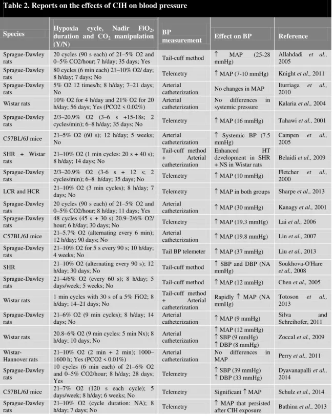
acute BP increase than hypocapnic IH (Bao et al., 1997). Similarly, Lesske et al. showed comparable changes in BP between two groups submitted to IH with or without hypercapnia (Lesske et al., 1997). On the other hand, based on the results of different CIH experimental protocols in rodents, Kanagy concludes that the level of PaCO2 influences the magnitude of an increase in BP (Kanagy, 2009). Concretely, eucapnic hypoxia induces a faster and greater increase than hypocapnic hypoxia (Kanagy, 2009), through mechanisms that presently remain unknown. Moreover, the greatest increases in BP have been observed in studies in which hypocapnia was prevented by CO2 administration (Morgan, 2009). Likewise, Tamisier et al., in a study performed in humans, reported that hypercapnic hypoxia leads to greater sympathetic activation than hypocapnic hypoxia (Tamisier et al., 2009). In line with these findings, the presence of hypocapnic or eucapnic hypoxia conditions leads to an underestimated increase in BP that must be taken into account. In conclusion, although some data suggest that PaCO2 may influence physiological responses to IH, further studies are needed to evaluate the combined effect of IH and hypercapnia. Another drawback that could be attributed to the IH paradigm is the fact that it is not accompanied by sleep fragmentation and does not incorporate monitoring of sleep.Each group of researchers has applied its own specific paradigm and these discrepancies may compromise the straightforward comparison of the results. The several paradigms of CIH, which simulate the cyclical pattern of hypoxia experienced by patients with OSA, diverge in some respects, namely in the animal species involved, e.g., Sprague-Dawley rats (Fletcher et al., 1995; Kanagy et al., 2001; Tahawi et al., 2001; Allahdadi et al., 2005; Chen et al., 2005; Phillips et al., 2005; Lai et al., 2006), Wistar rats (Dunleavy et al., 2005; Lefebvre et al., 2006), C57BL/6J mice (Julien et al., 2003), and CF-1 mice (Rosa et al., 2011), the severity of hypoxia, the number of hypoxic episodes per hour of sleep, the number of days of hypoxic exposure (exposure duration), and CO2 manipulation. Table 2 summarizes the variability observed in the CIH models.
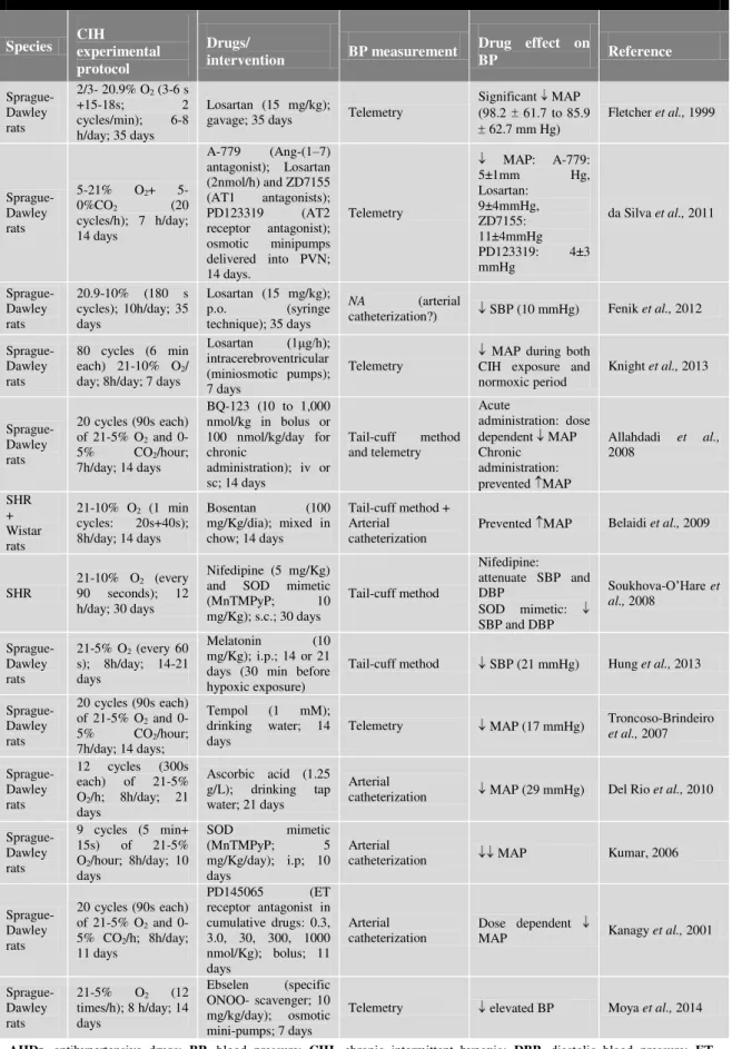
These models typically create moderate to severe oxygen desaturation, thereby mimicking severe forms of OSA and may therefore not be applicable to mild and moderate clinical OSA (Dematteis et al., 2009). CIH models with cycles of FiO2 of 5% or less usually mimic severe forms of OSA in humans and produce maximal changes in BP and heart rate (Dematteis et al., 2008). However, higher FiO2 (8–10%) has been used in rodent models of CIH
14
Table 2. Reports on the effects of CIH on blood pressure
Species Hypoxia cycle, Nadir FiOduration and CO 2,
2 manipulation
(Y/N)
BP
measurement Effect on BP Reference
Sprague-Dawley rats
20 cycles (90 s each) of 21–5% O2 and
0–5% CO2/hour; 7 h/day; 35 days; Yes Tail-cuff method
MAP (25-28 mmHg)
Allahdadi et al., 2005
Sprague-Dawley rats
80 cycles (6 min each) 21–10% O2/ day;
8 h/day; 7 days; No Telemetry MAP (7-10 mmHg) Knight et al., 2011 Sprague-Dawley
rats
5% O2 12 times/h; 8 h/day; 7–21 days; No
Arterial
catheterization No changes in MAP
Iturriaga et al., 2010
Wistar rats 10% O2 for 4 h/day and 21% O2 for 20 h/day; 56 days; Yes (PCO2 < 0.02%)
Arterial catheterization
No differences in
systemic pressure Kalaria et al., 2004 Sprague-Dawley
rats
2/3–20.9% O2 (3–6 s +15-18s; 2
cycles/min); 6–8 h/day; 35 days; No Telemetry MAP (16 mmHg) Tahawi et al., 2001
C57BL/6J mice 21–5% O2 (60 s); 12 h/day; 5 weeks; No
Arterial catheterization
Systemic BP (7.5 mmHg)
Campen et al., 2005
SHR + Wistar rats
21–10% O2 (1 min cycles: 20 s + 40 s); 8 h/day; 14 days; No
Tail-cuff method + Arterial catheterization
Enhanced HT development in SHR + NS in Wistar rats
Belaidi et al., 2009
Sprague-Dawley rats
2/3–20.9% O2 (3-6 s + 12 s; 2
cycles/min); 6–8 h/day; 35 days; No Telemetry MAP (10 mmHg)
Fletcher et al., 2000
LCR and HCR 21–10% O2 (3 min cycles); 8 h/day; 7
days; No Telemetry MAP in both groups Sharpe et al., 2013 Sprague-Dawley
rats
20 cycles (90 s each) of 21–5% O2 and 0–5% CO2/hour; 8 h/day; 11 days; Yes
Arterial
catheterization MAP (30 mmHg) Kanagy et al., 2001 Sprague-Dawley
rats
48 cycles (45 s + 30 s) 20.9–2/6% O2/
hour; 6 h/day; 30 days; No Telemetry MAP (19.3 mmHg) Lai et al., 2006 C57BL/6J mice 21–5.7% O2 (alternating every 6 min);
12 h/day; 90 days; No
Arterial
catheterization MAP (19.8 mmHg) Lin et al., 2007 Sprague-Dawley
rats
21–10% O2 for 5 s every 90 s; 10 h/day;
4 weeks; No Tail BP telemeter MAP (37 mmHg) Liu et al., 2013 SHR 21–10% O2 (alternating every 90 s); 12
h/day; 30 days; No Tail-cuff method
SBP and DBP (NA mmHg)
Soukhova-O'Hare et al., 2008 Sprague-Dawley
rats
21–4/6% O2 (every 60 s); 8 h/day; 5
days/week; 5 weeks; No Tail-cuff method MAP (12 mmHg) Chen et al., 2005
Wistar rats 1 min cycles with 30 s of a 5% FiO2; 8 h/day; 14–21 days; No
Tail-cuff method + Arterial catheterization
Rapidly MAP (NA mmHg)
Totoson et al., 2013
Sprague-Dawley rats
21–6% O2 (9 min cycles); 8 h/day; 14 days; No
Arterial
catheterization MAP (9 mmHg)
Silva and Schreihofer, 2011
Wistar rats 20.8h/day; 10 days; No –6% O2 (9 min cycles: 5 min Nx); 8 Arterial catheterization
MAP (12 mmHg) SBP (9 mmHg) DBP (8 mmHg)
Zoccal et al., 2009
Wistar- Hannover rats
21–10% O2 (2 min + 2 min); 1000– 1600 h; Yes (PCO2 < 0.01%)
Arterial catheterization
No differences in
MAP Perry et al., 2011 Sprague-Dawley
rats
10 cycles (6 min each) of 21–6% O2 and 0–5% CO2/hour; 8 h/day; 28 days; Yes
Telemetry SBP (39 mmHg) DBP (33 mmHg)
Dyavanapalli et al., 2014
C57BL/6J mice 21days/week; 8 h/day; 6 weeks; No –7% O2 (120 s each cycle); 5 Telemetry Significant MAP Schulz et al., 2014 Sprague-Dawley
rats
21–10% O2 (cycle duration: NA); 8
h/day; 7 days; No Telemetry
MAP that persisted
I
NTRO
DU
CTION
15
cycles are often used, reducing the number of cycles/h (Zoccal et al., 2007; Silva and Schreihofer, 2011) of daytime exposure, from 4 h/day (Kalaria et al., 2004), 6 h/day (Lai et al., 2006), 7 h/day (Fletcher et al., 1992a), 8 h/day (Chen et al., 2005; Belaidi et al., 2009; Zoccal et al., 2009; Knight et al., 2011; Silva and Schreihofer, 2011; Dyavanapalli et al., 2014; Schulz et al., 2014), 10 h/day (Liu et al., 2013) to 12 h/day (Lin et al., 2007). The exposure duration of 8 h/day seems to be that on which there is the greatest consensus (see Table 2). The duration of exposure seems to affect the study outcomes more than the hypoxic nadir or the rate of hypoxic cycling (Davis et al., 2013).An advantage of CIH models is they allow exposures that can be extended over months, enabling the investigation of chronic consequences that might occur in humans (Toth and Bhargava, 2013). The number of days necessary to induce an increase in BP seems to be dependent on the CIH paradigm. Some authors suggest that the BP increase triggered by CIH represents a time-dependent effect (Prabhakar et al., 2001; Hui et al., 2003; Dematteis et al., 2008; Zoccal et al., 2009). Moreover, both the time and severity of hypoxia have been shown to play an important role in the cardiovascular response (Li et al., 2007; Perry et al., 2007). It has recently been shown that a period of 14 days is not long enough to induce structural changes in cardiovascular structures, but these are already apparent after 35 days of incubation (Dematteis et al., 2008). Moreover, Iturriaga et al. report that the exposure of rats to CIH for 14 days enhanced the ventilatory response to hypoxia and produced a significant shift in heart rate variability, but these cardiorespiratory alterations occurred without noticeable changes in mean arterial BP until 21 days of CIH exposure (Iturriaga et al., 2010). Whereas some short-term protocols (7–14 days) cause a significant increase in BP (Belaidi et al., 2009; Knight et al., 2011; Silva and Schreihofer, 2011; Bathina et al., 2013), others show an increase in BP that occurs only after long-term exposure (35 days) to CIH (Prabhakar et al., 2001; Chen et al., 2005; Prabhakar et al., 2005; Zoccal et al., 2009) (see Table 2). Finally, most IH paradigms in rodents do not include CO2 supplementation (Lin et al., 2007; Fletcher et al., 2009; Iturriaga et al., 2010; Perry et al., 2011; Bathina et al., 2013). In fact, only some authors have manipulated the CO2 levels (Ooi et al., 2000; Kantores et al., 2006; Dyavanapalli et al., 2014) and fixed the values along the protocol (see Table 2).
Soukhova-16
O’Hare et al., 2008; Totoson et al., 2008; Belaidi et al., 2009), radiotelemetry (Fletcher et al., 2000; Tahawi et al., 2001; Lai et al., 2006; Knight et al., 2011; Bathina et al., 2013; Sharpe et al., 2013; Dyavanapalli et al., 2014; Schulz et al., 2014), and arterial catheterization (Kanagy et al., 2001; Kalaria et al., 2004; Campen et al., 2005; Lin et al., 2007; Belaidi et al., 2009; Zoccal et al., 2009; Iturriaga et al., 2010; Perry et al., 2011; Silva and Schreihofer, 2011; Totoson et al., 2013).
The tail-cuff method is the most frequently used indirect method for monitoring BP (Kurtz et al., 2005). In spite of this, in the context of chronic disease animal models, this method presents several disadvantages, such as the inability to assess the average level of BP throughout the day and night over the course of a study (Kurtz et al., 2005). Moreover, this method induces significant stress, disturbing multiple aspects of the cardiovascular system (Kurtz et al., 2005). Finally, its accuracy is dubious and most tail-cuff methods are not well suited to measuring diastolic pressure (Kurtz et al., 2005).
Regarding radiotelemetry, the availability of this technique has represented an enormous advance in HT research. This method allow BP measurements to be taken in conscious freely moving laboratory animals and also provides the ability to measure BP at any time of the day or night and for extended periods of time (Kurtz et al., 2005). The major limitations of radiotelemetry relate to the expense of acquisition and further maintenance of the telemetric devices, the need for surgical skills and training to perform the introduction of the catheter into the abdominal aorta or femoral artery, and also the risk of infection after the surgical intervention (Kurtz et al., 2005).
Arterial catheterization allows direct measurement of BP with a fluid-filled external catheter. Like telemetry, it is accurate and reliable and permits assessment of BP lability and diurnal variations in BP (Kurtz et al., 2005). This technique can be used effectively for acute studies in anesthetized animals or for long-term, continuous monitoring of arterial pressure in conscious animals (Kurtz et al., 2005). However, the maintenance of catheters in chronic studies can be labor intensive and meticulous care of the catheters is required to avoid infections and to prevent failure during long-term experiments (Kurtz et al., 2005).
I
NTRO
DU
CTION
17
BP increase in rats seems to be predominantly diastolic (Dematteis et al., 2008). The same finding was also reported in the early study of Fletcher et al. (Fletcher et al., 1992a).HUMAN MODELS
The variety of models of IH in healthy human subjects is much less impressive than that observed for animal models of sleep apnea. In terms of the exposure time, these models are usually divided into short-term and chronic (Foster et al., 2007). In short-term IH models, generally the exposure time (20–60 min) and the duration of the hypoxia or voluntary apnea period (30 s) are very limited. The protocols of Cutler et al. and Tamisier et al. are good examples of short-term models (Cutler et al., 2004; Tamisier et al., 2009). In contrast, Foster et al. made use of a chronic model, exposing healthy human volunteers to an hour of IH (5 min hypoxia alternating with 5 min normoxia) daily for two weeks (Foster et al., 2005). As in the animal models of IH, only some studies have controlled the level of CO2 (Foster et al., 2005), whereas others have not (Tamisier et al., 2009). Regardless of the protocol followed, exposing humans to CIH implies careful supervision.
In 2001, Xie et al. exposed nine healthy human subjects during wakefulness to 20 min of isocapnic hypoxia (arterial O2 saturation, 77–87%) and 20 minutes of normoxic hypercapnia (end-tidal PCO2, 15.3–8.6 Torr above eupnea) on two separate days. The subjects breathed through a leak-free nasal mask and the neurocirculatory and ventilatory responses to these two stimuli were further evaluated (Xie et al., 2001). These authors found that hypoxia induced a sympathetic activation that outlasted the chemical stimulus, whereas hypercapnia evoked a short-lived sympathetic activation (Xie et al., 2001). Years later, in a study performed with a larger sample (n=31), Cutler et al. used a model of IH induced by voluntary apnea (30 s of hypoxic apnea every 1 min – simulating an AHI of 60/h – for 20 min) to determine if the cessation of breathing is important in prolonged sympathetic activation (Cutler et al., 2004). This study also included two other groups that were exposed to intermittent hypercapnic hypoxia and to intermittent isocapnic/hypoxia, respectively (Cutler et al., 2004). Their results support the hypothesis that short-term exposure to intermittent hypoxic apnea results in sustained elevation of postganglionic muscle sympathetic nerve activity and that hypoxia is the primary mediator of this response (Cutler et al., 2004). The data reported by Leuenberger et al. one year later were in line with these results. They also found, in a study that enrolled 26 patients, a sustained sympathetic activation and also a transient elevation of BP following 30 min of voluntary end-expiratory apneas primed with a hypoxic gas mixture and lasting for 20 s in each minute (Leuenberger et al., 2005).