Antibiosis of
Trichoderma
spp strains native to northeastern Mexico
against the pathogenic fungus
Macrophomina phaseolina
José Luis Hernández Mendoza, María Isabel Sánchez Pérez,
Juan Manuel González Prieto, Jesús DiCarlo Quiroz Velásquez,
Jesús Gerardo García Olivares, Homar Rene Gill Langarica
Instituto Politécnico Nacional, Centro de Biotecnología Genómica, Tamaulipas, México.
Submitted: September 27, 2012; Approved: May 29, 2015.
Abstract
Sampling of agricultural soils from the Mexican northeastern region was performed to detect Trichodermaspp., genetically characterize it, and assess its potential use as a biologic control agent againstMacrophomina phaseolina.M. phaseolinais a phytopathogen that attacks over 500 species of cultivated plants and causes heavy losses in the regional sorghum crop. Sampling was performed immediately after sorghum or corn harvest in an area that was approximately 170 km from the Mex-ico-USA border. Sixteen isolates were obtained in total. Using colony morphology and sequencing the internal transcribed spacers (ITS) 1 and 4 of 18S rDNA, 14 strains were identified asTrichoderma harzianum, T. koningiopsis and T. virens. Subsequently, their antagonistic activity against M. phaseolinawas evaluatedin vitro, and 11 isolates showed antagonism by competition and stoppedM. phaseolinagrowth. In 4 of these isolates, the antibiosis phenomenon was observed through the for-mation of an intermediate band without growth between colonies. One strain, HTE808, was identi-fied asTrichoderma koningiopsisand grew rapidly; when it came into contact with theM. phaseolina colony, it continued to grow and sporulated until it covered the entire petri dish. Microscopic exami-nation confirmed that it has a high level of hyperparasitism and is thus considered to have high poten-tial for use in the control of this phytopathogen.
Key words:Macrophomina phaseolina, antagonism, hyperparasitism,Trichoderma.
Introduction
Trichoderma spp. is a fungus that can be isolated from soil and agricultural waste and can form opportunistic interactions with and cause hyperparasitism in other fungi (Monte, 2001; Samuels, 2006). This fungus is frequently used in the textile industry, and agricultural reports have in-dicated that it promotes vegetable growth and facilitates the absorption of water, mineral salts, nutrients and the use of carbohydrates. It also produces secondary metabolites such as antibiotics, mycotoxins, and phytotoxins (Lifshitzet al., 1986; Benhamou and Chet, 1996; Mukherjeeet al., 2006; Shoresh and Harman, 2008a, 2008b). These substances are involved in the antagonism phenomenon as a result of ei-ther competition or antibiosis or through hyperparasitism using structures called appresoria, which release enzymes
(glucanases, chitobioses and chitinases) or antibiotics (viri-din, gliotoxin or peptaibols) (Howell et al., 1993; Mukherjeeet al., 2006). These factors encourage the use of Trichoderma spp. in controlling agricultural crop phyto-pathogens (Benitezet al., 2004). The increase in the num-ber of its isolates, its high phenotypic variability, and its similarity withHypocreaandGliocladiumcomplicate the identification ofTrichoderma spp. through classic taxon-omy. With the recent application of nucleic acid-based identification techniques, the number of described Trichoderma species has tripled. Various methods have been used in these studies, including the following se-quences: (ITS) the 5 introns of the protein gene, which en-codes the elongation alpha 1 (tef1) translation factor; the gene that codes for actin (geneact); calmodulin (genecal);
Send correspondence to H.R.G Langarica. Instituto Politécnico Nacional, Centro de Biotecnología Genómica, Boulevard Del Maestro S/N esq. Elías Piña, Narciso Mendoza, Reynosa, 88710 Tamaulipas, México. E-mail: hgill@ipn.mx.
and one partial exon of the geneech42for chitinase (Bailey and Lumsden, 1998; Castleetal., 1998; Hermosa et al., 2000; Luet al., 2004; Veraet al., 2005; Druzhininaet al., 2006; Samuels et al., 2006). Macrophomina phaseolina (Tassi) Goid is a phytopathogenic filamentous fungus that belongs to the anamorphic Ascomycota Botryosphaeriaceaefamily (Crous et al., 2006; Arora et al., 2012) and produces both sclerotia and pycnidia. M. phaseolinais responsible for the plant disease called char-coal rot, which affects both roots and stems.M. phaseolina is widely distributed in tropical regions, specifically in ar-eas that are subjected to water stress, where it infects hun-dreds of different hosts (Songaet al., 1997) and causes se-vere economic losses (Smith and Carvil, 1997). In Mexico, M. phaseolinais found in both the northern region, where the climate is mostly hot and dry, and in the south, where the humidity is high and where the temperatures range from 30 °C in the summer to 5 °C in winter. This microorganism penetrates host tissues through mechanical pressure exerted by the spore germ tube and the sclerotia hyphae and through the dissolution of the cell wall via processes that are mediated by secreted enzymes (Ammonet al., 1974). Therefore, the present study was conducted to isolate native strains ofTrichodermaspp. that are present in northeastern Mexican agricultural soils and to evaluate theirin vitro an-tagonistic capacity againstM. phaseolina.
Materials and Methods
Origin of the strains
Forty-two samples were obtained from approxi-mately 2 kg of soil from the top 15 cm of lands recently cul-tivated with maize. To isolate the fungus, a technique developed in this laboratory was used, which consists of de-positing 150 g of soil into 180-mL Styrofoam cups, adding 10 maize seeds (Pioneer 3025) and soaking with sterile deionized water according to their needs. The cups were covered and observed for 15 days, after which they were checked to detect colonies with the typical morphology of Trichodermaspp. on the seeds and in the soil (Barnett and Hunter, 1998; Samuelset al., 2006). The isolations were performed on potato dextrose agar PDA (Difco, Sparks, MD), and only the colonies with different morphological characteristics were selected for further analysis. The HMP5 strain ofM. phaseolinaused in this study was iso-lated from bean plants (Phaseolus vulgarisL.) in Cotaxtla, México, and was provided by the Plant Biotechnology Lab-oratory of the Genomics Biotechnology Center of the IPN.
Antagonism
The fungi were cultivated on PDA (Difco, Sparks, MD) in petri dishes first individually and then in confronta-tion. The growth rate was measured every 12 h. Antago-nism tests were performed according to the methodology described by Acevedo (1995), which involves placing a
7-mm-diameter agar disc with a seven-day-old M. phaseolinamycelium and an agar disc with a four-day-old Trichodermamycelium at adjacent points of a PDA (Difco, Sparks, MD) petri dish. Each test was replicated three times, and statistical analysis (Mean comparison, Thukey p = 0.05) was performed using the SAS program. Graphs were made using Microsoft Excel XP and GraphPad Prism 4.00.
DNA extraction, ITS amplification and DNA sequencing
were compared with the reference sequences deposited in the NCBI GenBank and in the ISTH TricoOKEY data-bases. TheTrichodermaspecies was determined based on concordance with these databases.
Phylogenetic analysis
After sequencing, the ITS1-18S-ITS4 region of Trichoderma isolates were compiled SeqMan software (DNASTAR Inc., Madison, WI) and edited using the
BioEdit software
(http://www.mbio.ncsu.edu/BioEdit/bioedit.html). Multi-ple alignments were performed using the ClustalW soft-ware (http://www.ebi.ac.uk/clustalw/). Each sequence type was compared by a BLAST search with those available in GenBank (http://www.ncbi.nlm.nih.gov) and those in the ISTH TricoOKEY databases to determine approximate phylogenetic affiliations. Evolutionary distances were cal-culated using the Kimura two-parameter model. The
phylo-genetic tree was constructed with a neighbor-joining method using MEGA 4.0.
Results
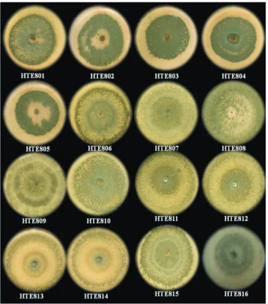
A total of 42 soil samples were obtained and pro-cessed, from which 16 strains were isolated based on their similarity of morphology toTrichodermaspp. (Figure 1). These isolates were coded HTE801 to HTE816. To identify the species, sequence typing was performed, and the se-quences obtained from each isolate were aligned and com-pared with the sequences of the GenBank database from NCBI and theTricoOKEY database from ISTH.
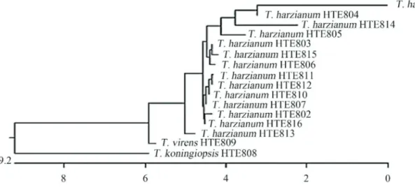
Isolate identification by this method showed that the strains coded from HTE801 to HTE807 and from HTE810 to HTE816 correspond toT. harzianumand that HTE808 corresponds toT. koningiopsisand HTE809 to T. virens (Figure 2). These results show that there is no association
among the geographical origins of the sample, the culti-vated species, growing condition and the isolated fungus.
Growth kinetics of assessed fungi:Trichodermaspp.
The data obtained from the tests performed when cul-tivating each of theTrichodermaspp. strains individually shows that generally between 96 and 120 h after seeding, the mycelium completely covers the petri dish surface (Fig-ure 3).
The growth rate curve is exponential up to 96 h and stabilizes thereafter. HTE801, HTE807 and HTE808 are considered fast growing because they cover the petri dish within 96 h after seeding, whereas a lower growth rate is observed for HTE809, with 132 h required post-inoculation for complete coverage. In most instances where the Trichoderma spp. is grown in the same petri dish asM. phaseolina, the fungus has the same developmental kinet-ics, except with strains such as HTE810, where a phenome-non that has not yet been reported in assays from the confrontation ofTrichodermaspp. with other fungi is ob-served. Therefore, there are several mechanisms involved inTrichoderma antagonism, namely antibiosis, whereby the antagonistic fungus produces antibiotics and competes for nutrients. In the case of mycoparasitism,Trichoderma directly attacks the plant pathogen by excreting lytic en-zymes such as chitinases,b-1, 3 glucanases and proteases (Haranet al., 1996). Because the skeleton of pathogenic fungi cell walls contains chitin, glucans and proteins, en-zymes that hydrolyze these components must be present in a successful antagonist to play a significant role in cell wall lysis of the pathogen. Filamentous fungal cell walls also contain lipids and proteins. It therefore was expected that antagonistic fungi synthesize proteases might act on the host cell wall (Loritoet al., 1994).
Growth kinetics of assessed fungi:M. phaseolina
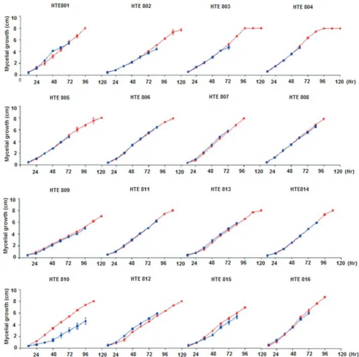
M. phaseolinais a fungus that has an exponential ki-netic growth in PDA (Difco, Sparks, MD) media and cov-ers the entire dish 120 h post-inoculation. The analysis of variance was performed using a Tukey test (p = 0.05), which was performed by comparing the growth data of the M. phaseolinastrain by itself and in antagonism with the different Trichoderma spp. strains, showed that at 60 h post-inoculation, the control and confrontation tests exhibit similar growth, except for strain HTE805, which develops more slowly. At the 72nd h, only nine of the evaluated strains (HTE801, HTE808, HTE813 and HTE816) came into contact with theTrichodermaspp. colonies (Figure 4). In all other treatments, contact was observed at the 84th h post-inoculation.
A statistical analysis of the growth kinetics between theM. phaseolinastrain (Figure 4) growing by itself and in confrontation showed that strains HTE801 and HTE803 of Trichodermaspp. increased the phytopathogen colony de-velopment rate. As observed in Figure 4, for strains HTE805, HTE809 and HTE813, immediately before the contact between the colonies of M. phaseolina and Trichodermaspp., the detected growth rate of the phyto-pathogen decreased, which modified the exponential growth line until it stabilized.
Antagonism tests
From the antagonism tests betweenTrichodermaspp. andM. phaseolina,the following four phenomena were de-tected: antagonism through competition, antibiosis, hyper-parasitism caused byTrichodermaspp. overM. phaseolina and the developmental delay of T. harzianum HTE810 when grown in competition withM. phaseolina. These tests were repeated three times, and the results are statistically significant. Competition is observed when the two fungi grow in the petri dish until their mycelia come into contact,
and theTrichodermaspp. initiates the formation of a bar-rier, which prevents the growth of M. phaseolina. Over time, the barrier consolidates, thereby preventing the ad-vance of the phytopathogen (Figure 5).
Antagonism through competition
All of theT. harzianumandT. virensstrains that were studied underwent antagonism by competition. Both Trichodermaspecies stopped the growth of the fungusM.
phaseolinaat the site of contact by forming a barrier that preventsM. phaseolinadevelopment.
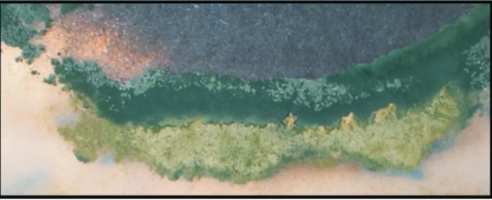
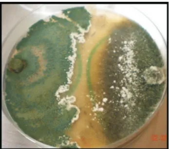
Antibiosis
From the confrontation between theTrichodermaspp HTE815 strain andM. phaseolina, it is observed that at 48 h and without contact between mycelia, a zone of growth in-hibition exists and the culture medium changes color, which could be due to secondary metabolite excretion. As
Figure 4- Mycelial growth (cm) ofM. phaseolinagrowing by itself and in confrontation with differentTrichodermaspp strains. The Percentage Inhibi-tion of Radial Growth ofM. Phaseolina(PIRGM) was determined as follows: PIRGM = [(R1-R2)/R1] x 100 where, R1 = radial growth of the pathogen and R2 = radial growth of the pathogen againstTrichodermaspp; The Percentage of Radial Growth Acceleration of Trichoderma (PRGAT) was deter-mined as follows: PRGAT= [(R2-R1)/R2] x 100 where, R1 = Radial growth ofTrichodermaspp. and R2 = radial growth ofTrichodermaspp. against the pathogen. Red lines indicate the exponential growth ofM. phaseolinaand blue lines indicate the growth ofM. phaseolinaagainstTrichodermaspp.
shown in Figure 6, a narrow band without growth can form between the fungi, even when they are far from physical contact.
Hyperparasitism
Mycoparasitism involves morphological changes, such as coiling and formation of appressorium-like struc-tures, which serve to penetrate the host. Differential antag-onistic activity has been observed for variousTrichoderma spp., which demonstrates semi-specificity in the interaction ofTrichodermawithM. phaseolina. Our results revealed that of the 16 strains tested, onlyT. koningiopsis (HTE808) shows antagonism by hyperparasitism. Microscopically,T. koningiopsis hyphae are rolled into phytopathogenic M. phaseolinahyphae. This is a desirable trait for agricultural purposes because this strain (HTE808) has potential for use in phytopathogen control.
Discussion
The isolates obtained showed the formation of con-centric rings that are typical ofTrichodermaspp. colonies, where the green color of the conidia is interleaved with the white of the mycelium, which is consistent with the charac-teristics previously described for this fungus (Barnett and Hunter, 1998; Druzhinina et al., 2006; Samuels, 2006). However, although the colony morphology serves to iden-tify fungi of this genus, it is insufficient to distinguish the species, which makes it necessary to confirm the species through molecular methods (Ospinaet al., 1999; Druzhi-ninaet al., 2005, 2006). The identification of the isolates in this study yielded three species, of whichT. harzianumwas the most frequently sampled. The presence ofT. harzianum had already been reported in this region of the country (Larralde-Coronaet al., 2008), and it is the species with the
widest distribution (Hermosaet al., 2000; Harmanet al., 2004). Additionally, theTichoOKEY database could only be used to compare the sequences of someT. harzianum,T. virensandT. koningiopsisstrains because, for sequencing the strains HTE801, HTE804, HTE805, HTE810 and HTE814, it was necessary to draw upon the NBCI GenBank database, where the identification was corrobo-rated. The antagonism phenomenon observed in these iso-lates, where the strains ofTrichodermaspp. did not allow the growth of theM. phaseolinacolonies, confirm the com-petition, antibiosis and hyperparasitism phenomena that have been reported for Trichoderma spp. (Hjeljord and Tronsmo, 1998; Hermosaet al., 2000; Benitezet al., 2004; Harman, 2006; Hoitinket al., 2006). Two phenomena that have not been previously reported were also observed; the first involves a strong decrease in the growth of the T. harzianumcolony strain HTE810 when cultivated in con-frontation tests withM. phaseolina(Figure 3). The other phenomenon detected is counter to the previous model of interaction, namely the induction rather than repression, of M. phaseolina colony growth in the presence of the T. harzianumstrain HTE801 and, to a lesser extent, HTE803. In both cases, the acceleration is statistically significant (Tukey, p = 0.05; Figure 3).
Antagonism through antibiosis was detected in strains HTE807, HTE810, HTE815 and HTE809 of Trichodermaspp., which show growth-free spaces between both fungi, as well as a change in coloring in the culture me-dium. These strains correspond to T. harzianum and T. virens,respectively. The growth-free zone is possibly due toTrichodermaspp. producing metabolites such as viridin and its derivatives, which function as antimicrobials (Benitezet al., 2004; Harman, 2006; Hoitinket al., 2006; Vinale et al., 2008). Finally, it was observed in T. harzianum strain HTE810 that the M. phaseolina strain HMP5 has a statistically significantly decreased growth rate, which was the initial impetus of these trials. We have not found reports of this response againstT. harzianum.
Conclusions
This sampling shows thatTrichoderma is a fungus that can be isolated from soil and that there is no relation-ship between the species and the conditions under which agriculture is carried out in this region. Of the detected fungi,T. harzianum is the most frequent species, withT. koningiopsisandT. virens following in frequency. From the antagonism test results, T. harzianum and T. virens show antagonistic activity through space competition, while T. koningiopsis showed a high level of hyper-parasitism onM. phaseolina, demonstrating a strong poten-tial for use as a control agent. TheM. phaseolinaHMP5 strain decreased theT. harzianumHTE810 growth when cultivated in a confrontation test.
Acknowledgments
We would like to thank the Instituto Politecnico Na-cional and Patronato para la Investigacion Fomento y Sa-nidad Vegetal A.C. for financial support for this project. We would also like to thank the Laboratorio de Biotec-nologia Vegetal of CBG-IPN for providing theM. phaseo-linastrain used in this study. Hernandez-Mendoza (JSHM), J.M Gonzalez-Prieto (JMGP) and H.R Gill-Langarica (HRGL) are SNI fellows, and supported by COFAA and EDI-IPN scholarships.
References
Acevedo R (1995) Control biológico de la pudrición blanca del ajo (Sclerotium cepivorum Berk) utilizando el micopara-sitismoTrichodermaspp.San Cristóbal, Venezuela. Under-graduate Dissertation, Universidad Nacional Experimental de Táchira, 109 pp.
Ammon V, Wyllie TD, Brown Jr MF (1974) An ultrastructural in-vestigation of pathological alterations induced by Macrophomina phaseolina(Tassi) Goid in seedlings of soy-bean, [Glycine max(L.) Merrill.] Physiol Plant Pathol 4:1-2. Arora P, Dilbaghi N, Chaudhury A (2012) Opportunistic invasive
fungal pathogenMacrophomina phaseolinaprognosis from immunocompromised humans to potential mitogenic RBL with an exceptional and novel antitumor and cytotoxic ef-fect. Eur J Clin Microbiol Infect Dis 31:101-107.
Bailey BA, Lumsden RD (1998) Direct effects ofTrichoderma and Gliocladium on plant growh and resistance to patho-gens.In: Harman GE, Kubiek CP (eds)Trichoderma and Gliocladium, Vol. 2. Enzymes, Biological Control and Commercial Applications. Taylor and Francis, London, pp. 185-204.
Barnett HL, Hunter BB (1998) Illustrated Genera of Imperfect Fungi. 4ª ed. Aps Press, USA.
Benhamou N, Chet I (1996) Parasitism of sclerotia ofSclerotium rofsiibyTrichoderma harzianum: Ultrastructural and cyto-chemical aspects of the interaction. Phytopathology 86:405-416.
Benitez T, Rincón AM, Limon MCet al.(2004) Biocontrol mech-anisms ofTrichodermastrains. Int Microbiol 7:249-260. Castle A, Speranzini D, Rghel Net al.(1998) Morphological and
molecular identification ofTrichoderma isolates on North American Mushroom farms. Appl Environ Microbiol 64:133-137.
Crous PW, Slippers B, Wingfield MJ (2006) Phylogenetic lin-eages in theBotryosphaeriaceae. Stud Mycol 55:235-253. Druzhinina IS, Kopchinskiy AG, Komon Met al. (2005) An
oligonucleotide barcode for species identification in TrichodermaandHypocrea. Fungal Genet Biol 42:813-828. Druzhinina IS, Kopchinskiy AG, kubicek CP (2006) The first 100
Trichoderma species characterized by molecular data. Mycoscience 47:55-64.
Haran S, Schickler H, Chet I (1996) Molecular mechanisms of lytic enzymes involved in the biocontrol activity of Trichoderma harzianum. Microbiology 142:2321-2331. Harman GE (2006) Overview of mechanisms and uses of
Trichodermaspp. Phytopathology 96:190-194.
Harman GE, Howell CR, Viterbo Aet al.(2004)Trichoderma species opportunistic avirulent plant symbionts. Nat Rev Microbiol 2:43-56.
Hermosa MR, Grondona I, Iturriaga EAet al.(2000) Molecular characterization and identification of biocontrol isolates of Trichodermaspp. Appl Environ Microbiol 66:1890-1898. Hjeljord L, Tronsmo A (1998)TrichodermaandGliocladiumin
biological control: an overview.In: Harman GE, Kubicek CP (eds)Trichoderma andGliocladium, Vol 2. Enzymes, Biological Control and Commercial Applications. Taylor and Francis, London, pp 131-151.
Hoffman CS, Wriston F (1987) A ten-minute DNA preparation from yeast efficiently releases autoctonous plasmids for transformation ofEscherichia coli. Gene 57:267-272. Hoitink HAJ, Maden LV, Dorrance AE (2006) Systemic
resis-tance induced by Trichodermaspp.: Interactions between the host, the pathogen, the biocontrol agent, and soil organic matter quality. Phytopathology 96:196-189
Howell C, Stipanovic R, Lumsden R (1993) Antibiotic production by strains of Gliocladium virensand its relation to bio-control of cotton seedling diseases. Biobio-control Sci Technol 3:435-441.
Larralde-Corona CP, Santiago-Mena MR, Sifuentes-Rincon AM et al.(2008) Biocontrol potential and polyphasic character-ization of novel native Trichoderma strains against Macrophomina phaseolinaisolated from sorghum and com-mon bean. Appl Microbiol Biotechnol 80:167-177. Lifshitz R, Windhan MT, Baker R (1986) Mechanism of
biologi-cal control of preemergence damping-off of pea by seed treatment with Trichoderma spp. Phytopathology 76:720-725.
Lorito M, Hayes CK, Di Pietro Aet al.(1994) Purification, char-acterization and synergistic activity of a glucan 1, 3-b -glu-cosidase and an N-acetylglucosaminidase from T. harzianum. Phytopathology 84:398-405.
Lu BS, Druzhinina IS, Fallah P et al. (2004) Hypocrea/Trichodermaspecies withpachybasium-like co-nidiophores: Teleomorphs for T. minutisporum and T. polysporum and their newly discovered relatives. Mycologia 96:310-342.
Monte E (2001) UnderstandingTrichoderma: Between biotech-nology and microbial ecology. Int Microbiol 4:1-4. Mukherjee M, Horwitz BA, Sherkhane PDet al.(2006) A
second-ary metabolite biosynthesis cluster inTrichoderma virens evidence from analysis of genes underexpressed in a mutant of defective in morphogenesis and antibiotic production. Current Genet 50:193-202.
Ospina GMD, Royse DJ, Chen Xet al.(1999) Molecular phylo-genetic analysis of biological control strains ofTrichoderma harzianumand other biotypes ofTrichodermaspp. associ-ated with Mushroom Green Mold. Phytopathology 89:308-313.
Samuels GJ (2006)Trichoderma: Systematics, the sexual state, and ecology. Phytopathology 96:195-206.
Samuels GJ, Dodd SL, Lu BSet al. (2006) The Trichoderma koningiiaggregate species. Stud Mycol 56:67-133. Shoresh M, Harman GE (2008a) The molecular basis of shoot
Shoresh M, Harman GE (2008b) The relationships between in-creased grow and resistance induced in plants by root colo-nizing microbes. Plant Signal & Behavoir 3:737-739. Smith GS, Carvil ON (1997) Field screening of commercial and
experimental soybean cultivars for their reaction to Macrophomina phaseolina. Plant Dis 81:363-368. Songa W, Hillocks RJ, Mwango’mbe AWet al.(1997) Screening
common bean accessions for resistance to charcoal rot (Macrophomina phaseolina) in Eastern Kenya. Exp Agric 33:459-468.
Vera R, Moreno B, Acevedo Ret al.(2005) Caracterización de aislamientos deTrichodermaspp. por tipo de antagonismo y electroforesis de isoenzimas. Fitopatol Venez 18:2-8. Vinale F, Sivasithamparamb K, Ghisalbertic MLet al.(2008)
Trichoderma plant pathogen interactions. Soil Biol Biochem 40:1-10.
Associate Editor: Carlos Pelleschi Taborda