Joao Daniel da Silva Seixas
Development of CO-Releasing
Molecules for the Treatment
of Inflammatory Diseases
Joao
Daniel da Silva Seixas
Development of CO-Releasing
Molecules for the Treatment
of Inflammatory Diseases
Dissertation presented to obtain a Ph.D. degree (Doutoramento) in Chemistry at the Instituto de Tecnologia Quimica e Biol6gica da Universidade Nova de Lisboa
Academic Supervisor: Prof. Carlos C. Romao
Company Supervisor: Dr. Nuno Arantes e Oliveira
This Thesis was financially supported by Fundac;ao para a Ciencia e Tecnologia, European Social Fund grant number SFRH/BDE/15501/2004 and ALFAMA-Research and Development of Pharmaceutical Drugs Ltd.
FCT
Funda~ao
para a Ciencia e a Tecnologia
MINISTERIO DA CLENCIA, TECNOLOGIA E ENSINO SUPERIOR
~ alfama
First Edition: October 2010 Second Edition: January 2011
Cover Picture:
Design by Sérgio Costa.
Molybdenum hexacarbonyl crystals purified by sublimation. Crystals obtained by the author.
Agradecimentos
Em primeiro lugar queria expressar o meu profundo agradecimento à ALFAMA, por me ter dado a oportunidade de realizar o Doutoramento na empresa e por me ter apoiado e dado todas as condições para o efectuar.
Ao Nuno Arantes-Oliveira, CEO da Alfama, por ter acreditado em mim e me ter proporcionado as condições necessárias para o meu crescimento profissional e científico. Pela sua capacidade muito especial de gerir pessoas e conciliar opiniões diversas, fazendo-nos sentir que somos parte fundamental no desenvolvimento e crescimento da empresa.
Ao meu orientador, o Prof. Carlos Romão por ter sido o meu mentor desde o início da minha carreira cíentifica, por ter podido sempre contar com o seu apoio, incentivo, boa-disposição e pelas largas horas passadas em profícuas discussões cíentificas que não raras vezes ultrapassavam o âmbito da Ciência e se estendiam a outras areas de interesse, bem diversificadas.
À minha “família cíentifica” da Alfama. Aos colegas de laboratório, passados e presentes, Ana Gorgulho, Ana Margarida (Maggie), Ana Rita, Bruno Guerreiro, Catarina Rodrigues, Filipa Cruz, Gonçalo Bernardes, José Fernandes, Lukas Kromer, Marta Norton de Matos, Nuno Penacho, Sandra Rodrigues e Vasco Romão. Obrigado por terem contribuido para um ambiente de trabalho espectacular, com boa disposição e regado com doses q.b de maluqueira! Além de fazerem com que ir trabalhar todos os dias fosse um prazer, esta Tese tem um bocadinho de todos vocês por isso Muito Obrigado!
Ao nosso team do outro lado do Atlântico Leo Otterbein, Sherrie Otterbein, Dave Gallo and Rachel Ruggieri e sem esquecer o Chairman Stan Kugell, thank you all!
Aos cientistas “séniores” da Alfama – Werner Haas e Jan Andersson – que me fizeram olhar para a Biologia com outros olhos e perceber que a Química era muito mais interessante quando aliada a esta.
Ao Walter Blättler, que em pouco tempo conseguiu “revolucionar” a minha maneira de pensar e por ter contribuido para o meu crescimento como cientista. No ITQB, um obrigado especial ao Prof. Miguel Teixeira pela ajuda com os estudos de EPR. Obrigado também aos restantes colegas, em especial aos “vizinhos” do 7º piso, que fazem deste Instituto um sítio onde dá gosto trabalhar. Em especial à São, por nunca ter sido só um serviço (de análise elementar) mas sempre um ponto de apoio, fonte de boa disposição e amizade ao nosso dispor! À Isabel Tomaz (que entretanto viajou do IST para a Faculdade de Ciências), pela grande ajuda e sugestões que deu na interpretação dos resultados de Dicroísmo Circular.
Aos meus amigos de sempre, que sempre souberam compreender e desculpar a minha ausência em períodos mais complicados. As verdadeiras amizades são assim, não precisam de explicações e tudo se resolve com um beijo e um abraço…
E por fim, o mais importante:
À minha namorada, a Sandra, por me ter sempre apoiado e pela paciência e compreensão inesgotável mesmo quando eu deixava a nossa casa de pernas para o ar com artigos e papelada espalhada em todo o lado!
À minha família “verdadeira” por todo o apoio e carinho e a quem devo aquilo que sou: à minha mãe e ao Luis Filipe, à Tia Aurora, Tio Luis, Ricardo e Carolina OBRIGADO por serem o meu suporte para tudo na vida.
Summary
Carbon Monoxide, CO, has been recognized as an endogenously produced, potent
biological mediator involved in many defense mechanisms both in physiologic
and pathologic situations. As a result of these signaling processes, CO possesses a
strong therapeutic potential on a wide range of disease indications. However, the
hardly avoidable safety and practical problems associated with therapeutic
inhalation of toxic CO gas, led to the search for molecules capable of delivering
CO to tissues in a living organism in a controlled and therapeutically useful
manner. From all the areas of the chemical space where such CO-Releasing
Molecules (CO-RMs) can be found, Metal Carbonyls Complexes (MCCs) seems
to be the most versatile.
It is the purpose of this Thesis to provide an extensive characterization of the
behavior of MCCs in the presence of biological molecules and media, in order to
identify the chemical and structural parameters that are more relevant to define
the profile of a therapeutically effective metal-based CO-RM drug.
In pharmacological terms, CO-RMs are prodrugs which carry and appropriately
deliver molecular CO as the therapeutically active principle. This delivery
requires the chemical decomposition of the CO-RM. Such decarbonylation is
triggered by an interaction of the CO-RM with the biological entity or medium.
Ideally, a CO-RM should be targeted to the diseased tissue or organ in order to
minimize its effective dose and prevent toxicity issues resulting from the
indiscriminate, non-specific release of CO in the organism. Therefore, targeting of
the CO-RM is largely dependent on the matching between the chemistry of
decarbonylation and the chemical properties of the tissue or organ where it should
take place, to produce the desired therapeutic effect.
As mentioned above, MCCs were selected as the most versatile molecular
structures that can provide controlled, targeted CO delivery because of their ease
of decarbonylation compared to other organic functionalities. To give a meaning
tune the nature of both the inner coordination sphere of the MCC (wherefrom CO
will have to be released), and the nature of the outer sphere made up by the distal
substituents appended to the ligands which will mediate the interaction between
the CO-RM and the biological environment.
However, the anticipated complexity of such a delivery process, together with the
fact that at the outset of this work extremely little was known about the
interaction between MCCs and biological molecules and systems, required a
stepwise approach towards the understanding of the chemistry of MCCs in such
biological media.
The starting point is the unavoidable need of establishing the “islands of stability”
of MCCs in the aqueous, aerobic solutions that are needed for administration to
living species. It is very loosely assumed by the chemical community that the vast
majority of MCCs, that are metal complexes in low oxidation states, are unstable
in air and water. In practice, very few such complexes have been manipulated in
aqueous, oxic conditions and very little is indeed known about their actual
stability under such conditions. Regardless of mechanistic details to be discussed
later, CO release results from the decarbonylation of the MCC dissolved in the
biological medium. Initially in solution, free molecular CO gas can later escape
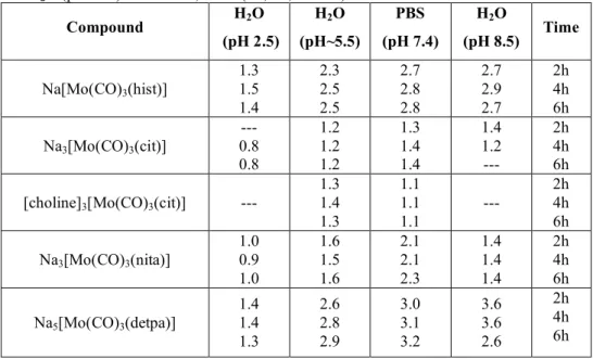
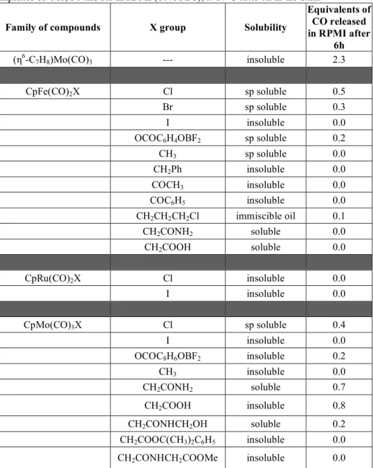
(diffuse) to the headspace of the in vitro experimental setup. Using a simple gas chromatography method, Chapter II describes the screening of the CO release
profile (extension and rate) of a large variety of organometallic carbonyls
dissolved in biologically relevant media/conditions. This mode of CO release is
called spontaneous because it ensues after simple dissolution in the biological medium at 37°C under air and in the dark. Most compounds tested were
octahedral carbonyl complexes and cyclopentadienyl containing carbonyl
complexes all of which with 18-electron configurations. The compounds are
based on Mo0, MoII, MnI, FeII and RuII with classical, N, O, P and S-donor ligands
or alkyl, acyl and halides. All the Mo0 compounds, ionic or neutral, showed a
high rate of CO dissociation unless a strong π acceptor like cyanide or phosphines
at high rates unless an halide (preferentially Cl-) is coordinated to the metal. Most
of the FeII and RuII compounds tested do not release CO in the conditions studied.
The influence of pH and O2 was also evaluated for a set of Mo0 ionic compounds
and it was established that O2 is the main trigger to promote CO release from
these complexes, leading to the formation of hydroxyl radical, which was detected
by ESR. The most remarkable fact was that ca. 2/3 of the compounds tested only
released between 0-9% of their total CO contents. Unexpectedly, the common
idea that MCCs are unstable in air and water is largely wrong. The need to study
the influence of pH derives from the need to see which CO-RMs are able to resist
the acidic environment of the stomach which is a determinant piece of
information for developing orally available CO-RMs. Moreover, acid sensitive
CO release may be particularly useful when driven to happen in the cellular
lyzosome following drug internalization through the cell membrane.
Since the most prominent biological activity of CO is anti-inflammatory, useful
CO-RMs should be targeted to inflamed tissues. Such tissues are rich in reactive
oxygen species (ROS), and therefore, the use of CO-RMs that are specifically
activated by ROS inside cells or in the intercellular space seems a reasonable and
promising strategy for targeting CO-RMs. Presently, very little is known about
the reactivity of organometallic carbonyls under any of these conditions. Chapter
III describes our studies on this type of CO release profile and concludes that: the
concept of oxidatively triggered CO release is valid for the overwhelming
majority of MCCs tested; this oxidation can be tuned by the choice of ancillary
ligands and the nature of the particular ROS (H2O2 or ROOH), and that ROS can
activate air stable MCCs turning them into active CO-RMs.
The specific mechanisms through which CO exerts its biological activity are still
not fully understood but is generally accepted that heme-proteins are the main
targets (if not the only) for the molecule in the organism. In Chapter III the rate of
CO release from several MCCs to Myoglobin (Mb) is evaluated by incubating
them with deoxy-Mb and following the rise of carboxy-myoglobin (CO-Mb). The
although they do not release CO to the headspace of their solutions. This effect
was named COdonation. Since MCCs are rather reduced, electron-rich species, they may engage in electron transfer processes with redox active proteins, namely
Myoglobin or Cytochrome C. A selected group of MCCs was also used to survey
this issue. Most of the compounds tested do not interfere with the redox state of
heme-proteins but almost all the Mo(0) complexes are strongly reducing agents
and this reduction is accompanied by CO transfer to the reduced Myoglobin to
give CO-Mb. This activity extends to Hemoglobin (Hb). Since animals have a
natural protection against high levels of CO, hemoglobin, the efficiency of the CO
delivery process requires that the CO-RM remains intact in the blood circulation.
Otherwise, Hb will scavenge free CO from the blood stream and transport it to the
lungs where it will be exhaled. The first studies of the behavior of MCCs and
CO-RMs in blood that we know of, revealed that most CO-CO-RMs do not release CO to
the erythrocytes, but those that do, are fully decarbonylated within a few minutes.
In fact, CO release from most Mo0 CO-RMs is much, much faster in blood than in
any other medium tested, including ROS rich media. We conclude that kinetic
stability towards CO substitution is necessary to achieve stability of CO-RMs in
blood. Ease of oxidation can be additive to substitutional lability but is not the
key factor to determine stability of transition metal CO-RMs in blood.
At the time this Thesis was initiated, very little work had been published on the
therapeutic activity of CO-RMs. The most promising results were obtained with
two Ruthenium complexes – CORM-2 and CORM-3. Chapter V focus the
attention on the development of a new series of Ru-based CO-RMs of the general
formula Ru(CO)3Cl2L, where L is a ligand with a N-, O-, P- or S- donor atom.
None of the compounds releases CO to the headspace of the GC apparatus but all
are able to transfer CO to deoxy-Mb. Interestingly, it was possible to show that
the amount of CO transferred can be tuned by the nature of L: from 0.2 equiv.
(when L is a strong ligand) to 1 equiv. CO (when L is a weak ligand).
The interaction between drugs and plasma proteins is one of the key aspects that
pharmacokinetic profile are just some of the parameters that may be determined
by the degree of interaction of a given drug with the several proteins. Essentially
nothing was known at the outset of this work with regard to the interaction of
plasma proteins with MCCs. In Chapter VI some exploratory studies using
Circular Dichroism and Absorbance Spectroscopy techniques are presented,
which aim to that determine the interaction between CO-RMs and the plasma
proteins Albumin and Transferrin. Although observed for all complexes with
substitutionally labile ancillary ligands it is not possible to ascertain the molecular
aspects of such interactions from the data available.
From all this work it can be concluded that MCCs are a suitable source of
CO-RMs because they can be tuned to resist attack by water, air and blood and,
therefore, may be equipped with the necessary properties to be administered to a
Resumo
O Monóxido de Carbono, CO, tem sido reconhecido como potente mediador
biológico produzido endogenamente e envolvido em diversos mecanismos de
defesa tanto em situações fisiológicas como patológicas. Como resultado destes
processos de sinalização o CO possui um forte potencial terapêutico numa vasta
gama de doenças. No entanto, é dificil contornar os problemas de segurança e
questões práticas associadas à administração por inalação deste gás tóxico, o que
desencadeou a procura de moléculas capazes de distribuir CO aos tecidos de um
organismo vivo de um modo controlado e terapêutico. De todas as áreas do
espaço químico onde tais Moléculas Libertadoras de CO (ML-CO) podem ser
encontradas, os Complexos Metálicos de Carbonilos (CMCs) parecem ser os mais
versáteis.
O objectivo desta Tese é fornecer uma caracterização extensiva do
comportamento dos CMCs na presença de moléculas e meios biológicos, de modo
a identificar os parâmetros químicos e estruturais mais relevantes para definir o
perfil de uma droga tipo ML-CO com acção terapêutica.
Em termos farmacológicos, as ML-CO são pró-drogas que carregam e distribuem
CO molecular como o princípio activo terapêutico. Esta distribuição requer a
decomposição química da ML-CO. Tal descarbonilação é despoletada por uma
interacção da CO com a entidade ou meio biológico. Idealmente, uma
ML-CO deve ser dirigida para um tecido ou orgão doente de modo a minimizar a sua
dose efectiva e prevenir a toxicidade resultante de uma libertação de CO
indiscriminada e não específica no organismo. Portanto, o direccionamento da
ML-CO depende da semelhança entre as condições em que ocorre a química da
descarbonilação e as propriedades químicas do tecido ou orgão onde deve ocorrer
para produzir o desejado efeito terapêutico.
Como mencionado acima, os CMC foram escolhidos como as estruturas
moleculares mais versáteis que permitem distribuir CO de forma controlada e
outras funcionalidades orgânicas. Para dar um significado à expressão
“distribuição controlada e direccionada de CO” é necessário ajustar e afinar ,tanto
a esfera de coordenação interna do CMC (de onde o CO irá ser libertado) como a
natureza da esfera externa, constituída pelos substituintes nos ligandos que irão
mediar a interacção entre a ML-CO e o ambiente biológico.
No entanto, a complexidade de tal processo de distribuição, juntamente com o
facto de no início deste trabalho, muito pouco ser conhecido sobre a interacção
entre CMCs e moléculas e sistemas biológicos, forçou a que se efectuasse uma
abordagem faseada de modo a entender a química dos CMCs em meio biológico.
O ponto de partida é a inevitável necessidade de identificar “ilhas de estabilidade”
dos CMCs em soluções aquosas, aeróbias necessárias para a administração a seres
vivos. É vagamente assumido pela comunidade química que a grande maioria dos
CMCs, que são complexos metálicos em baixos estados de oxidação, são
instáveis ao ar e na água. Na prática, muito poucos desses complexos foram
manuseados em condições aquosas, oxigenadas e muito pouco se sabe de facto
sobre a sua verdadeira estabilidade nas referidas situações. Independentemente de
detalhes mecanísticos que serão discutidos à frente, a libertação de CO resulta da
descarbonilação do CMC dissolvido no meio biológico. Inicialmente em solução,
o CO gás livre pode escapar (difundir) para a fase gasosa do apparatus experimental in vitro. Usando um método simples de cromatografia gasosa, (CG) o capítulo II descreve o escrutínio dos perfis de libertação de CO (quantidade e
velocidade) de uma grande variedade de complexos de carbonilos
organometálicos dissolvidos em meio/condições biológicas relevantes. Este modo
de libertação de CO é denominado espontâneo pois ocorre após dissolução no meio biológico a 37ºC, ao ar e no escuro. A maioria dos compostos testados são
complexos carbonílicos octaédricos e complexos carbonílicos contendo o ligando
ciclopentadienilo, todos com configuração electrónica de 18 electrões de valência.
Os compostos são baseados em Mo0, MoII, MnI, FeII e RuII com ligandos clássicos com átomos doadores N, O, P e S ou ligandos alquilo, acilo e halogenetos. Todos
dissociação de CO, a não ser que os co-ligandos sejam fortes aceitadores π como
cianeto ou fosfinas. Pelo contrário, os compostos de MnI não libertam CO em grande quantidade a não ser que um halogeneto (preferencialmente Cl-) esteja coordenado ao metal. A maior parte dos compostos de FeII e RuII testados não libertam CO nas condições estudadas.
A influência do pH e O2 também foi avaliada para um grupo de compostos iónicos de Mo0 e foi concluído que o O2 é o principal estímulo para promover libertação de CO destes complexos, levando à formação do radical hidroxilo, que
foi detectado por Ressonância Electrónica Paramagnética.
O facto mais impressionante é que ca. de 2/3 dos compostos testados apenas
libertaram 0-9% do total de CO possível. Inesperadamente, a ideia comum de que
os CMC são instáveis ao ar e água parece estar errada. A necessidade do estudo
da influência do pH advém da necessidade de perceber que ML-CO são capazes
de resistir ao ambiente acídico do estômago, que é uma informação vital para o
desenvolvimento de ML-CO para administração oral. Mais ainda, a libertação de
CO catalizada em meio acído pode ser útil se acontecer no lisossoma após
internalização da droga através da membrana celular.
Uma vez que o efeito biológico mais proeminente do CO é a sua acção
anti-inflamatória, ML-CO úteis devem ser direccionadas para tecidos inflamados. Tais
tecidos são ricos em espécies reactivas de oxigénio (ERO) e portanto o uso de
ML-CO que sejam especificamente activadas por ERO dentro das células ou no
espaço intercelular parece ser uma estratégia razoável e promissora para
direccionamento das ML-CO. Presentemente, muito pouco se sabe acerca da
reactividade dos complexos organometálicos contendo carbonilos, nestas
condições. No Capítulo III descrevem-se os estudos deste tipo de perfil de
libertação de CO e conclui-se que o conceito de libertação de CO por estímulo
oxidativo é válido para a grande maioria dos CMC testados; esta oxidação pode
ser afinada pela escolha dos ligandos auxiliares e pela natureza da ERO (H2O2 ou ROOH), e ainda que as ERO podem activar CMCs estáveis ao ar tornando-os
Os mecanismos específicos pelos quais o CO exerce a sua actividade biológica
ainda não estão totalmente compreendidos, mas é geralmente aceite que as
proteinas hémicas são o principal alvo (se não o único) para o CO no organismo.
No Capítulo IV a taxa de libertação de CO de diversos CMCs para a Mioglobina
(Mb) é avaliada por incubação destes com deoxi-Mb e seguindo a formação de
carboxi-mioglobina (CO-Mb).
Os complexos RuII(CO)3Cl(X)L apresentam as mais rápidas taxas de formação de CO-Mb apesar de não libertarem CO para a fase gasosa das suas soluções. Este
efeito foi denominado de doacção de CO. Uma vez que os CMCs são espécies reduzidas, ricas em electrões, podem participar em processos de transferência
electrónica com proteínas com actividade redox, nomeadamente Mb ou
Citocromo C. Um grupo selecccionado de CMCs foi também usado para abordar
este processo. A maioria dos compostos testados não interfere com o estado redox
das proteinas hémicas mas quase todos os complexos de Mo0 são fortes agentes redutores e esta redução é acompanhada pela transferência de CO para a
Mioglobina reduzida originando CO-Mb. Esta actividade é extensivel à
Hemoglobina. Uma vez que os animais têm uma protecção natural contra níveis
elevados de CO, a hemoglobina, a eficácia do processo de distribuição de CO
necessita que a ML-CO permaneça intacta na circulação sanguínea. De outro
modo a hemoglobina irá capturar o CO livre na corrente sanguínea e transportá-lo
ao pulmões onde será expelido.
Ao efectuar os primeiros estudos conhecidos sobre o comportamento de ML-CO
no sangue, mostrou-se que a maioria das ML-CO não libertam CO para os
eritrócitos mas os que o fazem, são completamente descarbonilados em poucos
minutos. De facto, a libertação de CO da maioria das ML-CO de Mo0 é muito mais rápida no sangue do que em qualquer outro meio testado, incluindo meios
ricos em ERO. Conclui-se que a estabilidade cinética em torno da substituição de
CO é necessária para obter estabilidade das ML-CO no sangue. A oxidação pode
ser um processo adicional à labilidade substitucional, mas não é o factor-chave
À altura do início desta Tese, muito pouco trabalho tinha sido publicado sobre a
actividade terapêutica das ML-CO. Os resultados mais promissores tinham sido
obtidos com 2 complexos de Ruténio – CORM-2 e CORM-3. No Capítulo V,
foca-se a atenção no desenvolvimento de uma nova série de ML-CO baseadas em
Ruténio, de forma geral Ru(CO)3Cl2L em que L é um ligando com átomo doador N-, O-, P- ou S. Nenhum dos compostos liberta CO para a fase gasosa do
apparatus experimental de CG, mas todos são capazes de o transferir para a deoxi-Mb. Curiosamente, foi possível demonstrar que a quantidade de CO
transferido pode ser ajustada pela natureza de L: desde 0.2 equiv. CO (quando L é
um ligando forte) até 1 equiv. CO (quando L é um ligando fraco).
A interacção entre drogas e proteínas do plasma é um aspecto-chave que
determina a sua acção farmacológica. A estabilidade metabólica, tempo de
meia-vida e perfil de fármaco-cinética são apenas alguns dos parâmetros que podem ser
modelados pelo grau de interacção de uma determinada droga com diversas
proteínas. No início deste trabalho muito pouco era conhecido a respeito da
interacção de proteínas do plasma com CMCs. No capítulo VI, são apresentados
alguns estudos exploratórios recorrendo a técnicas de Dicroísmo Circular e
Espectroscopia de Absorção que pretendem determinar a interacção entre ML-CO
e as proteínas do plasma Albumina e Transferrina. Apesar de tais interacções
terem sido observadas para todos os complexos com co-ligandos lábeis, não foi
possível determinar os aspectos moleculares envolvidos a partir dos dados
disponíveis.
De todo este trabalho pode-se concluir que os CMCs são uma fonte viável de
ML-CO pois podem ser modificadas para resistir a àgua, ar e sangue e portanto,
capazes de serem munidos das propriedades necessárias para serem administrados
List of Abbreviations:
Ac acetyl
acac acetylacetonate
ADME absorption, distribution, metabolism and excretion
AIP Alfama’s Intelectual Property
AUC area under curve
bipy bipyridyl
BMPO 5-tert-butoxycarbonyl 5-methyl-1-pyrroline N-oxide
bpa N-(p-carboxy-benzyl)bis(2-picolyl)amine
Bu butyl
BSA Bovine Serum Albumin
bw body weight
CD Circular Dichroism
CDs cyclodextrins
choline N,N,N – trimethylammonium cation
CHT 1,3,5-cycloheptatriene
cit Citrate
CMT cyclopentadienyl manganese tricarbonyl
CO carbon monoxide
CO-Hb carboxy-hemoglobin
CO-RM CO releasing molecule
Cp cyclopentadienyl
Cp* pentamethylcyclopentadienyl
Cp’ general abbreviation for a substituted Cp ring
Cy cyclohexyl
Cyst Cysteine
Cyt Cytochrome
DAB 1,4 – diazabutadiene
DAPTA 3,7-diacetyl-1,3,7-triaza-5-phosphabicyclo[3.3.1]nonane
DMSO dimethylsulfoxide
E.A. elemental analysis
Equiv. equivalent
ESI-MS electrospray ionization Mass Spectrometry
ESR Electron Spin Resonance (Spectroscopy)
Et ethyl
fac facial
FBS Fetal Bovine Serum
FID Flame Ionization Detector
GABA γ-aminobutyric acid
Gal-S-Me methyl-β-D-thiogalactoside
GC Gas Chromatography
GRM Gas Release Machine
GSH Glutathione
H2O2 hydrogen peroxide
Hb Hemoglobin
hist Histidinate
HO Heme-Oxygenase
HOMO Highest Occupied Molecular Orbital
HPLC High Performance Liquid Chromatography
HRP Horseradish Peroxidase
h-Tf Human apo-Transferrin
ICP-AES Inductively coupled plasma atomic emission
spectroscopy
Im Imidazole
Ind Indazole
IR Infrared
Kg kilogram
LD50 lethal dose 50%
LUMO Lowest Unoccupied Molecular Orbital
MCC Metal Carbonyl Complex
Me methyl
mg milligram
MMT Methylcyclopentadienyl manganese tricarbonyl
morph morpholine
MTD Maximum Tolerated Dose
MTO methyltrioxorhenium
NAC N-acetyl cysteine
NAD(P)H nicotinamide adenine dinucleotide (phosphate)
nita nitrilotriacetate
NMR Nuclear Magnetic Ressonance Spectroscopy
PBS phosphate buffered saline
PDA Photodiode Array
PEG polyethyleneglycol
Ph phenyl
pip piperazine
PPB plasma protein binding
ppm parts per million
PTA 1,3,5 - Triaza-7-phosphaadamantane
PTFE Polytetrafluoroethylene
p-tolyl 3–methyl toluene
py pyridine
RBC red blood cell
RCP Reducing Compound Photometer
ROS reactive oxygen species
RPMI RPMI-1630 supplemented with 10% FBS: culture
medium rich in aminoacids, inorganic salts,
vitamins and proteins
RT retention time
sp sparingly
TBHP tert-butylhydroperoxide
TCD Thermal Conductivity Detector
TRIMEB 2,3,6-tri-O-methyl-β-cyclodextrin
TTCN 1,4,7-Trithiocyclononane
Table of Contents
Agradecimentos
iSummary
iiiResumo
viiiList of abbreviations
xiiiChapter I. Introduction
1. The biological activity of Carbon Monoxide
12. CO and Heme Proteins
53. Plasma Binding Proteins and Interaction with Drugs
84. The Chemistry of CO release from Metal Carbonyl Complexes
115. The CO-RMs in the literature
256. References
26Chapter II. Evaluating spontaneous CO release from Metal
Carbonyls by Gas Chromatography
1. Summary
31
2. Introduction
323. Experimental Section
333.1 Methodology 33
Gas Chromatography 35
High-Pressure Liquid Chromatography 36
Electron-Spin Resonance 37
3.2 Technical Details 37
General Considerations 37
Synthetic Work 38
4. Results and Discussion
434.1 CO release profiles of octahedral [M(CO)xL6-x]± complexes 47
4.2 CO release profiles of the polyene and polyenyl carbonyl
complexes 65
4.3 Toxicity of cyclopentadienyl metal carbonyl complexes 72 4.4 CO release profile from cyclodextrin-encapsulated complexes 73 4.5 The influence of O2 in the CO release from metal carbonyl
complexes 76
5. Final Remarks and Conclusions
816. References
83Chapter III. Evaluating ROS induced CO release from Metal
Carbonyls by Gas Chromatography
1. Summary
872. Introduction
872.1 Introductory notes on reactive oxygen species and biologic
oxidative processes 87
2.2 Introductory notes on oxidative decarbonylation of metal
carbonyl complexes 91
3. Experimental Section
923.1 Methodology 92
Gas Chromatography 93
Reactions of CpMo(CO)3CH2CONH2 with [Cp2Fe]BF4, AgNO3
and AgBF4 (IR and GC) 93
3.2 Technical Details 94
Synthetic Work 94
4. Results and Discussion
94
4.1 The Manganese compounds 96
4.2 The Molybdenum compounds 99
4.3 The Iron compounds 110
4.4 Complementary observations relevant for the understanding
of ROS induced CO release processes 116
4.4.1 Methane formation in TBHP assays 116
4.4.2 Oxygen formation in TBHP and H2O2 assays 118
4.5.Oxidative Decarbonylation of CpMo(CO)3(CH2CONH2): a
case study 120
4.5.1 Reaction with 1-electron oxidants 121
4.5.2 Reaction with peroxides 125
5. Final Remarks and Conclusions
1326. References
1357. Acknowledgments
138Chapter IV. Study of reactivity between Metal Carbonyl
Complexes and Heme Proteins
1. Summary
139
2. Introduction
139
3. Experimental Section
1413.1 Methodology 141
CO release determination with the Mb assay 141 UV-VIS absorbance spectrophotometric measurement of the
reaction between Metal Carbonyl Complexes and Cytochrome
C 142
UV-VIS absorbance spectrophotometric measurement of the
reaction between Metal Carbonyl Complexes and Myoglobin 143
3.2 Technical Details 144
Synthetic Work 144
4. Results and Discussion
1444.1 Evaluating CO release through CO transfer from Metal
Carbonyls to Myoglobin 144
4.1.1 Discussion 157
4.1.2 Comparing CO release profiles by the Mb method 161 4.2. Redox interaction between Metal Carbonyl Complexes,
Cytochrome C and Myoglobin 163
4.2.1 Results 163
Cytochrome C 163
Myoglobin 166
4.2.2 Discussion 168
4.3 Behavior of CORMs in blood 169
4.3.1 Results and discussion 170
5. Final Remarks and Conclusions
175
6. References
178Chapter V. Chemical and Biological studies with
Ruthenium-based CO-RMs
1. Summary
1812. Introduction
1813. Experimental Section
1853.1 Methodology 185
Gas Chromatography 185
Quantification of CO release through the Mb assay 185 High-Pressure Liquid Chromatography 185
Horseradish Peroxidase Assay 185
3.2 Technical Details 186
General Considerations 186
Synthetic Work 187
4. Results and Discussion
1934.1 Identification of the species present in DMSO solutions of
[Ru(CO)3Cl2]2 (CORM-2) 193
4.1.1 Chemical and spectroscopical studies 193
a. Reactivity in DMSO 200
b. Summary 206
4.1.2 CO release profiles of CORM-3, CORM-2 and its
derivatives in DMSO 209
a. CO release to the headspace in aqueous media 209
b. CO transfer to deoxy-Myoglobin 209
4.1.3 Final Remarks 211
4.2 Development of new Ruthenium tricarbonyl compounds
4.2.1 Synthesis and Characterization 212 4.2.2 CO release profiles of the new [Ru(CO)3Cl2 L] complexes 217 a. CO release to the headspace in aqueous media 217
b. CO transfer to deoxy-Myoglobin 222 c. Deactivation of the CO transfer capacity 224
4.2.3 Conclusions 230
4.3 Anti-oxidant activity of RuII carbonyl complexes 231
5. Final Remarks and Conclusions
2406. References
2417. Acknowledgments
245Chapter VI. Interaction of Metal Carbonyl Complexes with
Serum Proteins
1. Summary
2472. Introduction
247
3. Experimental Section
2473.1 Methodological remarks on the Circular Dichroism and UV/VIS studies with Bovine Serum Albumin and Human Apo
-Transferrin 250
Circular Dichroism 252
UV/Vis absorbance spectrophotometry 252
3.2 Technical Details 253
Synthetic Work 253
4. Results and Discussion
253
4.1 Interaction with Bovine Serum Albumin 253
4.1.1 Study of the interaction of [Et4N][Mo(CO)5Br] and BSA 255
Circular Dichroism 255
UV-VIS absorbance 257
4.1.2 Study of the interaction of CpMo(CO)3CH2CONH2 and
BSA 259
Circular Dichroism 259
UV-VIS absorbance 261
4.1.3 Study of the interaction of Ru(CO)3Cl(glycinate) and
BSA
262
Circular Dichroism 262
UV-VIS absorbance 263
4.1.4 Study of the interaction of Na3[Mo(CO)3(cit)] and BSA 264
Circular Dichroism 264
UV-VIS absorbance 268
4.2 Interaction with Human Apo-Transferrin 270 4.2.1 Study of the interaction of [Et4N][Mo(CO)5Br] and h-Tf 271
Circular Dichroism 271
UV-VIS absorbance 272
4.2.2 Study of the interaction of CpMo(CO)3CH2CONH2 and
h-Tf
Circular Dichroism 273
UV-VIS absorbance 274
4.2.3 Study of the interaction of Ru(CO)3Cl(glycinate) and h-Tf 275
Circular Dichroism 275
UV-VIS absorbance 277
4.2.4 Study of the interaction of Na3[Mo(CO)3(cit)] and h-Tf 278
Circular Dichroism 278
UV-VIS absorbance 281
4.3 Discussion 282
5. Final Remarks and Conclusion
2876. References
2887. Acknowledgments
290Chapter VII. Conclusions and prospects
291Annex I
Chapter I: Introduction
1. The biological activity of Carbon Monoxide
Carbon monoxide (CO) is a colorless, odorless and tasteless gas that has always
been tagged as a noxious and dangerous molecule. It is a product of incomplete
combustion of organic compounds and the most common sources are car exhaust
fumes, smoke from fires, gas-powered engines and wood-burning fireplaces.[1] Historically, this poisonous reputation was built upon countless episodes of CO
exposure with fatal outcome; since CO binds to hemoglobin with more affinity
than O2 it drastically decreases the oxygen transport capacity of the
cardiovascular system. Moreover, when CO is bound to hemoglobin the
oxy-hemoglobin dissociation curve changes. If CO is bound to two heme sites in the
hemoglobin molecule, oxygen affinity increases and therefore it is no longer
available to be released in the tissues.[2]
Nevertheless, the binding between CO and hemoglobin is reversible and CO can
be replaced with oxygen applying common methodology like substitution
hyperbaric chambers[3] since the ratio between oxy-hemoglobin and CO-Hb is determined by the relative partial pressure of CO and O2. This is not a
straightforward process and the same partial pressures of CO and O2 give
different results in different species and individuals, as demonstrated in the
extensive work of Haldane and co-workers.[4] Thus, the toxic effects are largely
dependent on CO concentration and duration of the exposure.
The current methodology to assess CO levels in the body is the measurement of
the percentage of carboxy-hemoglobin (CO-Hb) in the blood. Table 1 lists
examples of several CO concentrations, the calculated CO-Hb levels and related
Table 1: Ambient CO concentration, % CO-Hb and human health symptoms associated (adapted from references [5] and [6]).
%CO-Hb in blood Concentration/
ppm Symptoms
2
10
20
30
40-40
60-80
10
70
120
220
350-520
1950
10 000
Asymptomatic
Exhaustion in healthy people and headaches
Dizziness, nausea, dyspnea
Visual disturbance
Confusion, syncope, seizures and coma
Cardiopulmonary dysfunction and death
Lethal in minutes
Human levels of CO-Hb differ depending on the external source they have been
exposed to, and, obviously, different individuals may experience different
symptoms. For instance, regular smokers present CO-Hb baseline levels
significantly higher (up to 9%) than non-smokers (typically below 2%).[7]
Despite being common sense that CO is a harmful poison for the organism[8, 9] several scientific discoveries forced scientists to look at this molecule from a
different angle. It was already in the middle of the 20th century that Sjöstrand made a discovery that was one of the most important milestones in CO history,
when he reported the Endogenous production of carbon monoxide in man under normal and pathological conditions.[10]
Sjöstrand, and later Coburn[11, 12] found that CO is endogenously generated in the
body through the degradation of senescent red blood cells. It was only 30 years
after Sjöstrand’s original paper that Tenhunen and co-workers[13-15] characterized the enzyme responsible for breaking down the hemoglobin with concomitant CO
release – Heme Oxygenase (HO).
The heme degradation is a concerted action catalyzed by HO with
NADPH-cytochrome c (P-450) reductase and oxygen. During the heme degradative
biliverdin-IX α (blue-green pigment) that is rapidly converted into bilirubin-IX α
(yellow pigment) by biliverdin reductase.[16-19]
This process is usually observed in bruises. During the injury, a dark red/purple is
observed, which arises from deoxygenated hemoglobin released from lysed red
blood cells. The released heme is then oxidized with formation of biliverdin,
which is responsible for a green tinge. Later, biliverdin is reduced into bilirubin
and a yellow coloration typical of this molecule is observed.
What at the first sight could look as a pure catabolic process results in the
production of biological active elements.[20-22] Iron regulates several genes
expression, including HO as well as transferrin receptors, ferritin (Fe
homeostasis) and NO synthase. Bilirubin and biliverdin are antioxidants and are
essential for the maintenance of the redox imbalance inflicted on cells and tissues
by oxidative and nitrosative stress.[23]
Therefore, HO has both anabolic and catabolic functions inside the cell. In its
catabolic functions it reduces the levels of intracellular heme concentration and
hemeprotein and therefore inactivates the most potent catalyst for free radicals
formation, the heme. In its anabolic functions HO produces the bilic pigments,
CO and iron, all biological active elements.
So far three isoforms of HO were identified but only two were studied in detail[24]
(reviewed in reference [25]). HO-2 is constitutively expressed in tissues such as
brain, liver and endothelium and regulates the basal levels of free heme, acting as
a neurotransmitter and regulator of vascular tone. HO-1 is an inducible isoform
that represents a pivotal defense against stressful stimuli like
ischemia-reperfusion damage, endotoxic shock, UVA radiations and other stressful insults
derived from oxidative and nitrosative stress.
HO-3 is so far a very poorly studied isoform, whose functions are not clearly
understood.[19] It has been found in the brain, heart, kidney, liver and spleen of rats but it does not possess any heme degrading activity.
Characterization of constitutive HO-2 and inducible HO-1 isoforms of heme
enzymes revealed the importance of this pathway in the physiological degradation
of heme and this production, under some pathophysiological conditions could
even be enhanced.[26-28]
Indeed, oxidation of heme by HO accounts for ca. 86% of the total CO production
in the body, ca.79% directly from RBC breakdown[27, 29] and the remaining 21%
from the turnover of other hemeproteins like myoglobin, cytochromes and
others.[14] The remaining CO comes from other non-heme sources like lipid peroxidation (e.g. NADPH-dependent oxidation of microssomal lipids[30])
photo-oxidation, Fe3+-ascorbate catalyzed oxidation of microssomes,[31] bacterial activity[32, 33] and xenobiotics.
For instance, methylene chloride which is often inhaled from paint strippers,
mixtures used for degreasing machinery and pesticide products, is converted to
CO by hepatic enzymes in the liver.[34]
Under normal physiological conditions, the rate of CO production in human body
is 16.4 µmol.h-1[35] but these basal levels may increase under stress or pathological conditions. Activation of HO-1 can lead to a local increase of CO in a determined
tissue and this is evidenced by the increase levels of CO in exhaled breath of sick
individuals.[36-41]
Taken altogether these results challenged CO toxic reputation and the scientific
community became more interested in its physiological profile rather than the
widely described toxic effects.
The discovery that CO activates soluble guanylyl cyclase (sGC)[42] was one of the first evidences that this molecule could act as an intracellular messenger.[43, 44]
Currently, a wide range of biological effects attributed to CO are already well
documented showing anti-inflammatory, anti-apoptotic and anti-proliferative
properties both in vitro and in vivo (reviewed in references [5, 20, 45, 46]). Also, the protective effects of exogenously applied CO have been demonstrated in
several models such as organ ischemia/reperfusion injury, hepatitis, vascular
injury, inflammatory lung disease and organ transplantation (for reviews see
levels between 10-250 ppm led the pharmaceutical company IKARIA® to test CO gas as a therapeutical molecule in on-going clinical trials in humans
(http://clinicaltrials.gov/ct2/results?term=carbon+monoxide).
With CO inhalation therapy stable CO-Hb levels (typically 12-20%) are
maintained during a desired period of time (usually 1h). However, the need for a
controlled hospital environment and appropriate machinery favor the quest for
better alternatives. CO-releasing molecules emerge as a suitable alternative to
deliver CO to tissues in a living organism in a controlled and therapeutically
useful manner, without increasing CO-Hb levels to dangerous limits.
2. CO and Heme Proteins
Endogenously produced CO as well as exogenously administered CO (as a gas)
both depend on hemoglobin transportation to reach the cellular targets. CO binds
hemoglobin in the heme center[48, 49] and is transported across the body until it
reaches the tissues. The affinity for heme is the driving force that leads to
formation of CO complexes with other hemeproteins. Plenty of evidence exists
for the formation of such adducts with myoglobin, soluble guanylyl cyclase,
inducible nitric oxide synthase, cytochrome P-450, cytochrome-c oxidase,
NADPH oxidase and the heme-oxygenase complex[43, 50-56] which leads hemeproteins to be considered the most likely (if not the only) target for CO in
biology.[47, 57] As a consequence of heme binding, intracellular CO potentially
influences the activity of these hemeproteins and these changes in activity may
result either in activation or inhibition of the metabolic functions. Indeed, the
most well-known and documented effect is undoubtedly CO “poisoning” that
arises from CO competition against O2 for binding to the four heme iron centers
of hemoglobin and producing carboxy-hemoglobin the formation of which is
favored by the 245 times higher affinity of CO over O2.
still under debate. For instance, coordination of CO to cyt c oxidase may prevent oxygen activation giving protection to reperfusion injury.
Myoglobin is a heme protein mainly found in muscle tissues whose primary
function is to buffer the oxygen concentration in these tissues.[58] The protein is a monomer, containing a single five-coordinate heme center (iron protoporphyrin
IX) that reversibly binds O2, without auto-oxidation. It stores O2 in muscles and
allows the consumption of the stored O2 in aerobic metabolism, providing “extra
O2” to small metabolic bursts, which wouldn’t be possible if only the circulating
O2 in hemoglobin was available. The crystal structure of Myoglobin was the first
protein structure to be revealed at atomic level. It was determined in the late
1950s by Kendrew and co-workers[59, 60] and since then new resolution structures were published in several ligation states.[61, 62]
Like other iron-based heme proteins, Mb also binds a broad range of ligands
depending on the redox state of the metal. The ferric form reacts readily with
water and anions like F, Cl, CN, N3 and OH. The ferrous form, in addition to
dioxygen also binds CO, NO, several alkyl isocyanides and nitroso aromatic
compounds.[63]
The heme site is “lodged” between two helices and the spatial design of the
environment (steric effect) as well as the pocket polarity determines several
important functions such as an effective discrimination of CO over O2 and the
prevention of irreversible aerobic auto-oxidation. This prevention is achieved by
enhancing stability of the oxy-heme form, favored by globins, against heterolytic
cleavage of the Fe-O bond, formed in peroxo-bridged hemes that would result in
the release of superoxide.
These complicated heme ligand affinities are regulated by polar, hydrophobic and
steric interactions between heme, ligands and distal aminoacid residues. The high
number of variables involved makes the kinetics of ligand binding a complex
process that involves multiple stereochemical constrained steps, that include (O2
is presented as an example): 1) displacement of a non-coordinated water molecule
non-covalent binding of O2 in the distal pocket and hydrophobic internal pockets; 4)
covalent bond formation through the in-plane movement of the iron atom forming
the hexacoordinate species; 5) relaxation of the protein and formation of a new
electrostatic interaction with the distal pocket, typically hydrogen bonding.[56]
Despite sharing the same general “architectural displacement”, some differences
are observed between O2 and CO binding forms. Similar iron-to-ligand bond
lengths are observed (around 1.8 Å) but CO binds normal to the heme plane while
the O2 adduct is intrinsically bent. This results in a higher localization of the
negative charge at the distal O of oxy-heme (often viewed as
ferriheme-superoxide adduct) contrary to the little charge separation in the heme-CO adduct.
Also the distal groups, through steric, electrostatic and H-bonding effects
contribute to the CO vs O2 discrimination.
These binding differences and conformational changes are reflected in the
spectroscopic properties of the different states that strongly vary according to the
ligand.
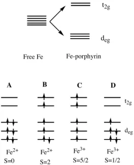
In the ferrous state the iron has six 3d electrons whereas in the ferric ion it has
five. The tetrapyrrole ring of protoporphyrin-IX divides the d orbitals into three
deg (dxy, dyz and dzx) and two t2g (dx2-y2, dz2) degenerated orbitals. In the ferrous
liganded (O2 and CO) forms, the energy difference between deg and t2g orbitals is
high so the three orbitals are filled with six electrons of opposite spin giving a low
spin state of S=0.
In ferrous deoxy state, the energy difference between the two set of orbitals is low
so that all the orbitals are filled with electrons according with Pauli’s exclusion
principle and Hund’s rule. In result, a high-spin species with S=2 ( ½ + ½ + ½ +
½ ) is obtained.
Ferric met-Mb has five 3d electrons and the energy difference between the two
Figure 1: Electronic configuration and spin state of the iron metal in Hb: Top: Degenerescence of the 3d electrons in free Fe in the presence of the ligand field of the porphyrin ring into three deg (dxy, dyz, dzx) and two t2g (dx2-y2, dz2) orbitals. Bottom: A- Fe atom in ferrous low spin state (S=0), e.g. Oxygen and carbonmonoxy Hb; B- Fe atom in ferrous high spin state (S=2), e.g. deoxy Hb; C- Fe atom in ferric high spin state (S=5/2), e.g. fluoromet and aquomet Hb; D- Fe atom in ferric low spin state (S=1/2), e.g. cianomet and azidemet Hb.
These electronic properties are reflected in different spectroscopic profiles which
allow the determination of different ligation states between “free” deoxygenated
heme and CO adducts.
3. Plasma binding proteins and interactions with
drugs
Most pharmaceutical drugs depend on bloodstream to reach the diseased tissues
or organs. Different delivery routes like oral dosing (per os), intraperitoneal (IP),
intravenous (IV), intramuscular (IM), subcutaneous (SC) or even transdermal rely
on blood to transfer the drug to targets.
Free Fe Fe-porphyrin deg
t2g
t2g
deg
Fe3+ Fe2+ Fe2+
S=5/2
S=0 S=2
C
A B
Fe3+ S=1/2
When drugs are administered to living systems there are several barriers that
reduce the amount of dosed compound that reaches the target. The barriers
encountered are diverse and include physicochemical and biochemical processes
such as cell membranes, metabolic enzymes, local pH variations, efflux
transporters and binding blood constituents.[64] The sequence of barrier events found by the molecules strongly depends on the route of administration and when
such barriers are met the drugs’ behavior is determined by the physicochemical
properties of the molecules. Their binding and reactivity with specific enzymes,
binding to transporters and plasma proteins as well as non-specific binding to
macromolecules, affects their absorption, distribution, metabolism and excretion
(ADME).[65-67] In the bloodstream, three barriers affect free drug availability:
enzymatic hydrolysis, red blood cell binding and plasma protein binding.
The hydrolytic enzymes in bloodstream include diverse enzymes like lipase, acid
and basic phosphatases, aldolase, dehydropeptidase, cholinesterase,
glucuronidase, phenol phosphatase and dehydrogenase. The concentration of such
enzymes and substrate specificity vary with factors like age, gender, race or
disease state.
Red blood cell binding is another factor affecting free unchanged drug
concentration since the cell membrane may bind drug molecules through
lipophilic interactions. Nevertheless, the ratio of RBC binding observed is very
small compared with plasma protein binding (PPB). Indeed, the major cause
leading to a decrease of free drug concentration in solution is PPB.
Approximately 6% to 8% of the plasma content are proteins, being the vast
majority transporters for natural occurring compounds. Their concentration in
plasma may vary with age or even disease states[68-70] but a more relevant fact is that drugs are also able to reversibly bind to these proteins. The affinity of
binding determines the ratio of bound and unbound drug in solution and a
stronger affinity will obviously decrease the amount of free drug in circulation.
Unless very high drug concentrations are used, the total protein binding is a
range due to the high capacity of drug binding in plasma which is never fully
saturated. The high binding capacity is provided by three types of binding
proteins: albumin, α1-acid glycoprotein and lipoproteins.
Human serum albumin is the main carrier in the human organism and possesses
six binding sites with high specificity which may carry different products such as
fatty acids, bilirubin or drugs like warfarin and ibuprofen.[71]
The concentration of α1-acid glycoprotein in blood (15 µM) is much lower than
that of Human Serum Albumin (HSA) (500-800 µM). It has only one binding site
and binds to basic drugs such steroids (e.g. disopyramide and lignocaine)
essentially by non-specific hydrophobic interactions.[72, 73]
Lipoproteins (very-high-density lipoprotein [VHDL], high-density lipoprotein
[HDL], low-density lipoprotein [LDL] and very-low-density lipoprotein [VLDL])
are particles constituted by non-polar lipids surrounded by more polar lipids and
protein that work as natural transport for cholesterol and triacylglycerols. Drug
binding to these proteins includes non-specific lipophilic interactions.
For most drugs, PPB has different effects some of which even contradictory.
However, the interpretation of these effects for CO-RMs is not so straightforward.
For most drugs binding prevents that the desired pharmaceutical effect is
maximized since in order to reach a therapeutic concentration in the tissues, the
drug must be unbound to permeate the membranes. In the case of CO-RMs, CO –
the active principle – is able to permeate the membranes so binding to proteins
may not have a deleterious effect, as the binding interaction itself may lead to
disruption of metal-carbonyl bonds and favor the release of the therapeutic agent.
On the other hand, half-life in circulation may also be increased since bound
drugs do not permeate into the liver and kidney for clearance. This will depend on
4. The Chemistry of CO release from Metal Carbonyl
Complexes
Metal Carbonyl Complexes (MCCs) are coordination compounds of the general
type LxM(CO)y where M is a transition metal in various oxidation states, Lx
represents a set of ancillary ligands and y ≥1.
The vast majority of the MCCs have an 18-electron count in the valence shell of
the central metal atom. This is called the Effective Atomic Number (EAN) or
18-electron rule, first stated by Nevil Sidgwick in 1927.[74] In this electronic situation, the bonding between ligands and metal is maximized by the
involvement of all the s, p and d orbitals of the metal. The 18 electrons
correspond to the full occupancy of the nine bonding molecular orbitals generated
by the nine metal valence orbitals (one ns orbital; three np orbitals and five (n-1)d
orbitals) and the appropriate ligand based orbitals. The nine corresponding
anti-bonding orbitals remain empty. Exceeding the 18-electron count requires the
occupation of an empty anti-bonding orbital which in metal carbonyl complexes
lies usually at rather high energies, that is, they have high HOMO-LUMO gaps.
Exceptions to the 18-electron rule are usually found at the earlier (Groups 3-5)
and later groups (Groups 9-10) of the transition metal series in the periodic table,
and in some particular cases like the [Fe(II)(porphyrin)] complexes of which
hemes are an example. However, the exceedingly high reactivity of low-valent
CO derivatives of group 3-5 metals on the one hand, and the high toxicity
associated with the sulphophilic metals of groups 9-10 on the other hand,
preclude their pharmacological use.
The carbonyl group is one the most studied ligands in organometallic transition
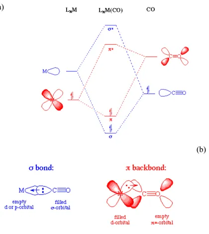
metal chemistry due to its particular bonding mode.
The bonding of CO to a metal consists of two contributions: a σ bond is formed
through a two-electron donation of the lone pair on carbon into a vacant and
suitably directed d-orbital on the metal. This electron donation increases the
filled metal d-orbital backdonates electrons to an empty π* orbital on the carbonyl
ligand. The second effect which delocalize electron density over the ligands is
known as π-backdonation or π-backbonding.[75] The 2 components of M-CO
bonding are illustrated in Figure 2.
(a)
(b)
Figure 2: (a) Molecular orbital diagram of a M(d2)-CO complex showing the σ and π components contribution to the bond formation. (b) Schematic representation of the orbitals overlap in M-CO bonding. Blue: σ overlap and donation from the lone-pair on C into a vacant (hybrid) metal orbital to form a σ M-C bond; Red: π overlap and donation from a filled d orbital on M into a vacant antibonding π* orbital on CO to form a π M-C bond.
CO is the archetypal π-acceptor ligand, a class that also includes C≡NR, N≡O and
C≡N- all of which possess empty π* orbitals with suitable energies to accept π
donation from another ligand, immediately increases the π-backbonding to CO to
alleviate the excess charge on the metal. An increase in backbonding leads to a
decrease in C≡O bond order which is reflected in a slight increase of CO
interatomic distance from 112.8 ppm in free CO to higher values in many
complexes. The longer CO bond is also reflected in a lower stretching frequency
in the IR spectra, changing from 2143 cm-1 in free CO to 2125-1850 cm-1 for terminal carbonyls in neutral metal carbonyl complexes. In fact, IR spectroscopy
provides important information on the structural identity and geometry of
carbonyl metal complexes, since carbonyl groups afford very distinct and intense
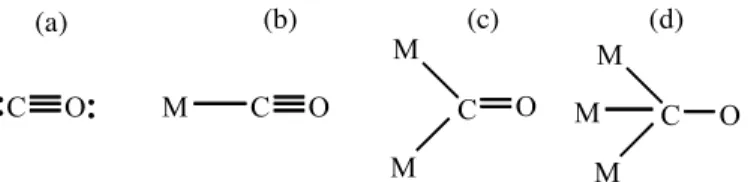
bands, depending on their bonding mode. Carbon monoxide typically binds in an
end-on fashion through carbon, although some extremely rare cases of
coordination through oxygen have already been reported, namely for
Aluminium[76] and Europium(III).[77] Apart from binding as a terminal ligand, CO
may also act as a symmetrical or unsymmetrical bridging ligand between two(µ2)
or three (µ3) metal centers. These are exemplified in Figure 3 and Table 2 reports
their IR stretching frequencies.
Figure 3: Typical CO bonding modes to metal center: a) “free” b) terminal; c) doubly bridging; d) triply bridging.
M C O
M
C O
M
C O
M
C O
M M
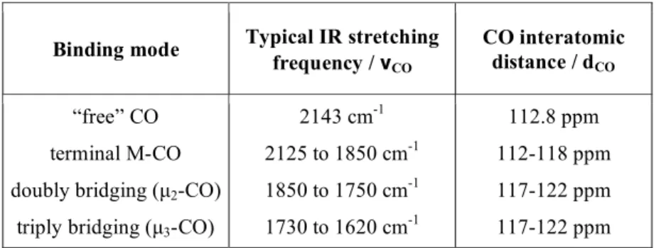
Table 2: Carbonyl group IR symmetric stretching frequencies and interatomic distances depending on the binding mode in neutral metal complexes.
As can be observed from Table 2, the position of the carbonyl bands in the IR
spectrum depends mainly on the bonding mode of the CO (terminal, bridging) but
also on the amount of electron density on the metal being backdonated to the CO.
As the number (and intensity) of the carbonyl bands observed depend on the
number of CO ligands present and the symmetry of the metal complex,
introduction of a strong σ donor or a worse π acceptor will lead to a decrease in
CO stretching frequency. Accordingly, Lewis base substituents, whose donor
atoms are mainly phosphines or sulphides possess energetically accessible vacant
dπ orbitals which can also enter into π bonding with the metal. However, almost
all the ligands that replace CO in substitution reactions are poorer π acceptors,
when compared to CO. Therefore, successive replacement of CO from M(CO)n
by other types of incoming ligands leads to progressive lower CO stretching
frequencies in the resulting complexes M(CO)n-xLx since the carbonyls accept a
grater share of the metal electronic density in comparison with the other
substituents.
The rupture of one or more of the M-CO bonds of a MCC by physical or chemical
processes leads to the liberation of CO and, therefore, CO release. The physical
processes are heat and light. Warming a MCC will eventually provide enough
energy to break one of the CO bonds and thermally induced CO release will take
place. The classical example is the thermal decomposition of Ni(CO)4 into
metallic nickel and gaseous CO that takes place at 40°C or that of Pd(CO)4 which
Binding mode Typical IR stretching frequency / νCO
CO interatomic distance / dCO
“free” CO
terminal M-CO
doubly bridging (µ2-CO) triply bridging (µ3-CO)
2143 cm-1 2125 to 1850 cm-1 1850 to 1750 cm-1 1730 to 1620 cm-1
112.8 ppm
112-118 ppm
117-122 ppm
happens even below room temperature. Compounds of this kind cannot be
considered for pharmacological uses because of the small gap that exists between
the physiological temperature of living organisms (37°C) and room temperature.
If they decompose at 37°C they surely have a very short shelf life at room
temperature.
Interaction of a MCC with UV or more rarely with visible light causes the
labilization of M-CO bonds, and promotes photo-induced CO release. This is a
general characteristic of MCCs. However, with the exception of topical uses on
the skin, or local photodynamic methods this kind of activation of CO release has
no other practical pharmacological use.
Besides these two strictly physical processes that lead to M-CO bond breaking
even in vacuum, ligand substitution reactions are the most common chemical
pathway to CO release. Ligand substitution is a chemical process whereby one
molecule reacts with a MCC and replaces CO within the coordination sphere of
its central metal atom. CO is then liberated to the reaction medium from which it
may escape to the atmosphere or enter into another chemical reaction. In the case
of biological applications this free CO will be taken up by an appropriate target
and initiate a biological cascade of events of therapeutic significance.
From the previous we have to conclude that in order to use MCCs as CO-RMs for
biological/pharmacological applications, we need to control their reactions with
the biological molecules in the medium, which will eventually replace CO in the
coordination sphere of the metal in a reaction called ligand substitution.
The hundreds of studies published on the topic of ligand substitution in metal
carbonyl complexes were carried out under inert atmosphere because most MCCs
have low valent metal centers that are sensitive to oxidation by atmospheric
oxygen.[78, 79] Moreover, with very few exceptions all these studies were carried
out in organic solvents of low polarity (e.g. toluene, cyclohexane
1,2-dichloroethane) due to the lipophilic nature of most MCCs and the need to
simplify the kinetic models of reactivity. Studies carried out in water or protic
![Table 1: Ambient CO concentration, % CO-Hb and human health symptoms associated (adapted from references [5] and [6])](https://thumb-eu.123doks.com/thumbv2/123dok_br/15766091.640517/28.765.93.622.147.402/table-ambient-concentration-health-symptoms-associated-adapted-references.webp)
![Figure 5: Left: HPLC trace of a water solution of Na 3 [Mo(CO) 3 (cit)] under air at room temperature over 75 min](https://thumb-eu.123doks.com/thumbv2/123dok_br/15766091.640517/89.765.169.641.574.760/figure-left-hplc-trace-water-solution-under-temperature.webp)
![Table 10: Equivalents of CO released by the complexes Mn(CO) 5 Br, CpFe(CO) 2 Cl, [Et 4 N][Mo(CO) 5 Br], CpMo(CO) 3 Cl and (Ac-Cp)Mo(CO) 3 Cl and their encapsulated forms in TRIMEB and also CpFe(CO) 2 Cl encapsulated in β-CD, in RPMI after 2h,](https://thumb-eu.123doks.com/thumbv2/123dok_br/15766091.640517/101.765.140.699.204.523/table-equivalents-released-complexes-cpfe-encapsulated-trimeb-encapsulated.webp)
