O R I G I N A L P A P E R
Benefits of membrane electrodes in the electrochemistry
of metalloproteins: mediated catalysis of
Paracoccus pantotrophus
cytochrome
c
peroxidase by horse cytochrome
c
: a case study
P. M. Paes de SousaÆS. R. PauletaÆD. RodriguesÆM. L. Simo˜es Gonc¸alves Æ
G. W. PettigrewÆ I. MouraÆ J. J. G. MouraÆM. M. Correia dos Santos
Received: 19 December 2007 / Accepted: 10 March 2008 / Published online: 26 March 2008
ÓSBIC 2008
Abstract A comparative study of direct and mediated
electrochemistry of metalloproteins in bulk and membrane-entrapped solutions is presented. This work reports the first electrochemical study of the electron transfer between a bacterial cytochrome c peroxidase and horse heart cyto-chrome c. The mediated catalysis of the peroxidase was analysed both using the membrane electrode configuration and with all proteins in solution. An apparent Michaelis constant of 66±4 and 42±5 lM was determined at pH 7.0 and 0 M NaCl for membrane and bulk solutions, respectively. The data revealed that maximum activity occurs at 50 mM NaCl, pH 7.0, with intermolecular rate constants of (4.4±0.5)9106and (1.0±0.5)9106 M-1 s-1 for membrane-entrapped and bulk solutions, respectively. The influence of parameters such as pH or ionic strength on the mediated catalytic activity was analysed
using this approach, drawing attention to the fact that careful analysis of the results is needed to ensure that no artefacts are introduced by the use of the membrane configuration and/or promoters, and therefore the dependence truly reflects the influence of these parameters on the (mediated) catalysis. From the pH dependence, a pKof 7.5 was estimated for the mediated enzymatic catalysis.
Keywords Membrane electrodeMediated catalysis
Bacterial cytochrome cperoxidaseHorse cytochromec
Abbreviations
AUME Gold membrane electrode
BCCP Bacterial cytochrome cperoxidase ME Membrane electrode
Introduction
The use of membrane electrodes (ME), with the protein imprisoned between a dialysis membrane and the electrode surface, has some important advantages. These are (1) the use of very small volumes of protein—2ll (to overcome the limitation of sample availability), (2) the ease of elec-trode preparation and low cost, (3) the rapid investigation of various experimental parameters, such as pH and ionic strength and (4) the thin layer configuration, which easily allows quantitative information about the redox processes to be obtained [1,2].
Moreover, the membranes used in this type of electrode configuration are charged, offering a convenient way to modulate the electric environment close to the electrode surface, and thus ascertain the role of electrostatic inter-actions in the efficiency of the electron transfer. It is also P. M. Paes de SousaS. R. PauletaI. MouraJ. J. G. Moura
ReQuimte, Centro de Quı´mica Fina e Biotecnologia, Departamento de Quı´mica,
Faculdade de Cieˆncias e Tecnologia, Universidade Nova de Lisboa, 2829-516 Caparica, Portugal e-mail: patricia.sousa@dq.fct.unl.pt
D. RodriguesM. L. Simo˜es Gonc¸alves
M. M. Correia dos Santos (&) Centro de Quı´mica Estrutural, Instituto Superior Te´cnico, Av. Rovisco Pais, 1049-001 Lisbon, Portugal e-mail: mcsantos@ist.utl.pt
G. W. Pettigrew
Royal (Dick) School of Veterinary Studies, University of Edinburgh,
Summerhall,
important to point out that this configuration is an inter-esting strategy when adsorption of a protein or an enzyme to the electrode surface is not successfully achieved, hindering the use of protein film voltammetry [3]. Addi-tionally, the entrapment of proteins between the membrane and the electrode surface enables the achievement of thin-layer conditions and avoids diffusional problems [4].
Studies reporting the benefits of the ME in protein electrochemistry and its role in modulating the redox behaviour focused on the direct electrochemistry of small electron transfer proteins. In these works, a critical com-parison was made with data obtained with non-ME [5–7]. More recently ME were used to study multiredox cofactor-containing enzymes and their interaction with both physi-ological and non-physiphysi-ological redox partners [8, 9]. In both studies, although the direct electrochemistry and the mediated catalysis of the proteins were analysed using that strategy, with the proteins in solution or adsorbed on a modified electrode, no comparison was made among the different strategies.
The mediated catalysis of the bacterial cytochrome c
peroxidase (BCCP) from Paracoccus pantotrophus by
P. pantotrophus pseudoazurin examined at a gold ME (AUME) was recently reported by us [10]. Cyclic voltam-metry was used to analyse the direct transfer to pseudoazurin and to probe its interaction with BCCP. The results obtained showed that this small copper protein is a competent elec-tron donor to BCCP, in agreement with solution steady-state kinetics performed spectrophotometrically [11]. The pH and ionic strength effect on the voltammetric signal of pseudo-azurin as well as on the intermolecular rate constant were also easily studied at the ME.
Horse heart cytochrome c, a commercially available smallc-type haem protein, has been previously used as an electron donor to Paracoccus BCCP in solution steady-state kinetic studies performed spectrophotometrically [12]. Although it is not a physiological partner of BCCP, this small protein was shown to be kinetically competent as an electron donor to the Paracoccus enzyme. However, horse cytochromecexhibits a distinct kinetic and binding behaviour from the physiological partners pseudoazurin, a type I copper protein, and cytochromec550, which is also a smallc-type haem protein. The physiological partners bind at the same site on the surface of BCCP near the electron-transferring haem of the enzyme [13], while horse cyto-chromec has been proposed to have two binding sites, a looser binding site near the electron-transferring haem and an additional tight one in-between the two haems of the enzyme [14]. Below 50 mM NaCl, there is little change in the activity when the electron donor is either cytochrome
c550or pseudoazurin. In contrast, when horse cytochromec is used as an electron donor there is a threefold decrease in activity as the ionic strength is lowered, and this correlates
with an increased binding affinity [15, 16]. It was con-cluded that the higher-affinity binding was, in fact, non-productive and that only when that attachment was loosened by raised ionic strength could the horse cyto-chromecmigrate to the true electron transfer site.
This work reports the first electrochemical study of the electron transfer between BCCP and horse cytochrome c. The mediated catalysis of BCCP was analysed using the ME configuration and with all proteins in solution. Our aim is to compare the results obtained with these two strategies in order to gain a better insight into the interactions that occur at the ME surface and to highlight the effects of this strategy on the direct and mediated electrochemistry of metalloproteins.
Materials and methods
Proteins and chemicals
Horse heart cytochrome c was obtained from Sigma and used without further purification. Pseudoazurin and BCCP were isolated and purified as described before [17]. The concentration of the proteins was determined spectropho-tometrically using the extinction coefficient at 409 nm, e=250 mM-1cm-1, and at 550 nm, e=29.5 mM-1 cm-1, for fully oxidized BCCP and reduced horse cyto-chromec, respectively [17,18].
4,40-Dithiodipyridine and CaCl2 were obtained from Sigma and all other chemicals were pro analysis grade. All solutions were prepared with deionized water from a Milli-Q water purification system.
Apparatus and procedures
The cyclic voltammograms were obtained using an EG&G-PAR model 273A potentiostat/galvanostat controlled via the 270 software. The scan rate varied between 5 and 200 mV s-1. Throughout this article, all potential values are referred to the standard hydrogen electrode.
A conventional three-electrode configuration cell was used, with a platinum auxiliary electrode and an Ag/AgCl reference electrode (BAS MF-2052; 205 mV vs. the stan-dard hydrogen electrode). The working electrode was a gold disk electrode from BAS (MF-2014) with a nominal radius of 0.8 mm. The effective surface area of the elec-trode was determined from its response in a known concentration of the ferrocyanide/ferricyanide couple (D=7.84910-6cm2s-1 [19]) and was found to be
0.0195 cm2.
sonicated for 5 min, rinsed well with Milli-Q water and finally dipped into 1 mM 4,40-dithiodipyridine solution for 5 min. The membrane configuration was prepared as pre-viously described [6] using a negatively charged Spectra/ Por MWCO 3500 membrane.
In typical experiments, the supporting electrolyte, as well as the working solution, contained 10 mM phosphate buffer pH 7.0±0.1, 0.5 mM 4,40-dithiodipyridine and 1 mM
CaCl2. Horse cytochromecwas present in a concentration of 100lM and the concentration of BCCP varied between 0.25 and 1lM. In the experiments with a saturating con-centration of substrate, 100–350lM H2O2was present in the electrolyte solution. The effect of substrate concentra-tion was determined by varying the H2O2 concentration between 10 and 200lM. The pH of the electrolyte was varied from 5.4 to 10.5 by adding 5 M HCl or 2 M NaOH to a mixed-buffer system containing 10 mM 2-morpholinoe-thanesulfonic acid, sodium phosphate,N-(2-hydroxyethyl) piperazine-N0-ethanesulfonic acid, tris(hydroxymethyl) aminomethane and 3-cyclohexylamino-1-propanesulfonic acid. The effect of the ionic strength was studied by adding increasing amounts of NaCl (up to 500 mM).
It should be pointed out that in order to have repro-ducible results for some BCCP concentrations more than one ME had to be prepared, to cover the whole range of experimental variables. This is due to the fact that the enzyme exists in a monomer–dimer equilibrium in which only the dimer is active. Dilution of the enzyme shifts the equilibrium towards the monomer and therefore the activ-ity is lost with time [14], which corresponds to a decrease of the catalytic current.
All solutions were deaerated for 10 min with high-purity nitrogen, and all measurements were performed at least in duplicate in a temperature-controlled room at 20±1°C.
Results and discussion
Catalytic activity ofP. pantotrophusBCCP with horse heart cytochromecas an electron donor
ME cyclic voltammetry
The electrochemical behaviour of horse cytochrome c is well known either in bulk or entrapped on a ME. Indeed, Eddowes and Hill [20] found that essentially reversible voltammetry of cytochromecin solution could be observed at a 4,40-dipyridyl-modified gold electrode. Latter, Lojou and Bianco [5] showed that fast electrochemical response was observed when a thin layer of protein solution was entrapped between a negatively charged dialysis membrane and a non-modified gold electrode surface. In both cases favourable conditions for electron transfer to occur were
achieved, which accounts for the similarity of the values determined for the formal potentials.
In this work, the direct electrochemistry of cytochromec
was revisited at a AUME, but this time in the presence of 4,40-dithiodipyridine (data not shown). The promoter was used in order to have similar experimental conditions for the studies both in solution and with the membrane-con-figuration electrode (further reasons will be explained below).
Thin-layer behaviour will be observed as long as the entrapped layer thickness l is smaller than the diffusion-layer thickness for a given experimental time scale,t:
l\ð2DtÞ1=2 ð1Þ
where D is the diffusion coefficient of the species [21]. Under our experimental conditions (cyclic voltammetry with scan rates between 5 and 200 mV s-1), this condition was verified for the lowest scan rates (v\100 mV s-1),
with ip varying linearly with v in this range. From this variation [21], an entrapped-solution volume, V, of 1.2910-5cm3 was estimated, which corresponds to an entrapped-layer thickness l=V/A=6.2910-4cm.
Since the diffusion coefficient of cytochrome c is
D=1.2910-6cm2s-1 (see below), thin-layer condi-tions occur forv\160 mV s-1, as verified.
The ratio of the cathodic (ipc) and anodic (ipa) peak currents was close to 1 and the peak-to-peak separation, DEp=Epa-Epc, was close to 20 mV for the lowest scan rates. In the same range, the width at half height (DEp,1/2) was also constant for the cathodic and anodic peaks, with a value close to 90 mV. AlthoughDEpincreased withv, the average of the reduction and oxidation peak potentials remained almost constant for all scan rates and a formal reduction potential E°0=(Epc+Epa)/2=250 ±5 mV was estimated at pH 7.0, in agreement with other reported values (e.g. 280 mV, ME at pH 7.6 [5], and 255 mV, bulk solution at pH 7.0 [20]).
The catalytic activity ofP. pantotrophusBCCP towards horse cytochrome cwas then investigated at the AUME, with working solutions of 100lM cytochrome c and different BCCP concentrations (0.25–1lM) in the pres-ence of a saturating substrate concentration (125–350lM, depending on the enzyme concentration) or with a working solution of 100lM cytochrome cand 0.25lM BCCP in the presence of increasing H2O2 concentrations. These solutions and the electrolyte contained 1 mM CaCl2 (for enzyme activation [12,22]).
increases with H2O2concentration in the electrolyte, and a sigmoidal wave develops when a saturating concentration of substrate is reached. Voltammograms from solutions containing only BCCP either in the presence or in the absence of H2O2 were indistinguishable from the back-ground current (data not shown).
The half-wave potential of the sigmoidal waves (E1/2
=275 ±10 mV=E°0) shows that the actual transfer
process is the catalysed reduction of cytochrome c. The catalytic current is scan rate independent up to 50 mV s-1 and increases for increasing BCCP concentrations. This behaviour is consistent with a reaction mechanism involving an initial heterogeneous electron transfer reac-tion at the electrode (Fig.2, step 1), followed by homogeneous chemical reactions: the oxidized form of cytochrome c is regenerated by BCCP (Fig. 2, step 2)
which, in turn, is recycled by H2O2(Fig.2, step 3). This mechanism can be simplified to
OxþeRed
RedþZ!Ox ð2Þ
provided that the following conditions are obeyed: (1) the heterogeneous electron transfer (Fig.2, step 1) is a reversible reaction; (2) the homogeneous chemical reaction (Fig.2, step 2) is irreversible; (3) the reaction between cytochrome c and BCCP is pseudo first order with a reaction rate constant given byk0=kCBCCP, wherekis the second-order rate constant [10,25].
The first condition is well obeyed, as previously dem-onstrated. The second condition is fulfilled because BCCP is being reoxidized in the catalytic cycle owing to the excess of H2O2 in solution (Fig.2, step 3), and not by transferring electrons back to horse cytochrome c. As to requisite 3, it would imply that BCCP is present in large excess, which is not the case. However, under saturating concentrations of H2O2, this requisite is obeyed when the rate of recycling the oxidised BCCP by H2O2is not rate-limiting. If this is the case, BCCP will always be available to react with cytochrome c and pseudo-first-order condi-tions are met.
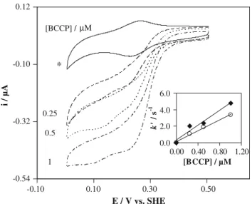
Cyclic voltammograms at 20 mV s-1were obtained for 100 lM cytochromecand increasing BCCP concentrations (0.25–1lM), in the absence and in the presence of a sat-urating concentration of H2O2(125–350lM, depending on the enzyme concentration), and are shown in Fig.3.
From these recordings the catalytic current (icat) was determined as the difference between the current in the presence and in the absence of substrate, both measured at the same potential. The data was treated according to La-viron’s theory for diffusionless electrochemical systems [4, 10], where the catalytic current is given by
icat ¼n Fk0VCCytc ð3Þ In this equation,Vis the entrapped-solution volume,Cis the concentration and the other terms have the usual meaning.
From the variation of the pseudo-first-order rate con-stant,k0, with BCCP concentration, an intermolecular rate -0.32
-0.19 -0.06 0.07
-0.05 0.15 0.35 0.55
-0.05 0.15 0.35 0.55
E / V vs. SHE
E / V vs. SHE
i / µA
[H2O2] / µM
0
25
50
100 0.00
0.06 0.12 0.18
0 100 200 300
[H2O2] /µM
i
ca
t
/ µ
A
a
-0.26 -0.14 -0.02 0.10
i / µA
[H2O2] / µM
0
50
100 25
0.00 0.04 0.08 0.12
0 100 200 300
[H2O2] /µM
i
ca
t / µ
A
b
Fig. 1 Cyclic voltammograms (v=20 mV s-1) at the gold
mem-brane electrode (a) or bulk solution (b) of 100lM horse heart cytochromecand 0.5lM bacterial cytochromecperoxidase (BCCP) in the presence of increasing concentrations of H2O2. The medium consisted of 10 mM phosphate buffer pH 7.0, 0.5 mM 4,40 -dithiodi-pyridine and 1 mM CaCl2.SHEstandard hydrogen electrode
H2O2
H2O
Electrode
e
-Cytcox
Cytcred
BCCPox
BCCPred
Step 1 Step 2
k’
Step 3
Fig. 2 Mediation scheme for BCCP: the electrode reduces horse
constant k=(3.2±0.5)9106M-1s-1 was estimated
(Fig.3, insert, circles).
For assays where the H2O2concentration was varied, for a constant cytochromecand BCCP mixture, the catalytic current was plotted as a function of the substrate concen-tration (Fig.1a, insert). It is clear that the catalytic current decreases after 150lM H2O2and so the data were fitted to the Michaelis–Menten kinetics for concentrations up to that value. Since thin-layer conditions are verified, the Michaelis–Menten equation has the form
icat¼ imaxCH2O2 CH2O2þKM
¼nFk
0VCCyt
cCH2O2
CH2O2þKM
ð4Þ
Using the CERN library Fortran program MINUIT algo-rithm, we estimated an apparent Michaelis–Menten constantKM=66±4 lM and an intermolecular electron transfer rate constantk=(3.4±0.3)9106M-1s-1.
This electron transfer rate constant is higher than the value determined for a physiological donor, pseudoazurin ((1.4±0.2)9105M-1s-1[10]). The more likely
expla-nation for the difference between the rate constants would be a difference in the redox potential of these two electron donors, but the values determined at pH 7.0 are very similar.
Therefore, this behaviour must be related to another factor that influences the donor–enzyme interaction. In the case of these small electron donors the global charge sur-face is high and asymmetrically distributed, creating a significant dipole moment. Actually, the dipole moment of
pseudoazurin is 651 D, and that of horse cytochromecis 299 D [11]. This twofold difference in the dipole moment probably results in a stronger association of pseudoazurin with BCCP, and consequently the dissociation of the complex is disfavoured, explaining the lower rate constants observed.
Solution cyclic voltammetry
The mediated catalysis of BCCP by horse cytochrome c
was also analysed at the gold electrode from solutions containing 100lM horse heart cytochrome c, 0.5lM BCCP and 1 mM CaCl2. Again, the peak current increases with H2O2concentration in the electrolyte, and a sigmoidal wave develops when a saturating concentration of substrate (100 lM) is present in the electrolyte, which is scan rate independent up to 50 mV s-1(Fig.1b).
The theory describing such a mechanism for diffusion-controlled processes was developed by Nicholson and Shain [26] and applied to several kinetic studies of reac-tions between mediators and redox proteins [25]. The intermolecular rate constant can also be calculated from the value of the H2O2 saturated limiting current using Eq.5 [27,28]:
icat ¼n F A D1=2CCytcðk0Þ1=2 ð5Þ In this equation, D is the diffusion coefficient of cytochrome c. The valid value for our experimental conditions, D=(1.2±0.1)910-6cm2s-1, was
comp-uted from the dependence on the scan rate of either the cathodic or the anodic peak currents of the cyclic voltammograms for solutions containing both cytochrome
c and BCCP in the absence of H2O2, using the Randles– Sˇevcˇı´k equation [21]. Calculations using Eq.5 gave an intermolecular rate constant of k=(4.0±0.5)9105 M-1s-1.
The catalytic current for the different H2O2 concentra-tions (Fig.1b, insert) was also fitted to Michaelis–Menten kinetics for concentrations up to 100lM, and
KM=42±5 lM and k=(3.9±0.3)910 5
M-1s-1 were obtained.
Similar apparent Michaelis–Menten constants were estimated for the proteins in bulk and entrapped solution, indicating that the membrane does not hamper the substrate diffusion. A higher value for the intermolecular rate con-stant was obtained for the proteins using the membrane configuration, reflecting a more favourable domain for the molecular interactions that must be established for electron transfer to occur between BCCP and cytochromec. In both situations an inhibition effect by the substrate is observed for higher H2O2 concentrations, as verified in the case of the catalysis mediated by pseudoazurin [10]. A similar
-0.54 -0.32 -0.10 0.12
-0.10 0.10 0.30 0.50 E / V vs. SHE
i / µA
[BCCP] / µM
0.25
0.5
1 ∗
0.0 2.0 4.0 6.0
0.00 0.40 0.80 1.20
[BCCP] / µM
k'
/ s
-1
Fig. 3 Cyclic voltammograms (v=20 mV s-1) obtained at the gold
membrane electrode for 100lM cytochromecand increasing BCCP concentrations (0.25–1lM), in the absence (asterisk) and in the presence of a saturating H2O2concentration (125–350lM, depending on the enzyme concentration).Insert: Variation of the pseudo-first-order rate constant with BCCP concentration for 0 mM (circles) and
50 mM (diamonds) NaCl. The medium consisted of 10 mM
phos-phate buffer pH 7.0, 0.5 mM 4,40-dithiodipyridine and 1 mM CaCl
inhibitory effect was observed in the study of other per-oxidases [29–31].
Ionic strength and pH effects
One of the advantages of the ME approach is the ability to rapidly investigate effects on the redox behaviour of vari-ous experimental conditions, such as the pH or the supporting electrolyte composition. Another advantage of the approach is the small amount of protein needed for a series of measurements. This is so because once the ME has been mounted in the cell, it does not need to be removed or exchanged while changing any of those variables.
The same approach can also be applied to study the effect of such experimental variables on the kinetic activity of a more complex system, such as in mediated catalysis. Changes in the pH and/or the ionic strength of the medium may influence the rate of association, pre-orientation within the encounter complex, stability of the reactive complex or/and rate of dissociation of the prod-ucts. However, the interpretation of ionic strength (or pH) dependences of the kinetic activity is complex and the behaviour observed should not be masked by the influ-ence of those variables on the direct electrochemistry of the redox partners.
The effect of the ionic strength on the intermolecular rate constant for BCCP and horse cytochromecwas ana-lysed with the proteins entrapped at the AUME and in bulk solution with a working solution containing 100lM horse cytochrome c, 0.5lM BCCP and 1 mM CaCl2, in the presence of increasing amounts of NaCl (in the range 0– 500 mM) and a saturated H2O2concentration.
In both situations, the ionic strength dependence of the mediated catalysis has a bell-shaped curve, with a maxi-mum activity at 50 mM NaCl (Fig.4). The decrease of activity with increasing NaCl concentrations is consistent with the electrostatic character of the interaction between BCCP and the electron donor, cytochrome c. Indeed, cytochromechas an asymmetric charge distribution with a positive surface that surrounds the exposed haem edge, which is the region proposed to interact with the negative surface around the exposed electron-transferring haem of the peroxidase [12, 14]. The increase of activity at low NaCl concentrations can be explained considering that for the lowest ionic strength the encounter complex formed has an orientation that is not so favourable for electron transfer. Therefore, the increase in NaCl concentration enables the lateral search at the peroxidase surface for the competent electron transfer orientation [11,32]. Another explanation for this effect is that at low ionic strength cytochrome c
reduction at the electrode might be affected by the lower
koffrate constant of the complex with BCCP.
Using the approach described above (Eq.3), one can use the analysis of the catalytic current variation as a function of BCCP concentration (Fig.3, insert, diamonds) to esti-mate an intermolecular rate constant for maximum activity
k=(4.4±0.5)9106M-1s-1 for the membrane con-figuration. In the case of the bulk solution, a rate constant for maximum activity k=(1.0±0.5)9106M-1s-1
was determined using Eq.5.
Similar behaviour was observed by solution steady-state kinetics performed spectrophotometrically, when this small cytochrome is used as an electron donor, with a maximum turnover also occurring at 50 mM NaCl [11], as can be observed in Fig.4. However, in the voltammetric experi-ments with the membrane the variation in activity with ionic strength is less pronounced than that found with the proteins in bulk solution (either in the voltammetric assays or in solution steady-state kinetics performed spectropho-tometrically). In fact, from 50 to 500 mM NaCl the activity decreases by 64% at the ME, while in the other two cases the decrease is 98%.
A similar effect was observed for the mediated catalysis ofP. pantotrophusBCCP byP. pantotrophuspseudoazurin, where similar profiles were obtained with the membrane configuration and solution steady-state kinetics performed spectrophotometrically, though the maximum activity was observed for higher NaCl concentration in the former case. The differences may be due to the charge of the membrane, which somehow favours the formation of the encounter complex, making it less dependent on the ionic strength, in spite of the electrostatic forces that govern the interaction.
0.0 1.6 3.2 4.8
0 125 250 375 500
[NaCl] / mM
k
/ M
-1 s
-1
( × 1
0
6 )
0 1.8 3.6 5.4
Tu
rn
o
v
er
/ s
-1
( × 1
0
3
)
Fig. 4 Effect of ionic strength on the intermolecular rate constant for
100lM horse heart cytochrome c, 0.5lM BCCP and saturating H2O2(150lMdiamondsor 100lMcircles) at the gold membrane electrode (diamonds) or in bulk solution (circles) and on the activity of BCCP with horse cytochromecas an electron donor from solution steady-state kinetics performed spectrophotometrically (triangles) [11]. The medium consisted of 10 mM phosphate buffer pH 7.0 and
This may be also the reason why higher rate constants were determined for horse cytochrome c using the membrane configuration, although values of the same order of magnitude were obtained for maximum activity [k=(4.4±0.5)9106M-1s-1 for the membrane
con-figuration and k=(1.0±0.5)9106M-1s-1 for bulk
solution] as illustrated in Fig.4.
The behaviour observed in the voltammetric experi-ments truly reflects the effect of ionic strength on the interaction between the enzyme and the electron donor rather than on the direct electrochemistry of horse cyto-chromecdue to the presence of the promoter. The horse cytochrome c peak current was shown to be NaCl-inde-pendent when 4,40-dithiodipyridine is present in the medium (data not shown). Behaviour different from that observed at a bare gold electrode in the membrane con-figuration was evidenced, where a sharp decrease in current was observed at increasing NaCl concentrations [5]. Similar behaviour was also observed for the direct elec-trochemistry of pseudoazurin at a dithiodipyridine gold modified electrode [10].
The pH behaviour of horse cytochromecis well known to present a pK close to 9.5 associated with the disap-pearance of the 695 nm absorption band, which is believed to be due to the replacement of the iron-coordinated methionine residue by a nitrogenous ligand, assigned as Lys73 [33,34].
Previous bulk voltammetric studies of horse cytochrome
c at a 4,40-bipyridyl-modified gold electrode in the pH range 4.5–10.5 showed that while the peak potential remains essentially constant until pH 9, the peak current profile has a bell shape [35]. The maximum current was observed at pH 7 and two pKvalues around 9 and 5 were computed from that variation. The former is in agreement with the value obtained by other techniques, as well as with the observedEpvariation. The latter was attributed by the authors to the protonation of the promoter that disrupts the interaction between the gold surface and the protein, leading to a decrease of the electrochemical current [35].
For the reasons presented above, the pH dependence of the intermolecular rate constant for BCCP and horse cytochromecwas analysed at the bare AUME, i.e. in the absence of 4,40-dithiodipyridine.
In order to accurately interpret the results of this pH dependence, the effect of this parameter on the electro-chemical signal of horse cytochromecalone was studied between pH 5 and 10 in the same conditions. As expected, the redox potential remained constant for pH\9 and a
pronounced decrease was observed for higher pH values (data not shown). A pK of 9.1±0.1 was determined, in accordance with reported values [33–35]. For the peak current, a bell-shaped profile was obtained (Fig.5, circles), behaviour similar to that observed for bulk solution in the
presence of the promoter [35]. The peak current pH dependence was fitted to the following equation [36]
icat ¼ iopt.
1þ10ðpKa1pHÞþ10ðpHpKa2Þ
ð6Þ
and pKvalues of 5.3±0.3 and 9.5±0.3 were estimated.
The latter is in clear agreement with the characteristic alkaline pKof this protein.
Several electrochemical studies using different approa-ches have observed a similar behaviour for the dependence on the pH of the horse cytochromeccurrent, and in some cases an acidic pK with an identical value, within the experimental errors, has been determined. However, depending on the authors and the conditions employed in the studies, the pKwas assigned differently, namely to the proton-dissociation constant of the promoter [35], the Coulombic properties of the electrode surface [37, 38], the competitive adsorption of protons on the clay-modified electrodes [39] or the protein denaturation induced by the applied electric field at the electrode–solution interface [40]. In the present work a ME was employed, and so another possible cause for the profile observed at low pH is the degradation of the membrane–protein–electrode inter-action, given that in the absence of a promoter charge effects stand out as being most important, as previously mentioned.
In the present study the same behaviour was observed either in the presence (data not shown) or in the absence of 4,40-dithiodipyridine, eliminating the promoter protonation as the possible cause. Moreover, considering the fact that
0.00 0.10 0.20 0.30
5 7 9 11
pH ica
t
/ µ
A
0.00 0.02 0.04 0.06
i
p
c / µ
A
Fig. 5 pH dependence of 100lM horse heart cytochromecat the
gold membrane electrode (circles) and catalytic current for 100lM
horse heart cytochrome c, 1.0lM BCCP and saturating H2O2
the profile obtained at the bare ME is similar to that observed for the several bulk conditions, it seems that the acidic pKis not due to an artefact of the membrane.
In fact, this acidic pK may be the result of an acidic residue or a group of residues involved in the interaction with the electrode, but not affecting the haem environment and consequently not changing the redox potential. Denaturation of the protein would also lower the con-centration of horse cytochrome c responding at low pH, and must be considered. Indeed, the analysis of the vol-tammograms of horse cytochrome c at decreasing electrolyte pH shows that the redox signal is at the same potential, but with a decreasing current which does not return to the initial value when the pH is raised back to 7.0 (data not shown).
The pH effect in the mediated catalysis at the AUME was studied in the range 5–10, using solutions containing 100lM horse cytochrome c, 1lM BCCP, 1 mM CaCl2 and a saturated H2O2 concentration (350lM). The catalytic current presents a bell-shape profiled (Fig.5, diamonds) with no activity being observed above pH 9, which can be attributed to the inactivation of the enzyme at high pH [10]. This curve can be fitted to Eq.6withioptat pH 6.5 and two pKvalues, 5.6±0.1 and 7.5±0.1.
Taking into account the above results, we cannot con-clude if the acidic pKestimated for the mediated catalysis is also related to the protein–protein interaction or only due to horse cytochromecalone. However, preliminary solu-tion steady-state kinetics performed spectrophotometrically of Paracoccus BCCP using horse cytochrome c as an electron donor (Pauleta et al., unpublished results) revealed an acidic pK at the same value, and similar values were observed for the enzymatic activity of Pseudomonas aeruginosa (5.2) [41] and Rhodobacter capsulatus (6.1) [42] BCCP.
Moreover, binding studies using microcalorimetry showed that the formation of this electron transfer complex involves the release of 0.46 protons at pH 6.0, which also suggests the existence of an acidic pK, at around pH 6.0 [16]. Therefore, this pKreflects a proton-dissociation pro-cess associated both with the horse cytochromecand with the interaction/activity of this protein with BCCP.
A more basic pK was also observed for the solution steady-state kinetics determined spectrophotometrically of
P. aeruginosa(6.8) andR. capsulatus(7.9) BCCP [41,42], as now estimated for theParacoccusenzyme. This pKcan be clearly attributed to the mediated enzymatic catalysis, since the current profile of the horse cytochromecitself is invariable in this pH region (Fig.5). As several authors have suggested, these pK values can be assigned to the propionates of the BCCP haems, histidine ligands or proposed residues involved in the catalysis such as Glu128 [32, 42, 43]. However, the identity of the residue(s)
responsible for this proton dissociation can only be disen-tangled through site-directed mutagenesis.
Conclusions
This work highlights the use of ME in the study of direct and mediated electrochemistry of metalloproteins. The results reported in the present work show that ME are very attractive for the study of complex metalloproteins sys-tems, particularly with regard to the very small amounts of protein needed and the ease with which experimental variables can be changed.
Mediated catalysis of BCCP by horse cytochromecwas easily analysed, with minimum consumption of proteins, just varying the H2O2 concentration in the electrolyte solution. While the proteins stay entrapped in close vicinity to the electrode surface, small ions can diffuse through the membrane. For the same reasons, the effect on the catalytic activity of experimental conditions, such as pH and ionic strength, can be rapidly analysed.
The fitting to Michaelis–Menten kinetics of the experi-mental data, from studies with the proteins either entrapped in the membrane or freely diffusing in solution, led to similarKMvalues, showing that no artefacts are introduced in the study by the use of a ME. Higher values for the intermolecular rate constants for horse cytochrome c and BCCP were obtained with the membrane configuration, which shows that the membrane does not disrupts the formation of the encounter complex and can even favour it. Since electrostatic interactions are fundamental for the formation of the complexes between P. pantotrophus
BCCP and its electron donors, this configuration seems to be an adequate option for the electrochemical study of the mediated catalysis in biological systems, where electro-static forces play an important role.
To draw conclusions about the influence of variables, such as pH or ionic strength, on the mediated catalytic activity, one must first pay attention to the behaviour of the mediator, i.e. the protein that actually exchanges the electrons with the electrode. For this reason, the ionic strength effect was analysed using a promoter to render the cytochrome c–electrode–membrane interaction more hydrophobic in nature, so the variation observed for the catalytic activity could only be due to changes in the binding between BCCP and the redox partner. On the other hand, the use of promoters might mask the pH effect depending on their acid–base behaviour and a possible interference must be verified.
Acknowledgments This work was within the research project
References
1. Haladjian J, Bianco P, Nunzi F, Bruschi M (1994) Anal Chim Acta 289:15–20
2. Haladjian J, Thierry-Chef I, Bianco P (1996) Talanta 43:1125– 1130
3. Armstrong FA (2002) In: Bard AJ, Stratmann M (eds) Encyclo-pedia of electrochemistry, vol 9. Wiley-VCH, Weinheim, pp 13– 28
4. Laviron E (1979) J Electroanal Chem 101:19–28 5. Lojou E, Bianco P (2000) J Electroanal Chem 485:71–80 6. Correia dos Santos MM, Paes de Sousa PM, Simo˜es Gonc¸alves
ML, Krippahl L, Moura JJG, Lojou E, Bianco P (2003) J Elec-troanal Chem 541:153–162
7. Santos M, Correia dos Santos MM, Simo˜es Gonc¸alves ML, Costa C, Roma˜o JC, Moura JJG (2006) J Inorg Biochem 100:2009– 2016
8. Lojou E, Cutruzzola F, Tegoni M, Bianco P (2003) Electrochim Acta 48:1055–1064
9. Ferapontova EE, Ruzgas T, Gorton L (2003) Anal Chem 75:4841–4850
10. Paes de Sousa PM, Pauleta SR, Simo˜es Gonc¸alves ML, Pettigrew GW, Moura I, Correia dos Santos MM, Moura JJG (2007) J Biol Inorg Chem 12:691–698
11. Pauleta SR, Guerlesquin F, Goodhew CF, Devreese B, Van Beeumen J, Pereira AS, Moura I, Pettigrew GW (2004) Bio-chemistry 43:11214–11225
12. Gilmour R, Goodhew CF, Pettigrew GW, Prazeres S, Moura JJG, Moura I (1994) Biochem J 300:907–914
13. Pauleta SR, Cooper A, Nutley M, Errington N, Harding S, Guerlesquin F, Goodhew CF, Moura I, Moura JJG, Pettigrew GW (2004) Biochemistry 43:14566–14576
14. Pettigrew GW, Prazeres S, Costa C, Palma N, Krippahl L, Moura I, Moura JJG (1999) J Biol Chem 274:11383–11389
15. Pettigrew GW, Pauleta SR, Goodhew CF, Cooper A, Nutley M, Jumel K, Harding SE, Costa C, Krippahl L, Moura I, Moura J (2003) Biochemistry 42:11968–11981
16. Pettigrew GW, Goodhew CF, Cooper A, Nutley M, Jumel K, Harding SE (2003) Biochemistry 42:2046–2055
17. Goodhew CF, Wilson IB, Hunter DJ, Pettigrew GW (1990) Biochem J 271:707–712
18. Bowden EF, Hawkridge FM, Chlebowski JF, Bancroft EE, Thorpe C, Blount HN (1982) J Am Chem Soc 104:7641–7644 19. Kakihana M, Ikeuchi H, Satoˆ GP, Tokuda K (1980) J Electroanal
Chem 108:381–383
20. Eddowes MJ, Hill HAO (1977) J Chem Soc Chem Commun 21:771–772
21. Bard AJ, Faulkner LR (eds) (2001) Electrochemical methods, fundamentals and applications. Wiley, New York
22. Gilmour R, Goodhew CF, Pettigrew GW, Prazeres S, Moura I, Moura JJG (1993) Biochem J 294:745–752
23. Vandewalle PL, Petersen NO (1987) FEBS Lett 210:195–198 24. LoE` tzbeyer T, Schuhmann W, Schmidt HL (1996) Sens
Actua-tors B 33:50–54
25. Lopes H, Besson S, Moura I, Moura JJG (2001) J Biol Inorg Chem 6:55–62
26. Nicholson RS, Shain I (1964) Anal Chem 36:706–723
27. Coury LA, Oliver BN, Egekeze JO, Sosnoff CS, Brumfield JC, Buck RP, Murray RW (1990) Anal Chem 62:452–458
28. Save´ant JM, Vianello E (1965) Electrochim Acta 10:905–920 29. Kim KL, Kang DS, Vitello LV, Erman JE (1990) Biochemistry
29:9150–9159
30. Scott DL, Bowden EF (1994) Anal Chem 66:1217–1223 31. Dequaire M, Limoges B, Moiroux J, Save´ant JM (2002) J Am
Chem Soc 124:240–253
32. Pettigrew GW, Echalier A, Pauleta SR (2006) J Inorg Biochem 100:551–567
33. Moore GR, Pettigrew GW (eds) (1991) Cytochromes c: evolu-tionary, structural and physicochemical aspects. Springer, Berlin 34. Assfalg M, Bertini I, Dolfi A, Turano P, Mauk AG, Rosell FI,
Gray HB (2003) J Am Chem Soc 125:2913–2922
35. Eddowes MJ, Hill HAO (1979) J Am Chem Soc 101:4461–4464 36. Cornish-Bowden A (ed) (2001) Fundamentals of enzyme
kinet-ics. Portland, London
37. Harmer MA, Hill HAO (1984) J Electroanal Chem 170:369–375 38. Armstrong FA, Cox PA, Hill HAO, Lowe VJ, Oliver BN (1987)
J Electroanal Chem 217:331–366
39. Sallez Y, Bianco P, Lojou E, (2000) J Electroanal Chem 493:37– 49
40. Fedurco M, Augustynski J, Indiani C, Smulevich G, Antalik M, Bano M, Sedlak E, Glascock MC, Dawson JH (2005) J Am Chem Soc 127:7638–7646
41. Soininen R, Ellfolk N (1972) Acta Chem Scand 26:861–872 42. Koh M, Meyer TE, De Smet L, Van Beeumen JJ, Cusanovich
MA (2003) Arch Biochem Biophys 410:230–237
43. Echalier A, Goodhew CF, Pettigrew GW, Fu¨lo¨p V (2006) Structure 14:107–117