A C-TYPE LECTIN FROM Bothrops jararacussu VENOM CAN ADHERE TO EXTRACELLULAR MATRIX PROTEINS AND INDUCE THE ROLLING OF
LEUKOCYTES
ELÍFIO-ESPOSITO S. L. (1), HESS P. L. (1), MORENO A. N. (2), LOPES-FERREIRA, M. (3), RICART C. A. O. (4), SOUZA M. V. (4),
HASSELMAN-ZIELINSKI F. (1), BECKER J. A. (1), PEREIRA L. F. (1)
(1) Laboratory of Animal Physiology, Center of Biological and Health Sciences, Pontifical Catholic University of Paraná (PUCPR), Curitiba, Paraná State, Brazil; (2) Laboratory of Immunology, PUCPR, Curitiba, Paraná State, Brazil; (3) Laboratory of Immunopathology, Butantan Institute, São Paulo, São Paulo State, Brazil; (4) Brazilian Center for Services and Research on Proteins, Department of Cellular Biology, University of Brasília, Brasília, Federal District, Brazil.
ABSTRACT: Purification of a lectin from Bothrops jararacussu venom (BjcuL) was carried out using agarose-D-galactose affinity gel. MALDI-TOF gave a major signal at m/z 32028, suggesting the presence of a dimmer composed of two identical subunits. Divalent cations were required for the lectin activity, as complete absence of such ions reduced hemagglutination. BjcuL was more effective at neutral pH and showed total loss of activity at pH values below 4.0 and above 9.0. Its agglutinating activity remained stable at 25°C until 60min, but increased when at 35°C for at least 15min. Adhesion assays to extracellular matrix (ECM) glycoproteins showed that the biotinylated lectin (0.039–5.0µg/100μl) was capable of binding to fibronectin and vitronectin in a dose-dependent manner. The binding was partially inhibited in the presence of D-galactose. BjcuL (1.25–10μg/30μl) potential was investigated for leukocyte rolling and adhesion to endothelial cells in living microvessels using intravital microscopy, which showed that it induced a dose-dependent increase in rolling and adherence of leukocytes, acting directly on endothelial cells of postcapillary venules. The specific association between lectins and their ligands, either on the cell surface or on the ECM, is related to a variety of biological processes. The complementary characterization of BjcuL, shown here, is useful to further understand the venom effects and as a background for future investigation for therapeutic strategies.
KEY WORDS: venoms, fibronectins, vitronectin, snakes, intravital microscopy, leukocytes.
CONFLICTS OF INTEREST: There is no conflict.
CORRESPONDENCE TO:
INTRODUCTION
Bothrops species, known as jararacas, are widely found from the South to the
Northeast of Brazil and also in Bolivia, Paraguay and Northern Argentina (2). In
Brazil, they are responsible for approximately 70% of the snakebites in humans,
which are lethal in 0.3% cases. Bothrops jararacussu venom is composed of a
complex mixture of proteins and bioactive peptides, especially PLA2, proteases, and
C-type lectins (Ca2+-dependent), which play different roles in local and systemic
injury processes that characterize bothropic envenomation (25).
Lectins are non-enzyme and non-immune proteins capable of binding specifically and
non-covalently to carbohydrates (13), rendering them important in cell and
cell-molecule recognition processes. Most of the C-type lectins of Viperidae venoms have
been described as disulfide-linked dimers of two homologous polypeptides of
~14kDa. They have erythrocyte-agglutinating activity and other properties such as
adhesion to plasmatic proteins (28), mitogenic activity on lymphocytes (24), binding
to platelet receptors inducing either activation or inhibition of platelet aggregation
(22), and reduction of renal flow and glomerular filtration rate (16). The lectin from B.
jararacussu venom (BjcuL) has been purified and characterized as a C-type
galactoside-binding lectin (4,29). Cells from human metastatic breast cancer and
human ovarian carcinoma were shown to adhere weakly to BjcuL, reducing the
viability of these and other tumor cell lines (5). BjcuL has been reported to induce
edema and increase vascular permeability in mouse hind paws, and was recognized
by commercial antivenom raised against a pool of Bothrops venoms (29).
The activation of the immune response reported for BjcuL is expected to be due to an
interaction of the lectin with glycoconjugates, provoking important specific cellular
responses in the formation of innate or acquired immunity. Snakebites are known to
cause characteristic local reactions such as severe edema and redness, followed by
necrosis (15) and accumulation of leukocytes at the bite site (8,9), which are
attributed to metalloproteinases and phospholipases (6). Nevertheless, other
components of the venom can also contribute to such stimulation, resulting in an
inflammatory response (26).
In the current study, we report an improvement to the BjcuL isolation method and a
capacity of the lectin to interact in vitro with fibronectin and vitronectin and to promote
in vivo migration of leukocytes. MATERIALS AND METHODS Venom and Reagents
Bothrops jararacussu venom was obtained from Butantan Institute, São Paulo, Brazil.
Fibronectin and vitronectin from human plasma were gifts from Dr. Sílvio Sanches
Veiga (18,32).
Lectin Purification
BjcuL was purified as previously described (4), with modifications: crude venom was
suspended in CTBS buffer (NaCl 150mM, Tris-HCl 20mM, CaCl2 5mM), pH 7.5, to a
final concentration of 20mg/ml and applied to a 5ml agarose-D-galactose column
(Pierce, USA). After 4h agitation at 4°C, elution was carried out with the same buffer.
The retained material was eluted with 100mM lactose in the buffer and this fraction
was extensively dialyzed against distilled water for complete removal of the sugar.
Carbohydrate content in the dialysis water was tested by the method of Dubois et al.
(7). Protein concentrations were determined (21) using bovine serum albumin as
standard.
Sodium Dodecyl Sulphate-Polyacrylamide Gel Electrophoresis (SDS-PAGE)
Electrophoresis was performed (20) on 20% SDS-polyacrylamide gels (12X10cm,
0.75mm thick), under natural and reducing conditions, with β-mercaptoethanol. A low
molecular weight protein kit was used for standardization (Gibco BRL, USA).
MALDI-TOF Spectrometry
BjcuL was analyzed by MALDI-TOFy using a Reflex IV mass spectrometer (Bruker
Daltonics, Karlsruhe, Germany). The sample was applied onto the target plate using
the dry droplet technique, in which 1μl sample containing 1μg total protein in 0.1% trifluoroacetic acid (TFA) was mixed with 1μl matrix (20mg/ml sinapinic acid in 0.1% TFA, 40% acetonitrile). The sample was allowed to dry and was then washed twice
Hemagglutination Assays
Agglutination assays were conducted using untreated human erythrocytes. These
were washed three times with 0.9% aq. NaCl and suspended at a concentration of
1.0X10⁷cells/ml. Tests were carried out in hemolysis tubes by titration of the lectin
(50μg/ml) in 150mM aq. NaCl, followed by addition of the erythrocyte suspension at a
volume corresponding to 10% of that of the lectin solution. Tubes were gently shaken
and incubated at 22ºC for 1h. Agglutination was evaluated by the naked eye and
graded from 0 (negative) to +4. The hemagglutination titer is calculated as the
reciprocal of the multiple of the dilution giving a positive reaction. Controls for all
titrations were prepared by substituting lectin for saline solution. The test for divalent
cation requirements was carried out as described above; however, CaCl2, MgCl2 or
MnCl2 alone, at indicated concentrations, were added to NaCl solution. The pH range
within which the glycoprotein exhibits optimum binding to human erythrocytes was
determined by titration of the lectin samples in the following buffers: 50mM sodium
citrate-phosphate (pH 3.0–5.0), 50mM sodium acetate (pH 4.0–5.0), 50mM sodium
phosphate (pH 6.0–7.5) and 50mM Tris-HCl (pH 8.0–9.0), all containing 150mM NaCl
and 20mM CaCl2. The lectin thermal stability was estimated by incubating the
samples at increasing temperatures up to 80ºC for 15, 30, 45 and 60min, followed by
spontaneous cooling to 25°C and titration with saline solution (150mM NaCl/20mM
CaCl2).
ECM Protein Adhesion Assay
BjcuL (2mg) was diluted in 50mM NaCO3 (1ml), pH 8.6, and mixed with 74µl of
Sulfo-NHS-LC-biotin (Pierce Chemical Co., Rockford, IL, USA) solution (1mg/ml). After
40min incubation in the dark, the mixture was ultrafiltrated through a Centricon YM3
membrane (Amicon Division, W.R. Grace & Co., Beverly, MA). For adhesion assays,
wells of 96-well flat bottom plates (MaxiSorp, FluoroNunc, Roskilde, Denmark) were
coated with 100μl of fibronectin or vitronectin (0.25μg/well) in 0.1M NaHCO3/Na2CO3,
pH 9.5, overnight at 4°C. Excess protein was removed by washing the wells three
times with phosphate buffered saline (PBS) containing 0.05% Tween-20 (washing
incubation, wells were washed (3X) and biotinylated lectin was added (2.5–
0.04µg/well). For a negative control, lectin was replaced by the washing buffer; for a
positive control, KM+ (a lectin isolated from Artocarpus integrifolia seeds) was used
(10µg/well). After 2h incubation, wells were washed (5X) followed by 30min
incubation with avidin-peroxidase (0.4μg/well). Then, wells were washed again (7X),
and lectin adhesion to immobilized proteins was revealed after 30min incubation with
benzenediamine O-phenylenediamine substrate, 50µl/well, in the dark. The reaction
was stopped by addition of 2M H2SO4 (50µl/well) and colorimetrically quantified by
absorbance reading at 490nm on a plate reader (Tecan, Sunrise Remote, Austria).
For inhibition assays, BjcuL was preincubated with different concentrations of
carbohydrates for 1h at room temperature and allowed to adhere to immobilized
proteins. Results were expressed as the mean ± standard deviation. Statistical
analyses were carried out using ANOVA and Tukey test.
Microcirculatory Alterations
Observations of leukocyte interactions in the mouse cremaster muscle venules were
performed as previously described (27,31). Mice were anesthetized with an ip
injection of sodium pentobarbital (50mg/kg body weight), placed on a water-heated
bed (at 37°C), and their cremaster muscle was exposed for topical application of
lectin (1.25, 2.5, 5 and 10μg/30μl). Control experiments were performed by applying
30μl PBS under otherwise identical conditions. Muscle preparations were observed in
a triocular microscope (Axioskope, Carl-Zeiss) and analyzed with an image analyzer
software (KS 300, Kontron). Images were obtained using an X10/025 longitudinal
distance objective/numeric aperture and 1.6 optovar. A five-minute observation
period was recorded before the lectin application in order to analyze the dynamics in
the control tissue. Experiments were carried out for up to 30min. A 5μg dose was
used to evaluate rolling and adherent leukocytes at 10, 20, and 30min.
RESULTS
Purification and Characterization of B. jararacussu Venom Lectin
The purification procedure for BjcuL was monitored using agglutination assays with
with a single chromatoghaphic step, using a column of agarose-D-galactose affinity
gel, eluted with 100mM lactose in CTBS (Figure 1A). As shown in Table 1, in a
typical purification procedure from 100mg crude venom, ~5mg of purified lectin was
obtained with increased hemagglutinating activity. It migrated on Coomassie-stained
SDS-PAGE gels as a single band of ~15kDa or ~30kDa in the presence or absence
of 2-mercaptoethanol, respectively (Figure 1B). This is consistent with mass
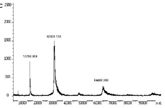
spectrometry results, which showed a major ion of 32028Da and minor ones of
~16000 and 64000Da (Figure 1C), suggesting that the lectin is mainly a dimer
composed of two identical subunits linked by cystine bonds, but other aggregate
levels are possible.
Table 1. Properties of the fractions obtained during lectin purification (from 100mg of
crude venom). Fraction Volume (ml) Protein concentration
(mg.ml-1)
Total protein (mg) Agglutination titera Total activity (AU)b Specific activity (AU/mg)c
Crude venom 5 20 100 4 20 0.2
Purified
fraction 28 0.18 5.2 16 448 86
a
Titer is recorded as the reciprocal highest dilution showing a +4 agglutination degree. b
Volume X agglutination titer c
Figure 1. Lectin purification. (A) Elution profile of crude venom (100mg) on agarose-D-galactose column (5ml), collected in 2ml fractions. Total proteins were eluted with CTBS, pH 7.5, and adsorbed fraction with 100mM lactose solution in the same buffer (indicated by the arrow). (B) SDS-PAGE of the purified fraction. MM: molecular mass marker proteins (kDa);
CV: Bothrops jararacussu crude venom; D: denaturant conditions in the presence of
2-mercaptoethanol; N: natural conditions. (C) MALDI-TOF mass spectrometry of B.
jararacussu lectin. The sample was applied using the dry droplet technique. The spectrum was acquired in linear mode.
Hemagglutination Characterization
BjcuL agglutinated all types of human erythrocytes, but showed a high preference for
type B over type A (data not shown), which indicates blood-group specificity,
depending on the cell-surface carbohydrate moieties. Divalent cations such as Ca2+,
Mg2+, Mn2+ were required for BjcuL activity, as their complete absence reduced
hemagglutination (Figure 2). The best results were obtained with Ca2+ and Mg2+ at 40
and 50mM, respectively. The lectin was more effective at a neutral pH range,
showing a gradual increase in activity from pH 5.0 to 7.0, 7.5 then decreasing up to
pH 8.5. It showed total loss of activity when assayed at pH values below 4.0 and
above 9.0. Agglutinating activity remained stable up to 60min at 25°C, but increased
when the lectin was left at 35°C for at least 15min. Its activity also remained stable
from 40 to 50°C up to 30min, then drastically reduced. At 70°C, the lectin was
Adhesion Assays
Adhesion assays to ECM glycoproteins showed that the biotinylated lectin (0.039–
5.0µg/100µl) was capable of binding, in a dose-dependent manner, to fibronectin and
vitronectin (Figure 3A). Binding was partially inhibited in the presence of D-galactose.
Mannose, up to a concentration of 100mM, did not inhibit BjcuL binding to the
glycoproteins (Figure 3B).
Intravital Microscopy
To investigate BjcuL potential for leukocyte rolling and adhesion to endothelial cells
under the conditions prevailing in living microvessels, the cremaster muscle of mice
was exposed for topical application of lectin (1.25, 2.5, 5, and 10μg/30μl) and
evaluated after 30min. A few rolling leukocytes (velocity>30µm/s), but essentially no
firmly adherent cells, were observed in the postcapillary venules of control mice
(black bars, Figure 4). Numerous leukocytes interacted with the endothelium in the
cremaster of BjcuL-treated mice (Figure 4A), and the vast majority of these cells
adhered firmly to the vessel walls after lectin application. The increase in rolling and
adherent leukocytes, induced by lectin, was dose-responsive (Figure 4B). The
average number of rolling and adherent leukocytes was higher in the lectin group
than in the control group until 30min (Figure 5). Analysis of the recorded videotapes
did not show any evidence of accumulated platelets in postcapillary venules of
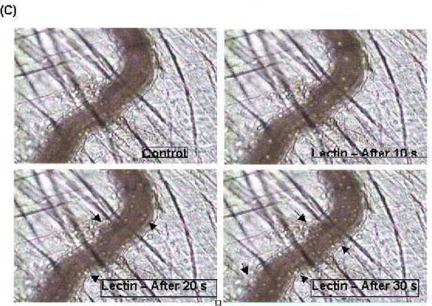
Figure 5. Evaluation of the kinectics of rolling and adherent leukocytes. Samples (30μl) containing 5μg lectin were topically applied on the cremaster muscle of anesthetized mice. The aspect of pre and postcapillary venules was observed every 10min during 30min and rolling (A) and adhesion (B) were recorded during 1min. The photos represent an intravital micrograph of cremaster muscle after 0, 10, 20 and 30s of topical application of lectin (C). Arrowheads indicate adhered leukocytes. * p<0.05, compared with control group.
DISCUSSION
Thrombolectin was the first lectin isolated from snake venom. It was discovered in the
venom of Bothrops atrox and identified as a β-galactoside-binding dimeric lectin of Mr
28000, similar in certain aspects to other Ca2+-dependent (C-type lectins; CTL) β
-galactoside-binding lectins of vertebrates (12). Soon after, hemagglutinins from
different snake venoms were discovered and characterized as a large family of
structurally homologous proteins. These are dimers of ~30kDa, composed of similar
disulfide-linked monomers with the CTL motif presenting saccharide-binding activity
(14).
BjcuL is a typical component of this group of molecules. Its purification was first
yield. Panunto et al. (29) isolated the lectin using a non-derivatized Sepharose 4-B
column and obtained a similar result. Our modification of the former method
consisted in a 4h agitation period at 4°C prior to elution, which allowed an ~5% yield.
Apparently, it permitted a better interaction between the venom-pool lectin molecules
and the gel matrix. The result of MALDI-TOF confirmed the purity of the
carbohydrate-free fraction: carbohydrate might have been present because of an
inefficient dialysis step or as column debris. Furthermore, this result confirms the
expected molecular weight of BjcuL monomers of ~16kDa (3). The amount of
recovered lectin is consistent with the analysis of B. jararacussu venom gland
transcriptome, which showed transcripts related to C-type lectin proteins to an order
of 5% (17).
BjcuL can adhere, although weakly, to human metastatic breast cancer and human
ovarian carcinoma cell lines, suppressing the viability of these tumor cells (5).
However, considering its action in envenomation, few speculations were presented.
BjcuL has been shown to be capable of producing edema and briefly increasing
vascular permeability in mouse hind paws (29), which are signs of inflammatory
processes. Extravasation of immune cells from peripheral blood through the vascular
endothelium into the ECM is also a common event in inflammatory manifestations.
We evaluated the cellular infiltration induced by lectin and our results, using intravital
microscopy, show that the lectin acts directly on endothelial cells of postcapillary
venules, creating an adhesive surface for the rolling of a great number of leukocytes.
Neutrophil influx induced by ip injection of B. jararaca venom into mice has been
shown to be related to the expression of adhesion molecules, responsible for rolling,
firm adhesion, and transmigration events associated with neutrophil migration (33).
Molecules in venom responsible for these effects have not been described yet, but it
is possible to anticipate that lectins are involved, as lectin-carbohydrate interactions
in leukocyte recruitment into inflammatory sites have become more evident (1).
Galectin-3 is chemotactic in humans, promoting adhesion of neutrophils as well as
other cell types to ECM proteins in an integrin-dependent manner (19,23). This
bridging of neutrophils to ECM due to galectin-3 appears to involve activation of the
cells via Ca2+ and Mg2+-dependent processes. Ganiko and collaborators (10,11)
artocarpin, with laminin, fibronectin and heparin makes possible the establishment
and maintenance of a KM+ molecular gradient, a necessary phenomenon to induce
the directed movement of neutrophils.
Here, we showed the interaction of BjcuL with ECM components and evaluated the
dependence of the carbohydrate recognition domain (CRD) in the interaction of BjcuL
with fibronectin and vitronectin. Snake venom lectins are known to bind mostly to
galactose and also to mannose (34). However, our results demonstrated that lectin
pre-incubated with 100mM D-galactose had its interaction capacity reduced by about
20%, whereas its incubation with 100mM D-mannose did not give rise to any
alteration. These results reinforce the hypothesis that CRDs are involved in the
inductive activity of migration (30) as well as the galactoside-binding preference of
BjcuL.
Hemagglutination assays were carried out in order to analyze the integrity of the
purified BjcuL as well as to establish the optimum conditions for biological studies.
Our results indicated the requirement for divalent cations other than Ca2+, such as
Mg2+, besides physiological conditions of pH and temperature for a better binding
activity, which was expected, as the venom must act on mammalian preys.
In conclusion, we demonstrated a further characterization of BjcuL with respect to its
hemaglutinating activity, which can be increased at neutral pH and temperature
around 37°C, in the presence of Mg2+, as a replacement for Ca2+. We showed that
BjcuL can adhere to fibronocetin and vitronectin and act directly on endothelial cells
of postcapillary venules, creating an adhesive surface for the rolling of a great
number of leukocytes. However, to consider its participation in inflammation events,
further investigation on its effect over leukocytes is needed.
ACKNOWLEDGEMENTS
We thank Butantan Institute for providing B. jararacussu venom, Nuno M. Domingues
(Brazilian Center for Protein Sequencing, University of Brasilia) for MALDI-TOF
analyses, and Dr. Silvio Sanches Veiga for the glycoproteins. This research was
Dr. M. Lopes-Ferreira is supported by The State of São Paulo Research Foundation
(FAPESP).
REFERENCES
1 BUTCHER EC. Leukocyte-endothelial cell recognition-three (or more) steps to
specificity and diversity. Cell., 1991, 67, 1033-6.
2 CAMPBELL JA., LAMAR WW. The venomous reptiles of Latin America. Ithaca;
London: Comstok Publ. Assoc., 1989, 425.
3 CARVALHO DD., MARANGONI S., NOVELLO JC. Primary structure
characterization of Bothrops jararacussu snake venom lectin. J. Prot. Chem., 2002,
21, 43-50.
4 CARVALHO DD., MARANGONI S., OLIVEIRA B., NOVELLO JC. Isolation and
characterization of a new lectin from the venom of the snake Bothrops jararacussu.
Rev. Biochem. Mol. Biol. Intern., 1998, 44, 933-8.
5 CARVALHO DD., SCHMITMEIER S., NOVELLO JC., MARKLAND FS. Effect of
BJcuL (a lectin from the venom of the snake Bothrops jararacussu) on adhesion and
growth of tumor and endothelial cells. Toxicon, 2001, 39, 1471-6.
6 DECLAUX C., DELACOURT C., D`ORTHO MP., BOYER V., LAFUMA C., HARF A.
Role of gelatinase B and elastase in human polymorphonuclear neutrophil migration
across basement membrane. Am. J. Respir. Cell. Mol. Biol., 1996, 14, 288-95.
7 DUBOIS M., GILLES KA., HAMILTON JK., REBERS PA., SMITH F. Colorimetric
method for determination of sugars and related substances. Anal. Chem., 1956, 28,
350-6.
8 FARSKY SHP., CRUZ JWMC., CURY Y., TEIXEIRA CFP. Leukocyte response
induced by Bothrops jararaca crude venom: in vivo and in vitro studies. Toxicon,
1997, 35, 185-93.
9 FLORES CA., ZAPPELLINI A., PRADO-FRANCESCHI J. Lipoxygenase-derived
mediators may be involved in in vivo neutrophil migration induced by Bothrops
erythromelas and Bothrops alternatus venoms. Toxicon, 1993, 31, 1551-9.
10 GANIKO L. Neutrophil haptotaxis induced by the lectin KM+. Glycoconj. J., 1998,
11 GANIKO L., MARTINS AR., FREYMULLER E., MORTARA RA.,
ROQUE-BARREIRA M-C. Lectin KM+-induced neutrophil haptotaxis involves binding to
laminin. Biochim. Biophys. Acta, 2005, 1721, 152-63.
12 GARTNER TK., OLGIVIE ML. Isolation and characterization of three Ca2+
-dependent b-galactoside-specific lectins from snake venoms. Biochem. J., 1984,
224, 301-7.
13 GOLDSTEIN IJ., HUGHES RC., MONSIGNY M., OSAWA T., SHARON N. What
should be called a lectin? Nature, 1980, 285, 66.
14 GUIMARÃES-GOMES V., OLIVEIRA-CARVALHO AL.,
JUNQUEIRA-DE-AZEVEDO IL., DUTRA DLS., PUJOL-LUZ M., CASTRO HC., HO PL., ZINGALI RB.
Cloning, characterization and structural analysis of a C-type lectin from Bothrops
insularis (BiL) venom. Arch. Biochem. Biophys., 2004, 432, 1-11.
15 GUTIÉRREZ JM., LOMONTE B. Local tissue damage induced by Bothrops snake
venoms. Mem. Inst. Butantan, 1989, 51, 211-23.
16 HAVT A., TOYAMA MH., NASCIMENTO NRFD., TOYAMA DO., NOBRE ACL.,
MARTINS AMC., BARBOSA PSF., NOVELLO JC., BOSCHERO AC., CARNEIRO
EM., FONTELLES MC., MONTEIRO HSA. A new C-type animal lectin isolated from
Bothrops pirajai is responsible for the snake venom major effects in the isolated
kidney. Intern. J. Biochem. Cell Biol., 2005, 37, 130-41.
17 KASHIMA S., ROBERTO PG., SOARES AM., ASTOLFI-FILHO S., PEREIRA JO.,
GIULIATI S., FARIA JÚNIOR M., XAVIER MAS., FONTES MRM., GIGLIO JR.,
FRANÇA SC. Analysis of Bothrops jararacussu venomous gland transcriptome
focusing on structural and functional aspects: I - gene expression profile of highly
expressed phospholipases A2. Biochimie., 2004, 86, 211-9.
18 KLEIMAN HK., MCGARVEY ML., HASSEL JR., STAR VL., CANNON FB.,
LAURIE GW., MARTIN GR. Basement membrane complexes with biological activity.
Biochemistry, 1986, 25, 312-8.
19 KUWABARA I., LIU FT. Galectin-3 promotes adhesion of human neutrophils to
laminin. J. Immunol., 1996, 156, 3939-44.
20 LAEMMLI UK. Cleavage of structural proteins during the assembly of the head of
21 LOWRY OH., ROSENBROUGH NJ., FARR IL., RANDALL RJ. Protein
measurement with the Folin phenol reagent. J. Biol. Chem., 1951, 193, 265-75.
22 LU Q., NAVDAEV A., CLEMETSON JM., CLEMETSON KJ. Snake venom C-type
lectins interacting with platelet receptors. Structure-function relationships and effects
on haemostasis. Toxicon, 2005, 45, 1089-98.
23 MASSA SM., COOPER DN., LEFFLER H., BARONDES SH. L-29, an
endogenous lectin, binds to glycoconjugate ligands with positive cooperativity.
Biochemistry, 1993, 32, 260-7.
24 MASTRO AM., HURLEY DJ., WINNING RK., FILIPOWSKI R., OLGIVIE ML.,
GARTNER TK. Mitogenic activity of snake venom lectins. Cell. Tissue Kinet., 1986,
19, 557-66.
25 MILANI R., JORGE MT., FERRAZ DE CAMPOS FT., MARTINS FP., BOUSSO
A., CARDOSO JLC., RIBEIRO LA., FAN HW., FRANÇA FOS., SANO-MARTINS IS.,
CARDOSO D., FERNANDEZ ICOF., FERNANDEZ JC., ALDRED VL., SANDOVAL
MP., PUORTO G., THEAKSTON RDG., WARRELL DA. Snake bites by jararacuçu
(Bothrops jararacussu): clinicopathological sudies of 29 proven cases in São Paulo
State, Brazil. Q. J. Med., 1997, 90, 323-34.
26 MOURA-DA-SILVA AM., LAING GD., PAINE MJI., DENNISON JMTI., POLITI V.,
CRAMPTON JM., THEAKSTON RGD. Processing of pro-tumor necrosis factor-alpha
by venom metalloproteinase: a hypothesis towards explaining local tissue damage
following snake bite. Eur. J. Immunol., 1996, 26, 200-5.
27 NORMAN KE., KATOPODIS AG., THOMA G., KOLBINGER F., HICKS AE.,
COTTE MJ., POCKLEY AG., HELLEWELL PG. P-Selectin glycoprotein ligand-1
supports rolling on E- and P-selectin in vivo. Blood, 2000, 96, 3585-91.
28 OZEKI Y., MATSUI T., HAMAKO J., SUZUKI M., FUJIMURA Y., YOSHIDA E.,
NISHIDA S., TITANI K. C-type galactoside-binding lectin from Bothrops jararaca
venom: comparison of its structure and function with those of Bothrocetin. Arch.
Biochem. Biophys., 1994, 308, 306-10.
29 PANUNTO PC., SILVA MA., LINARDI A., BUZIN MP., MELO SE., MELLO SM.,
PRADO-FRANCESCHI J., HYSLOP S. Biological activities of a lectin from Bothrops
30 SANTOS-DE-OLIVEIRA R., DIAS-BARUFFI M., THOMAZ SMO., BELTRAMINI
LM., ROQUE-BARREIRA MC. Neutrophil migration-inducing lectin from Artocarpus
integrifolia. J. Immunol., 1994, 153, 1798-807.
31 SPERANDIO M., SMITH ML., FORLOW SB., OLSON TS., XIA L., MCEVER RP.,
LEY K. P-selectin glycoprotein ligand-1 mediates L-selectin-dependent leukocyte
rolling in venules. J. Exp. Med., 2003, 197, 1355-63.
32 YATOHGO T., IZUMI M., KASHIWAGI H., HAYASHI M. Novel purification of
vitronectin from human plasma by heparin affinity chromatography. Cell Struct Funct.,
1988, 13, 281-92.
33 ZAMUNER SR., TEIXEIRA CFP. Cell adhesion molecules involved in the
leukocyte recruitment induced by venom of the snake Bothrops jararaca. Mediators
Inflamm., 2002, 11, 351-57.
34 ZHA HG., LEE WH., ZHANG Y. Cloning of cDNA encoding C-type lectins form
Elapidae snakes Bungarus fasciatus and Bungarus multicinctus. Toxicon, 2001, 39,