IN VITRO
ACTIVITY OF ANTIFUNGAL AND ANTISEPTIC AGENTS AGAINST
DERMATOPHYTE ISOLATES FROM PATIENTS WITH TINEA PEDIS
Maria Magali Stelato Rocha Soares
1*; Arlete Emily Cury
21
Instituto de Ciências Biológicas e Químicas, Faculdade de Ciências Farmacêuticas,
PUC de Campinas, Campinas, SP, Brasil.
2Faculdade de Ciências Farmacêuticas, Universidade de São Paulo,
São Paulo, SP, Brasil.
Submitted: February 02, 2001; Approved: May 28, 2001
ABSTRACT
The
in vitro
activity of antifungal and antiseptic agents were evaluated against dermatophytes isolated from
patients with tinea pedis. The antifungals studied were: ciclopirox olamine, cetoconazole, tolciclate and terbinafine,
and the antiseptics were: povidine iodine (PVPI), propolis, Fungol®, Andriodermol®, and boric acid. The minimum
inhibitory concentration (MIC) or the minimal dilution concentration (MDC) was determined by an agar dilution
method using modified yeast nitrogen agar base, and the minimum fungicidal concentration (MFC) or minimum
fungicidal dilution (MFD) was determined with subcultures on Sabouraud dextrose agar. All drugs studied were
active against the dermatophytes at lower concentrations than those used in products and/or pharmaceutical
preparations for topical use. Some antifungal agents, mainly terbinafine and tolciclate, presented higher efficacy
than the other drugs, with lower MICs and MFCs values. It was concluded that the use of these antiseptic drugs
represent an excellent alternative for the topical treatment of tinea pedis. For the treatment of severe cases these
are the antifungal agents of choice.
Key words: dermatophytes, antifungal agents, antiseptic agents, susceptibility testing, tinea pedis
INTRODUCTION
Tinea pedis is one of the most frequent mycoses; it occurs in
several classes of patients, but especially in immunosupressed
individuals (21).
Topical or systemic treatments have been empirically
administered, when, in general, specific antifungal agents are
employed (3,13,18). Although such drugs are widely used, some
of them have been reported as ineffective and even toxic to the
host (3,4,9,10,13,18). These factors have lead to the development
and marketing of new drugs, but most of them have the same
pharmacological active groups and the same mechanisms of action
as those previously commercially available (9,10,13). However,
some fungi are significantly different regarding susceptibility to
such drugs (5,8,12,20,23,24).
* Corresponding author. Mailing address: Instituto de Ciências Biológicas e Químicas, Laboratório de Microbiologia, PUC de Campinas, Av. John Boyd Dunlop, s/n, Jd. Ipaussurama, Caixa Postal 1111. 13059-900, Campinas, SP, Brasil. Tel.: (+5519) 729-8307. Fax: (+5519) 729-8517. E-mails microimu.acad.pucamp.com.br. or [email protected]
Therefore, despite the need of further studies focusing on the
solution of these medical problems, there has been increasing
interest in the usage of chemical substances with non-specific
activity in the treatment of mycoses, such as antiseptic drugs
(2,6,11,14,16).
The aim of this study was to evaluate the
in vitro
activity of
commercially available antifungal and antiseptic agents against
dermtophytes isolated from patients with tinea pedis.
MATERIALS AND METHODS
Patients
de Campinas) and 136 cadets and soldiers were from the military
academy clinic (Escola Prepatória de Cadetes do Exército
-EsPECx).
Mycological tests
Collection and direct examination of the clinical samples, as
well as isolation and fungal identification were performed according
to classic techniques (13,17).
Susceptibility testing
Microorganisms and Inoculum
Sixty six dermatophytes were isolated from the patients
under study, including 25 strains of
Trichophyton rubrum
, 36
strains of
Trichophyton mentagrophytes
,
and 5 strains of
Epidermophyton floccosum
. These strains were maintained on
Saboraud dextrose agar (SDA) and potato dextrose agar (PDA)
at 27-30ºC. They were subcultured at 3 months intervales, but
observed on a weekly bases. Just before use, the samples were
transferred to SDA and maintained for 7 days at 27ºC-30ºC
(17,19,20).
The strains
Candida pseudotropicalis
“Carshanton” and
C.
krusei
6258, both sensitive to cetoconazole (MIC range: 0.25 to 4
µm/ml), were used as controls. They had been obtained from the
Centro de Referência da Faculdade de Medicina, USP, SP. These
yeasts were maintained on SDA under the same conditions as the
dermatophytes. During this study, they were kept on SDA for 24
hours at 27ºC-30ºC (17,19,20).
The inoculum was prepared according to previous studies
(20), and attained aproximately 10
6cels/ml.
Antifungal and antiseptic agents
Commercially available products were utilized. The
antifungal drugs were cetoconazole and ciclopirox olamine
(Galena Química Farmacêutica Ltda), tolciclate (Carlo Erba S.A.)
and terbinafine cloridrate (Sandoz S.A.). The antiseptics were
Andriodermol® (Searle do Brasil Ltda), Fungol® (Laboratório
Silva Araujo Roussel), Propolis (Uniflora Apicultores
Associados Ltda), boric acid (Indafarma Indústria e Comércio
de Produtos Químicos Ltda) and povidine iodine (PVPI - Galena
Química Farmacêutica Ltda).
The antifungal agents were kept at 4ºC under vacuum, and the
antiseptic agents were maintained at room temperature. All the
drugs were kept away from light and moisture.
The stock solutions of cetoconazole, terbinafine and
tolciclate were prepared in dimethilsulphoxide (DMSO); the stock
solutions of ciclopirox olamine, PVPI and Andriodermol® were
prepared in absolute alcohol and the boric acid stock solution
was prepared in water (17,19,20). Organic solutions were left at
room temperature for 30 minutes in order to sterilize them. The
aqueous solutions were sterilized by filtration (0.22 µm filter)
(17,19,20). It was not necessary to prepare stock solutions of
Fungol® and Propolis.
Performance and reading
The susceptibility tests were performed as previously
described (20)
with modifications. The tests were performed in
duplicates and the final results were presented as the arithmetic
average of the values obtained.
Some fungi cultures were lost during the study, therefore not
all drugs were studied against all dermatophyte isolates.
Determination of minimum inhibitory concentration (MIC) or
minimum inhibitory dilution (MID)
From each antifungal agent stock solution, decimal dilutions
were prepared in modified yeast nitrogen base broth (YNBP). For
each dilution, 2 ml were added to 18 ml of YNBP before plating.
Under such conditions, the antifungal agent’s concentrations
ranged from 128 to 0.25 µg/ml for PVPI, cetoconazole and ciclopirox
olamine; from 16 to 0.031 µg/ml for tolciclate; from 4.0 to 0.007 µg/
ml for terbinafine; from 512 to 1.0 µg/ml for boric acid and from
2000 to 3.9 µg/ml for própolis. Since Andriodermol® and Fungol®
contain several active ingredients, they were studied at dilutions
ranging from 1:150 to 1:76800 and from 1:100 to 1:25600,
respectively.
In these tests, two additional plates were prepared as controls.
The first plate contained YNBP without any drug (B1) while the
second plate contained YNBP with the proper diluent
concentration for each specific drug studied (B2).
One µl of each dermatophyte and the control yeast
suspension was placed on each plate. The plates were incubated
at 27ºC-30ºC until visible growth of each fungus in the B1 control
(around 5 to 7 days).
The MIC or MID was defined as the lowest concentration or
highest dilution of the antifungal agent which resulted in plates
without visible colonies.
Determination of minimum fungicidal concentration (MFC)
or minimum fungicidal dilution (MFD)
Each inoculum which resulted in no growth in the previous
test and the growth on the control plates (B1 and B2) were
subcultured on SDA. The plates were incubated at 27ºC-30ºC
until growth of subculture on the control plates was visible.
The MFC or MFD was defined as the lowest concentration
or highest dilution of drug resulted in plates without visible
colonies.
RESULTS
Ciclopirox olamine
Terbinafine
The MICs of terbinafine for the strains were 0.007 µg/ml or
0.015 µg/ml. Most strains of
T. rubrum
(16; 72.7%),
T.
mentagrophytes
(24; 72.7%) and
E. floccosum
(2; 50%) were
inhibited at concentration of 0.007 µg/ml. The MFC ranged from
0.03 µg/ml to > 4 µg/ml. This antifungal agent was lethal to two
strains of
E. floccosum
at the concentration of 0.03 µg/ml, and at
0.5 µg/ml it was lethal to the other two strains. The fungicidal
concentration for 13 (59.1%) strains of
T. rubrum
was up to 0.25
µg/ml, and for 20 (60.6%) strains of
T. mentagrophytes
it was up to
0.5 µg/ml. Only 2 and 6 strains of
T.rubrum
and
T. mentagrophytes
,
respectively, were not killed by concentrations up to 4 µg/ml (Table 1).
Cetoconazole
The activity of cetoconazole, in terms of MIC, ranged from 1
µg/ml to 32 µg/ml. Yet, at concentrations up to 4 µg/ml it inhibited
most strains of
T. rubrum
(15; 68.2%) and
T mentagrophytes
(23;
67.6%). Among the five strains of
E. floccosum
, four were
inhibited at concentrations up to 2 µg/ml. The MFC of this
antifungal agent ranged from 4 µg/ml to >128 µg/ml. It was
observed that the lethal concentrations for most isolates of
T.
rubrum
(12; 54.5%) and
T. mentagrophytes
(20; 58.8%) were up
to 16 µg/ml and 32 µg/ml, respectively, while for 12 (35,3%)
isolates of
T. mentagrophytes
and two of
E. floccosum
it was 128
µg/ml (Table 1).
Tolciclate
For this antifungal agent, the MICs ranged from 0.03 µg/ml to
0.5 µg/ml. Most isolates of
T. rubrum
(18; 78.3%),
T. mentagrophytes
(19; 57.6%) as well as two strains of
E. floccosum
were inhibited at
concentrations of 0.06 µg/ml. The susceptibility, in terms of MFC,
ranged from 0.5 µg/ml to >16 µg/ml. The lethal concentration for
most strains of the three species studied was up to 4 µg/ml. Two
strains of
T. rubrum
were regarded as exceptions; for one of them,
the MFC was 8 µg/ml, and for the other, it was > 16 µg/ml (Table 1).
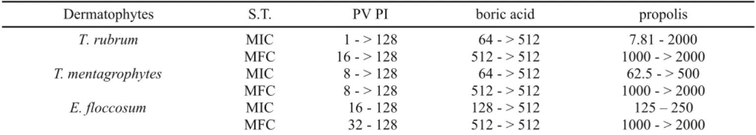
PVPI
The MICs for PVPI ranged from 4 µg/ml to > 128 µg/ml. However,
only one strain each of
T.rubrum
and
T. mentagophytes
was
inhibited at concentrations > 128 µg/ml. The MFCs ranged from 8
µg/ml a > 128 µg/ml. Concentrations up to 128 µg/ml were not
lethal to 3 strains of
T. rubrum
and one of
T. mentagrophytes.
For
two strains of
E. floccosum
the lethal concentration was 32 µg/ml
and for two other, it was 128 µg/ml (Table 2).
Boric acid
Boric acid’s MICs ranged from 64 µg/ml to 512 µg/ml. However,
most isolates of
T. rubrum
(14; 63.6%) and of
T.
mentagrophytes
(32; 91.4%) were inhibited respectively up to 256 µg/ml and 512
µg/ml. One of the
E. floccosum
strains was inhibited at 128 µg/ml,
while two others were inhibited at 512 µg/ml, but 2 of the strains
was not susceptible to this antifungal agent at the concetrations
studied. The MFCs ranged from 512 µg/ml to > 512 µg/ml against
the three species of dermatophytes studied. (Table 2).
Propolis
The MICs of this agent ranged from 7.81 µg/ml to > 2000
µg/ml. However, most strains of
T. rubrum
(12; 54.5%) were
Dermatophytes S.T. ciclopirox olamine terbinafine cetoconazole tolciclate
T. rubrum MIC 8 - 32 0.007 – 0.015 1 - 32 0.003 – 0.125
MFC 16 - 64 0.03 - > 4 4 - 128 1 - > 16
T.mentagrophytes MIC 16 - 32 0.007 – 0.015 1 - 32 0.003 – 0.5
MFC 16 - > 128 0.03 - > 4 8 - > 128 0.5 - 4
E. floccosum MIC – 32 0.007 – 0.015 1 - 8 0.003 – 0.25
MFC 16 - 32 0.03 - > 0.5 16 - 128 1 – 4
*
Table 1.
In vitro
susceptibility of dermatophytes to four antifungal agents.
*
S.T. = Susceptibilty testing range of activity of antifungal agents at different concentration (µg/ml); MIC = minimum inhibitory
concentration; MFC = minimum fungicidal concentration.
Table 2.
In vitro
susceptibility of dermatophytes to three antiseptic agents.
inhibited at the concentration of 125 µg/ml. For 22 (68.8%)
strains of
T. mentagrophytes
, the inhibition occurred at
concentrations up to 250 µg/ml of this antiseptic, and for only
one strain it was at the concentration > 2000 µg/ml. Two of the
E. floccosum
strains were inhibited by 125 µg/ml, and the two
others, were inhibited at 250 µg/ml. The MFCs of própolis, for
these three species, ranged from 1000 µg/ml to > 2000 µg/ml
(Table 2).
Fungol®
Most strains studied were susceptible to this product diluted
at 1:100. The exceptions were one strain of
T. rubrum
(4.6%),
two strains of
T. mentagrophytes
(5.9%) and one strain of
E.
floccosum
. At this dilution, Fungol® presented only
fungistactic activity against the other strains (5; 14.7%) of
T.
mentagrophytes
(Table 3).
Andriodermol®
All strains were inhibited by this product at concentrations as
low as 1:1200. Except for one isolate of
T. mentagrophytes
,
Andriodermol® was lethal to all strains studied at the dilution of
1:150 (Table 3).
DISCUSSION
In general, the clinical dermatophyte isolates were susceptible
to the tested antifungal and antiseptic agents in concentrations
that were lower than those incountered in commercial products
for topical use.
The MICs and MFCs obtained for the antifungal agents were
slightly lower than those obtained for the antiseptic drugs.
Terbinafine and tolciclate were the most potentially active
antifungal drugs, at low concentrations against the dermatophytes
studied. However, it should be noted that terbinafine at
concentrations of up to 4 µg/ml was not lethal to 2 (9.1%) of the
T.
rubrum
strains and to 6 (18.2%) of the
T. mentagrophytes
strains.
The same inactivity pattern was observed for tolciclate at
concentrations of up to 16 µg/ml against one of the 22
T. rubrum
strains. Although both drugs presented the same mechanism of
action (9,10,13), from the data that was obtained, it can be concluded
that there was not cross reactions between them. In the literature,
reports were found describing the use of terbinafine, that focused
the inhibitory activity of this drug. Except the work of the Arzeni
et al.
(1), who showed MIC up to 2,0 µg/ml, the MICs reported in
the literature ranged from 0.001 µg/ml to 0.1 µg/ml (5,12,22-23)
which were compatible with the results obtained in this work.
However, the data obtained for the MFCs, with values reaching >
4 µg/ml, diverged from the data presented by Fukuda
et al.
(7)
and
Arzeni
et al.
(1) to who reported values reaching up to 2.5 µg/ml
and 2.0 µg/ml, respectively.
The results obtained with tolciclate are in accordance to the
results of Zaror
et al.
(24). Those authors reported that all of the
dermatophytes studied by them were inhibited by tolciclate at
concentrations equal to or lower than 2.56 µg/ml. No reports were
found in the literature regarding the fungicidal activity of this
antifungal agent.
The drugs showing the highest MICs and MFCs were
própolis and boric acid (Table 2). Unlike the própolis, boric
acid was not lethal to most strains at the concentrations tested.
Although the literature lacked
in vitro
studies regarding the
effect of boric acid against dermatophytes, the papers
published with própolis solely included the determination of
the fungistactic action of this antiseptic. In general, data
presented in those studies are in accordance with the data
obtained in the work reported here. Some authors observed
that the própolis ethanol extract at 10% (15) or própolis at 5%
in hydroalcoholic gel or in propileneglicol gel (16) were able to
inhibit the growth of all strains of the dermatophytes studied.
In addition, comparing the activity of própolis to some specific
antifungal agents, some authors (16) found that there is a
similarity among the different drugs at the same concentrations
that are commercially available.
Although further studies are needed, including
in vivo
investigations, the data obtained indicate that antiseptic drugs
can be used as good alternatives in the topical treatment of
minor cases of tinea pedis, leaving the specific antifungal
drugs for use in the treatment of more severe cases of this
mycosis.
Table 3.
In vitro
susceptibility of dermatophytes to two antiseptic agents.
Dermatophytes S.T. FungolR AndriodermolR
T. rubrum MID 1/38400 - > 1/600
MFD 1/2400 - > 1/150
T. mentagrophytes MID 1/9600 - > 1/600
MFD 1/2400 - > 1/150
E. floccosum MID 1/9600 - > 1/1200
MFD
1/3200 - > 1/100 1/800 - > 1/100 1/800 - > 1/100 1/400 - > 1/100 1/400 - > 1/100
1/200 - > 1/100 1/2400 - > 1/150
RESUMO
Atividade in vitro de antifúngicos e
anti-sépticos frente a dermatófitos isolados
de pacientes com tinea pedis
A atividade
in vitro
de antifúngicos e anti-sépticos foram
avaliadas frente a dermatófitos isolados de pacientes com tinea
pedis. Os antifúngicos estudados foram: ciclopirox olamine,
cetoconazol, tolciclato e terbinafina, e os anti-sépticos foram:
iodo povidine (PVPI), própolis, Fungol®, Andriodermol®
e ácido
bórico. A concentração inibitória mínima (CIM) ou a diluição
inibitória mínima (DIM) foi determinada pelo método de diluição
em ágar utilizando “yeast nitrogen” base modificado, e a
concentração fungicida mínima (CFM) ou diluição fungicida
mínima (DFM) foi determinada por subcultura em Saboraud
dextrose ágar. Todas as drogas estudadas foram ativas frente
aos dermatófitos em concentrações menores do que as utilizadas
em produtos e/ou preparações farmacêuticas para uso tópico.
Alguns antifúngicos, principalmente a terbinafina e o tolciclato,
foram mais eficazes do que outras drogas estudadas,
apresentando CIMs e CFMs mais baixos. Concluíu-se que os
anti-sépticos representam uma ótima alternativa para tratamento
tópico de vários casos de tinea pedis sendo, entretanto, os
antifúngicos reservados para formas severas de tinea.
Palavras-chave:
dermatófitos, antifúngicos, anti-sépticos, teste
de susceptibilidade, tinea pedis
REFERENCES
1. Arzeni, D.; Barchiesi, F.; Compagnucci, P.; Cellini, A.; Simonetti, O.; Offidani, A.M.; Scalise, G. In vitro activity of terbinafine against clinical isolates of dermatophytes Med. Mycol.,
36:235-237, 1998.
2. Azevedo, R.V.; Komesu, M.C.; Candido, R.C.; Salvetti, C.; Rezende, F.H.C. Candida sp in the oral cavity with and without lesions: maximal inhibitory dilution of própolis and PERIOGARD. Rev. Microbiol.,
30:335-341,1999.
3. Bodey, G.P. Azole Antifungal agents. Clin. Infect. Dis., 14:S161-S169,
1992.
4. Carazo, J.L.S.; Losada, L.O.; Sanjuan, V.P. Tratamiento actual de las micosis superficiales. Rev. Iberoam. Micol., 16: S26-S30, 1999. 5. Clayton, Y.M. In vitro activity of terbinafine. Clin. Exp. Dermatol.,
14:101-103, 1989.
6. Cury, A.E. Atividade in vitro de alguns antissépticos bucais sobre Candida. Rev. Microbiol., 17:137-142, 1986.
7. Fukuda, T.; Naka, W.; Tajima, S.; Nishikawa, T. Neutral red assay in minimum fungicidal concentrations of antifungal agents. J. Med. Vet. Mycol.,34:353-356, 1996.
8. Furtado, M.S.S.; Minami, P.S. In vitro susceptibility tests of dermatophytes to griseofulvin and imidazole derivates, Rev. Microbiol.,
28:110-115, 1997.
9. Gupta, A.K.; Sauder, D.N.; Shear, N.H. Antifungal agents: An overview. Part I. J. Acad. American. Dermatol., 30:677-698, 1994.
10. Gupta, A.K.; Sauder, D.N.; Shear, N.H. Antifungal agents: An overview Part II. J. Acad. American. Dermatol., 30:911-933, 1994. 11. Hammer, K.A.; Carson, C.F.; Riley, T.V. Melaleuca alternifolia (tea
tree) oil inhibits germ tube formation by Candida albicans.Med. Mycol., 38: 355-362, 2000.
12. Jessup, C.J.; Ryder, N.S., Ghannoum, M.A. An evaluation of in vitro
activity of terbinafine. Med. Mycol., 38:155-159, 2000.
13. Lacaz, C.S.; Porto, E.; Martins, J.E.C. Micologia Médica:fungos, actinomicetos e algas de interesse médico. 8ª ed. Sarvier, São Paulo,
1991, 695p.
14. Lima, E.O. Estudo das Dermatofitoses em João Pessoa - Paraíba e da Atividade antifúngica de plantas medicinais da região contra alguns dos agentes isolados. São Paulo, 1996. (Ph.D. Thesis. Facudade de
Ciências Farmacêuticas. USP).
15. Milena, L.; Leifertova, I; Baloun, I. Fungistatic effect of própolis.
FoliaPharm. Univ. Carol., 13:29-44,1989.
16. Millet-Clerc, J.; Miclhel, D.; Simeray, J.; Chaumont, J.P. Étude préliminaire des propriétes fongistatiques de la própolis comparées à celles de queques produits commerciaux. Plant. Med. Phytother.,
21:3-7, 1987.
17. Murray, P.R.; Drew, W.L.; Kobayashi, J.S.; Thompson, J.H. Manual of Clinical Microbiology. ASM, Washington, 1992.
18. Roberts, D.T.; Cox, N.H.; Gentles, J.C.; Babu, K.K.R. Comparison of ketoconazole and griseiolfulvin in the treatment of tinea pedis. J. Med. Vet. Mycol., 25:347-350, 1987.
19. Shadomy, S.; Pfaller, M.A. Laboratory studies with antifungal agents: susceptibility tests quantitation in body fluids and biossays. In: Manual of Clinical Microbiology. 5ª ed. Ed. A.Balows, W.J.; Hausler Jr., K.L.; Herrmann, H.D.; Isenberg, H.J.; Shadomy. Washington, American Society for Microbiolgy, 1991, p.1173-83.
20. Soares, M.M.S.R.; Cury, A.E. Atividade de alguns antimicóticos contra fungos isolados de diferentes classes de pacientes. Rev. Bras. AN. Clin., 27: 3-11, 1995.
21. Soares, M.M.S.R.; Cury, A.E.; Schreiber, A.Z. Micose superficial da região podal em indivíduos considerados imunocomprometidos. An. Bras. Dermatol., 70:211-217, 1995.
22. Schuster, I.; Schaude, M.; Schatz, F.; Mieth, H. Preclinical characteristics of allylamines. In: Berg, D.; Plempel, M. (eds.) Sterol Biosynthesis Inhibitors: Pharmaceutical and Agrochemical Aspects.
Chichester. Ellis Horwood, 1988 : 449-470.
23. Venugopal, P.V.; Venugopal, T.V. Disk diffusion susceptibility testing of dermatophytes with allylamines. Int. J. Dermatol., 33:730-732, 1994.