197 197 197 197 197 Mem Inst Oswaldo Cruz, Rio de Janeiro, Vol. 92(2): 197-203, Mar./Apr. 1997
The First Instar Larva of
Lutzomyia longipalpis
(Diptera:
Phlebotomidae)
Antônio Cesar Rios Leite
+, Paul Williams
Departamento de Parasitologia, Instituto de Ciências Biológicas, Universidade Federal de Minas Gerais, Caixa Postal 486, 31270-920 Belo Horizonte, MG, Brasil
The morphology and chaetotaxy of the first instar larva of Lutzomyia(Lutzomyia) longipalpis are described based on observations made under scanning electron microscope. Because three-dimen-sional images were studied, some terminological changes are proposed to give a more realistic de-scription of the positions of the setae. On the larval body, the pairs of setae have the following number: 9 on the head, 12 on the prothorax, 8 on the meso- and metathorax, 6 on the first to eighth abdominal segments, and 8 on the ninth abdominal segment.
Key words: Lutzomyia longipalpis - morphology - first instar - larva - chaetotaxy - scanning electron microscopy
Lutzomyia (Lutzomyia) longipalpis (Lutz and Neiva, 1912) is the type species of the genus Lutzomyia França (Diptera: Phlebotomidae). This sand fly is of medical and veterinary interest be-cause it is the most important insect host of Leish-mania chagasi Cunha and Chagas, the causative organism of American visceral leishmaniasis. L. longipalpis has proved to be amenable to labora-tory colonization and some closed colonies have now been maintained for almost 20 years.
Although laboratory-reared material has been available for many years, little is known of the mor-phology of the immature stages of L. longipalpis. Guitton and Sherlock (1969), based on studies by optical microscopy, described the egg, fourth in-star larva and pupa. Accepting the limitations of their study methods, the descriptions of Guitton and Sherlock (1969) are inadequete.
Scanning electron microscope (SEM) was used by Ward and Ready (1975) to study the chorionic sculpture of sand fly eggs, including those of L. longipalpis. SEM studies have also been the basis of a description of the pupa of L. longipalpis (Leite et al. 1991). Herein, we provide illustrations of SEM studies on the first instar larva of L. longipalpis, like that already described for the fourth instar larva (Leite & Williams 1996).
This research was financially supported, in part, by CNPq.
+Corresponding author. Fax: +55-31-441.6909. E-mail:
[email protected]. Received 17 May 1996 Accepted 27 November 1996
MATERIALS AND METHODS
Specimens were obtained from a closed labo-ratory colony that has been maintained in Belo Horizonte since 1983. The colony originated from blood fed females collected in Abaetetuba, State of Pará, Brazil, by Marisa Cenizio dos Santos in collaboration with members of the Wellcome Para-sitology Unit, Belém in 1983.
Larvae were killed by dropping them in hot water (70°C). They were fixed in 70% ethyl alco-hol, dehydrated in a sequence of increasing con-centrations of ethyl alcohol, submitted to critical point drying in carbon dioxide, and spattered with colloidal gold (Leite & Williams 1996).
Apart from the head and the ninth abdominal segment, descriptions of setae are given in an an-terior-posterior sequence, beginning from the dorsal mid-line and working circumferentially in a latero-ventral direction. The nomenclature used here is based on that of Barretto (1941) and previ-ously adopted by Leite and Williams (1996).
RESULTS
The description is based on examination of 12 larvae.
198 198 198 198
198 First Instar Larva of Lutzomyia longipalpis ACR Leite, P Williams
Barbed setae on the head and on the lateral and ventral surfaces of segments have pointed tips.
Head - The head bears simple (bare) setae anteriorly and barbed (brush-like) setae posteri-orly (Fig. 2). Spinose hairs or finely pointed spines are implanted on dorso-, lateral- and ventro-pos-terior surfaces of the head (Figs 4,12). The mouth-parts (Fig. 3) are of a chewing type. The labrum is wide, with spiculate ridges. Each mandible has four strong teeth in a ventral position. The maxilla is spatulate; the maxillary palp is spinose and bears papiliform processes. The mentum bears four pairs of strong teeth, and the prementum has several denticles. The labio-hypopharyngeal complex (Fig. 7) lying between the mouth and dorso-mentum, is armed with lateral and ventral premental teeth, premental cusps, and premento-lingular teeth. The labio-hypopharynx has dorsal oral fringes. The antenna (Fig. 5) has a socket and two segments; the proximal segment is short and cylindrical, whereas the distal segment is ovoid, with an apical pyriform appendage. The egg breaker (Fig. 6) has the appearance of a sharp tipped pyramid. Exclud-ing the simple setae on the mandible (one large and two small setae) and maxilla (one large and one small setae), the numerical chaetotaxy of the head is as follows: anterior clypeal (1)!; posterior clypeal (2)!; anterior frontal (3)+; posterior frontal (4)*; dorsal vertical (5)*; lateral vertical (6)*; dor-sal genal (7)!; lateral genal (8)!; ventral genal (9)!. Prothorax - It has the appearance of two segments (Figs 11, 12). Anterior and posterior se-tae are separated by a transverse groove. The nu-merical chaetotaxy of the prothorax is as follows: intermediate anterior dorsal (1)*; external anterior dorsal (2)*; anterior lateral (3)!; external anterior ventral (4)!; intermediate anterior ventral (5)!, in-ternal posterior dorsal (6)*; intermediate posterior dorsal (7)*, external posterior dorsal (8)*; exter-nal posterior ventral (9)*; intermediate posterior ventral (10)*; small intermediate posterior ventral (11)!; internal posterior ventral (12)*. A small, lateral tubercle below seta 8 (Fig. 13) could be a rudimentary anterior spiracle.
Meso- and metathorax - These two segments (Figs 11, 12 and 14) have the same arrangement of setae: internal dorsal (1)*; intermediate dorsal (2)*; lateral accessory (a)!; lateral (4)*; external ventral (5)*, intermediate ventral (6)*, small intermediate ventral (7)*; internal ventral (8)*. Fig. 11 shows an anomalous condition: two setae arising from the same tubercle.
First seven abdominal segments - The topog-raphy of each seta, and their name, are identical for each segment: internal dorsal (1)+; intermedi-ate dorsal (2)*; external dorsal (3)*; dorsal acces-sory (a)!; lateral (4)*; ventral lateral (5)*; ventral (6)!. Ventrally, each of these abdominal segments have pseudopodia (Fig. 18) bearing small papil-lae.
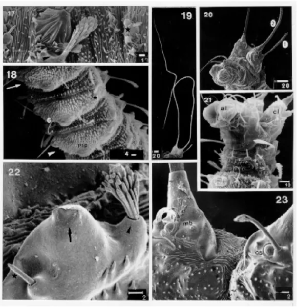
Eighth abdominal segment -This segment has the following arrangement of setae: internal dor-sal (1)*; intermediate dordor-sal (2)*; dordor-sal accessory (a)!; lateral (3)*; lateral accessory (b)!; external ventral (4)+; intermediate ventral (5)+; internal ventral (6)+. The posterior spiracle, with an ap-parently transverse opening, lies between setae 2 and a (Fig. 22).
Ninth abdominal segment - The last abdomi-nal segment is quite different from these lying an-teriorly (Figs 20, 21). There is a dorsal pair of prominent caudal lobes, with a single caudal seta arising from each lobe (Figs 19, 20). The follow-ing structures are visible (Fig. 23) at the base of each caudal lobe: a mammiliform button (or sensillum), a campaniform sensillum; the base of the caudal seta, and the posterior lobular seta. A caudal depression is rugose anteriorly and spicu-late posteriorly. Each caudal seta is 0.70 mm long. The anus lies between two ventral lobes. The arrangement of setae is as follows: anterior lobu-lar caudal (1)+; caudal (2)!; posterior lobulobu-lar cau-dal (3)+; intermediate post anal (4)!; external post anal (5)!; internal anal (6)!; intermediate pre-anal (7)!; internal pre-pre-anal (8)!.
DISCUSSION
The immature stages of phlebotomine sand flies are little known. This is because immatures are rarely encountered in the field and because most species are extremely difficult to rear in labora-tory conditions. The available descriptions of lar-vae are based on light microscopy studies. Most commonly, detailed descriptions have been given only of the fourth instar larva. Descriptions of the earlier instars have been much briefer and often only record the extent to which they differ from the fourth instar.
A handicap in preparing morphological de-scriptions of the larvae of phlebotomines arises from a lack of uniformity in the terminology used. The nomenclature proposed most recently for the larvae of New World sand flies (Ward 1976a) was devised to describe fourth instar larvae of species belonging to the subgenera Nyssomyia and Psychodopygus (Ward 1976b). These proposals proved to be inadequate for a description of the first stage of L. longipalpIs, a member of a dif-ferent subgenus. The descriptive terms used by ! : simple (bare) setae; + : small barbed setae; * : barbed
199 199 199 199 199 Mem Inst Oswaldo Cruz, Rio de Janeiro, Vol. 92(2), Mar./Apr. 1997
Scanning electron photographs of the first instar larva of Lutzomyia longipalpis. Fig. 1: hatching larva in lateral view. Fig. 2: head in latero-frontal view. Fig. 3: mouth parts in frontal view. Fig. 4: head in dorsal view. Fig. 5: antenna in posterior view. Fig. 6: egg breaker in anterior view. Bars in mm. Abbreviations: a: antenna, ap: antennal papilla, as: apical segment, aso: antennal
200 200 200 200
200 First Instar Larva of Lutzomyia longipalpis ACR Leite, P Williams
Scanning electron photographs of the first instar larva of Lutzomyia longipalpis. Fig. 7: labio-hypopharyngeal complex. Fig. 8: weakly barbed anterior frontal seta. Fig. 9: barbed posterior frontal seta with the spine-like branches. Fig. 10: simple lateral genal seta. Fig. 11: first (arrow) second and third thoracic segments, and the first abdominal segment (arrow head) in dorso-lateral view. Fig. 12: first (arrow) and second (arrow head) thoracic segments in latero-vental view. Fig. 13: probable anterior spiracle (arrow) in lateral view. Fig. 14: metathorax setae, showing the ventral external seta (black mark), intermediate ventral seta (asterisk), small intermediate ventral seta (star) and internal ventral seta (arrow head). Fig. 15: spinose tubercules on the dorsal and lateral sides of the abdominal segments (except on the last). Fig. 16: idem, on the ventral side. Bar in mm.
201 201 201 201 201 Mem Inst Oswaldo Cruz, Rio de Janeiro, Vol. 92(2), Mar./Apr. 1997
Scanning electron photographs of the first instar larva of Lutzomyia longipalpis. Fig. 17: dorsal intermediate seta (arrow head) with spatulate branches and tubercule (star) on the second abdominal segment. Fig. 18: first (arrow), second (star) and third (arrow head) abdominal segments in ventral view. Fig. 19: posterior end, showing the long caudal setae in ventro-lateral view. Fig. 20: idem, showing the ninth abdominal segment in latero-ventral view. Fig. 21: end of the eighth and ninth abdominal segment in ventro-lateral view. Fig. 22: posterior spiracle (arrow), showing the intermediate seta (arrow head) and dorsal acces-sory setae (star) in dorsal view. Fig. 23. Caudal lobe and depression, in posterior view. Bar in mm. Abbreviations: al: anal lobe,
cl: caudal lobe, cs: campaniform sensillum, mb: mamiliform button, psp: pseudopodium, 1: anterior lobular caudal seta, 2: caudal seta, 6: ventral seta.
Ward (1972) and Forattini (1973) were based on those introduced by Abonnenc (1956, 1972) to describe the larvae of Old World sand flies. All the aforementioned publications gave descriptions of fourth instar larvae, which are morphologically different from the larva of L. longipalpis. There-fore we reverted to the terminology of Barretto
(1941), who included descriptions of first stage larvae of several Brazilian species of phlebotomines.
202 202 202 202
202 First Instar Larva of Lutzomyia longipalpis ACR Leite, P Williams
to describe the larva of Bruchomyia argentina (Salchell 1953) and those of Nemapalpus nearcticus (Mahmond & Alexander 1992). Barretto’s terminology, thus, is applicable to both subfamilies (Bruchomyiinae and Phlebotominae) that Williams (1993) included in the family Phlebotomidae.
Studies by means of SEM revealed features of a first instar larva that were either overlooked or not seen in light microscope studies. An example is a number of setae on the head. Excluding setae on the mouthparts, Barretto (1941) recorded eight pairs of setae on the first instar larvae of the Bra-zilian species he studied. In dealing with Old World species, Perfil’ev (1968) recorded seven pairs of setae. In the present study, nine pairs of setae were seen on the head of the first instar larva of L. longipalpis.
Perfil’ev (1968) stated that first instar larvae of Old World phlebotomines have five teeth on the mandible but only four mandibular teeth in later instars. Other studies on larvae of both Old and New World sand flies have shown that there are four mandibular teeth in all larval instars. SEM observations on the first instar larva of L. longipalpis revealed the presence of only four teeth. This confirms the light microscope observations of Barretto (1941), Hanson (1968), Guitton and Sherlock (1969), and Abonnenc (1972).
Abonnenc (1956, 1972) and Perfil’ev (1968) considered that the antennae of sand fly larvae are composed of three segments. Other authors (Barretto 1941, Hanson 1968, Forattini 1973, Ward 1976b) have suggested that antenna of larvae has only two segments. SEM observations show that the first instar larva of L. longipalpis has an antenna with two segments: a small proximal segment and a much large, ovoid, distal seg-ment. The third (= basal) segment of Abonnenc and Perfil’ev can be better described as the anten-nal socket.
The integument of the head of the first instar larva of L. longipalpis is bare anterior but, posteri-orly, the dorsal, lateral and ventral surfaces bear minute, finely pointed spines. Such spines have been observed in Old World phlebotomines but their arrangement may differ from that seen in L. longipalpis. Perfil'ev (1968) recorded that such spicules occur over the entire head integument of Phlebotomus perfiliewi and P. chinensis; they lie lateral to egg breaker in P. major; in P. papatasi and P. caucasicus, they are arranged in small, iso-lated groups; and they are anterior and lateral to the egg breaker in Sergentomyia minuta.
The arrangement of barbed setae with spatu-late tips seen in L. longipalpis, has also been re-corded in two African species: P. freetownensis
sudanicus and S. schwetzi (Abonnenc 1956). Perfil'ev (1968) commented that the size and arrangement of spicules on the dorsal surface of the last abdominal segment seem to be character-istic for certain species of phlebotomines. P. papatasi, for example, has few spicules; such ar-ranged in a triangular-shaped area in P. sergenti, but in two triangular areas in P. perfiliewi. S. minuta has only a few spicules. In contrast to these Old World species, the first instar larva of L. longipalpis has a more extensive distribution of spicules on the dorsal surface of the ninth abdominal segment. A small tubercle below seta 8 on the prothorax is considered, herein, to be a rudimentary ante-rior spiracle or the primordium of this struc-ture. Mangabeira (1942a) figured the anterior spiracle of the first instar larva of Brumptomyia avellari. The certainty of the spiracle in B. avellari, examined by light microscopy, and the doubts after studies on L. longipalpis by SEM, could be an indication of morphological differences at generic level.
Differences between the larva of L. longipalpis and several first instar larvae of several Old World species have already been mentioned. The differ-ences between the first instar larvae of three spe-cies of Brumptomyia, described by Barretto (1941), Mangabeira (1942a, e) and Hanson (1968) and that of L. longipalpis deserves further study - by SEM, if possible. Hanson (1968) briefly described the first instar of Warileya rotundipennis, but a more detailed description is required before a valid com-parison can be made with the first instar larva of a species of Lutzomyia.
Within the genus Lutzomyia (which might be an invalid taxonomic concept), the first instar larva of L. longipalpis can be differentiated from all those described by Barretto (1941), Mangabeira (1942b-d) and Hanson (1968).
The foregoing discussion demonstrates that morphological features of first instar larvae can be distinctive at specific and generic levels, and can probably contribute to studies on the system-atics of phlebotomines which, hitherto, have been based on the morphology of adults.
ACKNOWLEDGMENTS
To the “Centro de Microscopia Eletrônica do ICB-UFMG” for the use of a scanning electron microscope.
REFERENCES
Abonnec E 1956.L’oeuf et les formes larvaires de trois 1933, P. freetownensis magnus Sinton, 1932 et P. schwetzi phlébotomes africains: P. freetownensis sudanicus Theodor Adler, Theodor er Parrot, 1929. Arch Inst Pasteur d’Alger 34: 540-549.
203 203 203 203 203 Mem Inst Oswaldo Cruz, Rio de Janeiro, Vol. 92(2), Mar./Apr. 1997
55: 1-289.
Barretto MP 1941.Morfologia dos ovos, larvas e pupas de alguns flebótomos de São Paulo. An Fac Med Univ São Paulo 17: 356-427.
Forattini OP 1973.Entomologia médica: Psychodidae. Phlebotominae. Leishmaniases. Bartonelose.
Editora Blucher Ltda., Editora da Universidade de São Paulo, São Paulo, 658 pp.
Guitton N, Sherlock IA 1969. Descrição das fases imaturas do Phlebotomus longipalpis Lutz e Neiva,
1912 (Diptera, Psychodidae). Rev Bras Biol 29:
383-389.
Hanson WJ 1968.The immature stages of the subfam-ily Phlebotominae in Panama (Diptera: Psychod-idae). Ph.D. thesis, University of Kansas,
Univer-sity Microfilms Inc., Ann Arbor, Michigan, 104 pp. + 27 pls.
Leite ACR, Williams P 1996. Description of the fourth instar larva of Lutzomyia longipalpis, under
scan-ning electron microscopy. Mem Inst Oswaldo Cruz 91: 571-578.
Leite ACR, Williams P, Santos MC 1991. The pupa of
Lutzomyia longipalpis (Diptera: Psychodidae
-Phlebotominae). Parassitologia 33: 477- 484.
Mahmond F, Alexander JB 1992. Immature stages of
Nemapalpus nearcticus (Diptera: Psychodidae). Flor Entomol 75: 171-178.
Mangabeira O 1942a. 8ª contribuição ao estudo dos
Phlebotomus (Diptera: Psychodidae) Phlebotomus (Brumptomyia) avellari Costa Lima, 1932. Mem Inst Oswaldo Cruz 37: 225-240.
Mangabeira O 1942b. 9ª contribuição ao estudo dos
Phlebotomus (Diptera: Psychodidae) Phlebotomus (Pressatia) triacanthus Mangabeira, 1942. Mem Inst Oswaldo Cruz 37: 241-250.
Mangabeira O 1942c. 10ª contribuição ao estudo dos
Phlebotomus (Diptera: Psychodidae) Phlebotomus longispinus Mangabeira, 1842. Mem Inst Oswaldo Cruz 37: 251-257.
Mangabeira O 1942d. 11ª contribuição ao estudo dos
Phlebotomus (Diptera: Psychodidae) Phlebotumus oswaldoi Mangabeira, 1942. Mem Inst Oswaldo Cruz 37: 287-295.
Mangabeira O 1942e.13ª contribuição ao estudo dos
Phlebotomus (Diptera : Psychodidae) Phlebotomus (Brumptomyia) travassosi Mangabeira, 1942. Mem Inst Oswaldo Cruz 37: 375-381.
Perfil’ev PP 1968. Fauna of U.S.S.R. Diptera Phlebotominae (sandflies). Akademiya Nauk SSSR.
Zoologicheskii Institut (English translation. Israel Program for Scientific Translations, Jerusalem), 363 pp.
Satchell GH 1953. On the early stages of Bruchomyia argentina Alexander (Diptera: Psychodidae). Proc R Entomol Soc Lond Ser A Gen Entomol 28: 1-12.
Ward RD 1972. Some observations of the biology and morphology of the immature stages of
Psychodopigus wellcome Fraiha, Shaw and Lainson,
1971 (Diptera: Psychodidae). Mem Inst Oswaldo Cruz 70: 15-28.
Ward RD 1976a. A revised numerical chaetotaxy of neotropical phlebotomine sandfly larvae (Diptera: Psychodidae). Syst Entomol 1: 89-94.
Ward RD 1976b. The immature stages of some phlebotominae sandflies from Brazil (Diptera: Psy-chodidae). Syst Entomol 1: 227-240.
Ward RD, Ready PA 1975.Chorionic sculpturing in some sandfly eggs (Diptera: Psychodidae). J Entomol 50: 127-134.
Williams P 1993. Relationships of phlebotomine sand flies (Diptera). Mem Inst Oswaldo Cruz 88:
204 204 204 204