Article
J. Braz. Chem. Soc., Vol. 24, No. 12, 1926-1932, 2013. Printed in Brazil - ©2013 Sociedade Brasileira de Química 0103 - 5053 $6.00+0.00
A
http://dx.doi.org/10.5935/0103-5053.20130240*e-mail: [email protected], [email protected]
Cyclodipeptides from Metagenomic Library of a Japanese Marine Sponge
Rui He,a,b,c Bochu Wang,*,b Toshiyuki Wakimoto,c Manyuan Wang,a
Liancai Zhub and Ikuro Abe*,c
aSchool of Traditional Chinese Medicine, Capital University of Medical Sciences, No. 10 Xitoutiao, You An Men, 100069 Beijing, P. R. China
bBioengineering College, Chongqing University, No. 174, Shanpingba Main Street, 400030 Chongqing, P. R. China
cGraduate School of Pharmaceutical Sciences, The University of Tokyo, 7-3-1 Hongo, Bunkyo-ku, 113-0033 Tokyo, Japan
A metagenômica independente de cultura é um meio atraente e promissor para explorar pequenas moléculas bioativas únicas de esponjas marinhas que abrigam micro-organismos simbiontes não cultiváveis. Foi realizada uma triagem funcional da biblioteca metagenômica da esponja marinha japonesa Discodermia calyx. O fracionamento bio-guiado do extrato cultivado em placas do clone bactericida pDC113 produziu onze ciclodipeptídeos: Ciclo(L-Thr-L-Leu) (1), Ciclo(L-Val-D-Pro) (2), Ciclo(L-Ile-D-Pro) (3), Ciclo(L-Leu-L-Pro) (4), Ciclo(L-Val-L-Leu) (5), Ciclo(L-Leu-L-Ile) (6), Ciclo(L-Leu-L-Leu) (7), Ciclo(L-Phe-L-Tyr) (8), Ciclo(L-Trp-L-Pro) (9), Ciclo(L-Val-L-Trp) (10) e Ciclo(L-Ile-L-Trp) (11). Eles são os primeiros ciclodipeptídeos isolados a partir de uma biblioteca metagenômica. A análise sequencial indicou que os ciclodipeptídeos isolados não foram sintetizados por peptídeo sintetases não ribossomais e não havia indícios significativos de sintetases ciclodipeptídicas.
Culture-independent metagenomics is an attractive and promising approach to explore unique bioactive small molecules from marine sponges harboring uncultured symbiotic microbes. Therefore, we conducted functional screening of the metagenomic library constructed from the Japanese marine sponge Discodermia calyx. Bioassay-guided fractionation of plate culture extract of antibacterial clone pDC113 afforded eleven cyclodipeptides: Cyclo(L-Thr-L-Leu) (1), Cyclo(L-Val-D-Pro) (2), Cyclo(L-Ile-D-Pro) (3), Cyclo(L-Leu-L-Pro) (4), Cyclo(L-Val-L-Leu) (5), Cyclo(L-Leu-L-Ile) (6), Cyclo(L-Leu-L-Leu) (7), Cyclo(L-Phe-L-Tyr) (8), Cyclo(L-Trp-L-Pro) (9), Cyclo(L-Val-L-Trp) (10) and Cyclo(L-Ile-L-Trp) (11). To the best of our knowledge, these are first cyclodepeptides isolated from metagenomic library. Sequence analysis suggested that isolated cyclodipeptides were not synthesized by nonribosomal peptide synthetases and there was no significant indication of cyclodipeptide synthetases.
Keywords: cyclodipeptides, diketopiperazines, metagenomics, marine sponge
Introduction
Marine sponges are rich and important sources for a broad range of secondary metabolites. Many of these biologically active compounds could be produced by symbiotic bacteria.1 However, the vast majority of
the sponge microbial community remains uncultured on laboratory conditions.2 Functional metagenomics,
exploring uncultured environmental microorganisms by
extracting genomic DNA directly from samples without any culture or isolation steps, has been proven to be a practical approach to search for unique bioactive small molecules from interesting resources, such as soil3,4 and
marine sponges.5 Therefore, searching for bioactive small
molecular compounds from metagenomic library of marine sponges is promising and attractive.
The marine sponge Discodermia calyx (D. calyx), containing calyculins6 as the major cytotoxic compounds
and calyxamides7 as the cytotoxic cyclic peptides, would be
He et al. 1927 Vol. 24, No. 12, 2013
screening of small molecules. Recently, four porphyrin pigments8 and three antibacterial β-hydroxyl fatty acids9
were identified from positive clones by functional screening from metagenomic library of this marine sponge. This implicated that the metagenomic library of this sponge would be worthy of further study. Therefore, we conducted the antibacterial screening of the metagenomic library of the marine sponge, D. calyx, which resulted in the detection of eleven cyclodipeptides (CDPs) from plate culture of active clone pDC113.
Results and Discussion
The metagenomic library of the marine sponge D. calyx, containing 2.5 × 10⁵ clones harboring ca. 40 kb insert DNA,
was constructed and screened for antibacterial activity using the two-layer overlay method. An active clone, pDC113, was detected by the clear inhibition zone against Bacillus cereus (B. cereus) on Luria-Bertani (LB) agar medium.
Bioassay-guided fractionation by Sephadex LH-20 column chromatography yielded two active fractions obtained from the EtOH extract of 50 plate (∅ 150 mm,
100 mL plate-1) cultures of pDC113, along with a
chloramphenicol containing active fraction. Both active fractions were further purified by reverse phase high performance liquid chromatography with diode array detector (RP-HPLC-DAD) to afford seven compounds (1-7) from F8 (Figure 1) and four compounds (8-11) from F14 (Figure 2). All other HPLC eluting fractions f1-f4 except for compounds 1-11 were collected and fractionated by time (0-10 min, 10-20 min, 20-30 min, 30 min) and showed no antibacterial activity against B. cereus. Therefore, antibacterial activities of both F8 and F14 can be ascribed to the isolated compounds. Besides, the plate culture of the negative control (strain EPI300 carrying the pCC1FOS fosmid vector) was also fractionated and the corresponding fractions showed no antibacterial activity, suggesting that active compoundsmight be specific to clone pDC113. In addition, comparison of the production of cyclodipeptides
1-7 from clone pDC113 and negative control showed that cyclodipeptides 1-7 were only produced by clone pDC113 (Supplementary Information Figure S1). This indicated that cyclodipeptides 1-7 wereclone-specific.
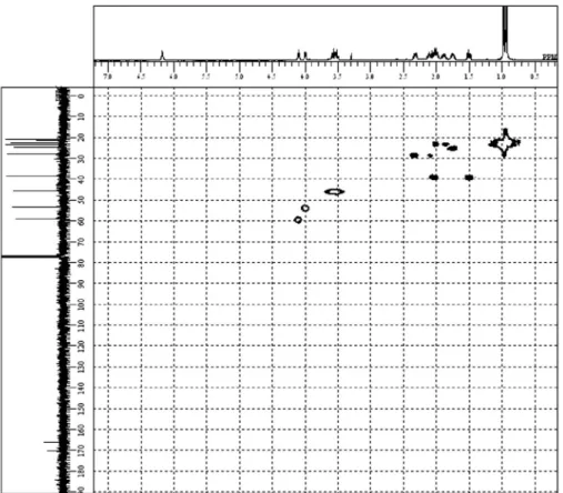
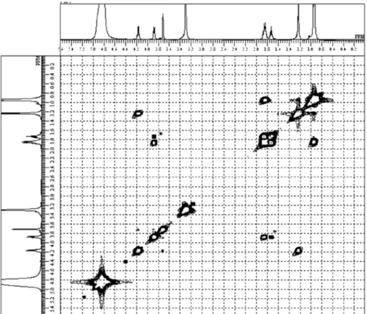
The identification of CDPs 1-11 (Figure 3)was based on the analysis of nuclear magnetic resonance (1H NMR,
13C NMR, 1H-1H COSY, HMQC, HMBC of compound 4
and 1H NMR, 13C NMR, and 1H-1H COSY of others)
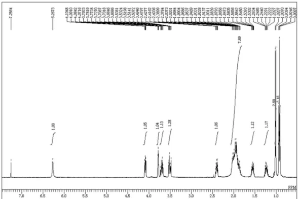
spectra (Figures S2-S37) and electrospray ionization mass spectrometry (ESI-MS) data (Table 1 and Figure S38). The dipeptide structures were evident from the observation of characteristic 13C signals of two amide
carbonyl groups (CONH, dC 165-172) and
1H signals of two α-protons (dH 3.5-4.2). Proline as a common counterpart
of compounds 2-4 and 9 was easily deduced from the presence of broad methylene multiplets (dH 1.7-3.7). The
NMR spectra clearly showed that valine, isoleucine, leucine and tryptophan were another counterpart in compounds 2-4
and 9, respectively. The presence of threonine, tyrosine and phenylalanine residues in other compounds was also clear based on the NMR data. To verify the diketopiperadine ring (DKP, Figure 4) formation, the HMBC spectrum of the major compound 4 (4.24 mg) (Table 1) was measured in CDCl3 (Figure 5 and Figure S8). The HMBC signals H-3
to C-1, H-8 NH to C-6 and C-7, H-6 to C-1 were strong evidences of the cyclic system of compound 4. The HMBC correlations of other CDPs were not detected due to the scarcity of materials. However, the NMR data in accordance with the MS data (Table 1) can elucidate the structures of 1-11 as Cyclo(Thr-Leu) (1),10 Cyclo(Val-Pro) (2),11
Cyclo(Ile-Pro) (3),12 Cyclo(Leu-Pro) (4),13 Cyclo(Val-Leu)
(5),11 Cyclo(Leu-Ile) (6),10 Cyclo(Leu-Leu) (7),10
Cyclo(Phe-Try) (8),14 Cyclo(Trp-Pro) (9),15
Cyclo(Val-Trp) (10) and Cyclo(Ile-Trp) (11).
Figure 1. Compounds (1-7) from active F8 of LH-20 by semi-preparative RP-HPLC-DAD. HPLC Conditions: linear gradient with a mixture of H2O
and MeCN, both containing 0.05% TFA. 0-20 min, 5-35% MeCN; 20-28 min, 35-56% MeCN; 28-29 min, 56-100% MeCN; and 29-32 min, 100% MeCN. Column: Cosmosil 5C18-PAQ-Waters, 10 × 250 mm. 2.5 mL min-1.
DAD profiles were measured with a Shimadzu HPLC System: LC-20AD and SPD-20A Prominence Diode Array Detector.
Cyclodipeptides from Metagenomic Library of a Japanese Marine Sponge J. Braz. Chem. Soc.
1928
The configurations of CDPs 2, 3, 4, and 5 were determined by chiral-phase gas chromatography (GC) analysis of amino acids. Retention times (min) of standard amino acids were as follows: L-Leu (10.0), D-Leu (11.3),
L-Val (6.2), D-Val (11.8), L-Ile (8.3), D-Ile (8.9), L-Pro
(8.8), D-Pro (9.2). Thus, after hydrolysis, the presence of
L-Val (6.2) and D-Pro (9.2) in compound 2, L-Ile (8.3) and D-Pro (9.3) in compound 3,L-Leu (10.0) and L-Pro (8.9) in
compound 4, L-Val (6.1) and L-Leu (10.0) in compound 5
were confirmed. Stereochemistry of other compounds was suggested by optical rotation values (Table 1) comparing with reported data: Cyclo(L-Thr-L-Leu) (1),10
Cyclo(L-Leu-L-Ile) (6),10 Cyclo(L-Leu-L-Leu) (7),10
Cyclo(L-Phe-L-Tyr) (8),16 Cyclo(L-Trp-L-Pro) (9),17
Cyclo(L-Val-L-Trp) (10)18 and Cyclo(L-Ile-L-Trp) (11).19
CDPs occur in numerous natural products and are often found alone or embedded in larger, more complex architectures in a variety of natural products from fungi, bacteria, marine sponges, plants, and mammals.20 Due
to their significant and diverse biological activities, such as antimicrobial,21,12 antitumor,21,22 antifouling,13
antiprion,23 antioxidant,10 Quorum sensing signals,24
immunosuppressive and anti-inflammatory activities, there has been an increasing interest in natural CDPs in recent years. Most CDPs isolated from natural sources were in the LL form. Interestingly, D-Proline existed in
compound 2 and 4. There were also some reports of DD
and DL enantiomers as natural products12,13 and showed very
strong activity against the pathogen Vibrio anguillarum
(MIC, 0.03-0.14 mg mL-1).12 There was no consistency
in the biological activity of the LL-enantiomers, which
depended on the assay systems.13,25,26
CDPs are catalyzed by two kinds of reported enzymes: nonribosomal peptide synthetase (NRPS) and small cyclodipeptide syntheases (CDPSs), a newly defined family of class-I aminoacyl-tRNA synthetase-like enzymes.27
Maiya and Li reported a bimodular NRPS enzyme FtmPS that used L-tryptophan and L-proline as substrates
to synthesize cyclodipeptide brevianamide F from the fumitremorgin gene cluster of Aspergillus fumigatus.28 Figure 3. Structures of isolated cyclodipeptides (1-11) from metagenomic library of the marine sponge D. calyx: Cyclo(L-Thr-L-Leu) (1), Cyclo(L-Val-D -Pro) (2), Cyclo(L-Ile-D-Pro) (3), Cyclo(L-Leu-L-Pro) (4), Cyclo(L-Val-L-Leu) (5), Cyclo(L-Leu-L-Ile) (6), Cyclo(L-Leu-L-Leu) (7), Cyclo(L-Phe-L-Tyr) (8), Cyclo(L-Trp-L-Pro) (9), Cyclo(L-Val-L-Trp) (10) and Cyclo(L-Ile-L-Trp) (11).
Figure 4. Structure of diketopiperazines.
He et al. 1929 Vol. 24, No. 12, 2013
Ding et al. also identified a bimodular NRPS named notE (2241 aa) based on the whole genome sequence of a marine-derived Aspergillus sp.29 However, sequence analysis of
clone pDC113 showed that there was no Adenylation (A) domain (required in an NRPS module), through blast research or NRPS predictor of 42 open reading frames (ORFs) encoded in 43.32 kb (Table 2). This indicated that the isolated CDPs were not synthesized by NRPS.
Subsequently, we compared the 42 ORFs to reported CDPSs to check whether there were any ORFs sharing homology with CDPSs. CDPSs used aminoacyl-tRNAs as substrates to synthesize the two peptide bonds of
various CDPs.30 Until now, there were nine CDPSs
using L amino acids reported.31 However, only three of
them (AlbC, Rv2275 and YvmC-Blic) have been fully elucidated including the crystallographic structures. AlbC (239 aa) was firstly reported to form cyclo(L-Phe-L-Leu)
in the biosynthesis of albonoursin from Streptomyces noursei32 through ping-pong catalytic mechanism.30
Rv2275 (289 aa) synthesized Cyclo(L-Tyr-L-Tyr) in the
first step of biosynthesis of mycocyclosin.33 YvmC (249
aa) formed LLcyclodileucine in the biosynthetic pathway
of pilcherrimin.34 Interestingly, the CDPSs shared only
moderate sequence similarity (19-27% sequence identity). Sequence alignment of nine reported CDPSs showed only seven conserved residues at positions Gly35, Ser37, Gly79, Tyr128, Tyr178, Glu182 and Tyr202 (AlbC numbering) and shared only three catalytic residues (Ser37, Tyr178and Glu182).30,31 Therefore, we aligned the 42 ORFs in clone
pDC113 with reported CDPSs to check whether any ORF contained the nine conserved regions or the three catalytic
residues (Ser37, Tyr178 and Glu182). Unfortunately,
there was no potential ORF candidate either sharing all conserved regions or the three catalytic residues of reported
CDPSs. Through sequencing alignments it was difficult to discover significant indications of potential candidate ORFs related to CDPSs involving in the biosynthesis of isolated cyclodipeptides.
The isolated CDPs were not biosynthesized by NRPS and there were no obvious potential CDPSs candidates through sequence analysis of the insert DNA of clone pDC113. It had high possibility that they were biosynthesized by new enzymes encoded by new genes. This result favors the most attractive theoretical potential of metagenomics – to be powerful for the finding of new genes with enhanced chances. The cyclodipeptides producing clone pDC113 were detected and the insert DNA of pDC113 was sequenced and analyzed. Although there were no indications of the potential CDPSs candidates, there is high possibility to discover the functional genes from 42 ORFs encoded in 43.32 kb by subcloning and mutation. The isolated CDPs 1-11 were combination of
L and Damino acids residues. Identification of the functional
genes involving in the biosynthesis of isolated CDPs is currently under investigation.
Conclusions
Eleven CDPs (1-11) were isolated by bioassay-guided fractionation from LB agar plate culture of positive clone pDC113 screened from metagenomic library of marine sponge D. calyx. To the best of our knowledge this is the first report of CDPs from metagenomic library. Based on the protein BLAST of the sequence, the biosynthesis of the isolated CDPs, some of which containing D-proline
residue, was not through NRPS. Sequencing alignments of 42 ORFs to reported CDPSs indicated that there was no significant potential ORF candidate related to CDPSs. It
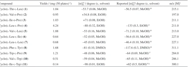
Table 1. Yields, optical rotation and ESI MS data of cyclodipeptides
Compound Yields / (mg (50 plates)-1) [α]
D25 / degree (c, solvent) Reported [α]D25/ degree (c, solvent) m/z [M]+
Cyclo(L-Thr-L-Leu) (1) 1.04 –53.7 (0.06, MeOH) –56.5 (0.07, MeOH)10 215.1
Cyclo(L-Val-D-Pro) (2) 0.95 +34.8 (0.08, EtOH) 197.0
Cyclo(L-Ile-D-Pro) (3) 1.03 +71 (0.08, EtOH) 211.1
Cyclo(L-Leu-L-Pro) (4) 4.24 –88 (0.32, EtOH) –133 (0.3, EtOH)11 211.0
Cyclo(L-Val-L-Leu) (5) 1.88 –53 (0.16, MeOH) –71.2 (0.10, MeOH)10 213.0
Cyclo(L-Leu-L-Ile) (6) 0.64 –52 (0.05, MeOH) –56.6 (0.10, MeOH)10 227.0
Cyclo(L-Leu-L-Leu) (7) 0.68 –45 (0.03, MeOH) –46.4 (0.10, MeOH)10 227.1
Cyclo(L-Phe-L-Tyr) (8) 1.68 –81 (0.10, DMSO) –117.6 (0.3, DMSO)16 311.1
Cyclo(L-Trp-L-Pro) (9) 1.21 –48 (0.06, MeOH) –64 (0.69, MeOH)17 284.0
Cyclo(L-Val-L-Trp) (10) 0.51 –59 (0.04, MeOH) –65 (0.11, MeOH)18 286.1
Cyclo(L-Ile-L-Trp) (11) 0.14 –98 (0.01, EtOH) +82 (0.5, EtOH)19,a 300.1
Cyclodipeptides from Metagenomic Library of a Japanese Marine Sponge J. Braz. Chem. Soc.
1930
Table 2. The enzyme homology analysisof pDC113 (18 ORFs encoded in 18.507 kb of 43.32 kb)
ORF Size / aa Enzyme E value Identity
(100%)
1 131 zinc finger protein [Syntrophus aciditrophicus SB]
conserved hypothetical protein [Stigmatella aurantiaca DW4/3-1] putative metal-binding protein [Eggerthella sp. YY7918]
9e-22 6e-21 5e-17 43 38 41 2-24a
25 302 hypothetical protein CHU_0606 [Cytophaga hutchinsonii ATCC 33406] 3e-59 39
26 392 ring-hydroxylating dioxygenase, large terminal subunit [gamma proteobacterium HIMB55]
phenylpropionate dioxygenase and related ring-hydroxylating dioxygenases, large terminal subunit [uncultured gamma proteobacterium HF0010_05D02]
Rieske (2Fe-2S) domain-containing protein [Parvibaculum lavamentivorans DS-1]
1e-107 2e-103 3e-86 41 40 38
27 348 5,10-methylenetetrahydromethanopterin reductase [Phenylobacterium zucineum HLK1] Luciferase-like, subgroup [Frankia sp. CN3]
F420-dependent oxidoreductase [Frankia sp. EuI1c]
8e-143 3e-120 6e-119 60 54 55
28 232 sensory box histidine kinase/response regulator [Synechococcus sp. JA-2-3B’a(2-13)] PAS fold family [Microcoleus chthonoplastes PCC 7420]
unnamed protein product [Desulfobacterium autotrophicum HRM2]
1e-41 2e-41 4e-41 42 40 36
29 573 PAS/PAC sensor hybrid histidine kinase [Opitutus terrae PB90-1] multi-sensor hybrid histidine kinase [Chthoniobacter flavus Ellin428] unnamed protein product [Desulfatibacillum alkenivorans AK-01]
1e-125 1e-122 2e-113 54 54 51
30 97 heme NO binding domain-containing protein [Nostoc punctiforme PCC 73102] unnamed protein product [Acaryochloris marina MBIC11017]
Chain A, Crystal Structure Of An H-Nox Protein From Nostoc Sp. Pcc 7120, L66wL67W DOUBLE MUTANT 3e-36 7e-31 8e-30 63 55 53
31 278 transposase, IS4 family protein [Roseiflexus sp. RS-1] 5e-67 45
32 59 transposase, IS4 family protein [Roseiflexus sp. RS-1] 1e-14 59
33 448 transposase [marine psychrotrophic bacterium Mst37]
IS element transposase [Pseudoalteromonas haloplanktis ANT/505] putative transposase [uncultured bacterium]
3e-79 9e-66 8e-58 35 31 38
34 86 heme NO binding domain-containing protein [Nostoc punctiforme PCC 73102] unnamed protein product [Cyanothece sp. PCC 7425]
unnamed protein product [Nostoc sp. PCC 7120]
9e-28 3e-25 2e-24 58 61 59
35 359 lipopolysaccharide heptosyltransferase II [Flexistipes sinusarabici DSM 4947] ADP-heptose:LPS heptosyltransferase II [Fusobacterium sp. 3_1_5R] glycosyl transferase family protein [Denitrovibrio acetiphilus DSM 12809]
1e-63 4e-63 1e-61 32 31 35
36 366 putative glycosyl transferase [Candidatus Cloacamonas acidaminovorans] glycosyltransferase [Leptospira borgpetersenii serovar Hardjo-bovis JB197]
5e-81 9e-65
40 34
37 346 glycosyl transferase family 9 [Caldithrix abyssi DSM 13497]
family 9 glycosyl transferase [Chloroherpeton thalassium ATCC 35110]
3e-63 7e-58
36 37
38 402 CDP-glycerol:poly(glycerophosphate)glycerophosph otransferase [Caldithrix abyssi DSM 13497] hypothetical protein CLOAM0422 [Candidatus Cloacamonas acidaminovorans]
1e-120 3e-115
49 47
39 75 transposase, truncation [Synechococcus sp. JA-3-3Ab] 1e-17 59
40 288 unnamed protein product [Meiothermus ruber DSM 1279] unnamed protein product [Truepera radiovictrix DSM 17093] IS605 OrfB family transposase [Nitrosococcus watsonii C-113]
4e-85 1e-79 1e-79 49 46 47
41 486 unnamed protein product [Geobacter metallireducens GS-15] unnamed protein product [Pelobacter propionicus DSM 2379]
D-glycero-D-mannoheptose-7-phosphate inase and D-glycero-D-mannoheptose-1-phosphate adenylyltransferase [Geobacter sulfurreducens KN400]
3e-152 4e-151 9e-149 51 52 52
42 85 membrane-bound nitrate reductase large subunit [uncultured bacterium] 0.017 57
He et al. 1931 Vol. 24, No. 12, 2013
was highly possible that they were biosynthesized by novel enzymes encoded by interesting genes. This result will surely be helpful for discovering new genes by attractive metagenomics. Subcloning and mutation are under investigation to search for the functional genes.
Experimental
General experimental procedures
1H and 13C NMR spectra were recorded on a JEOL
ECX-500 spectrometer in DMSO-d6, CD3OD and CDCl3. 1H and 13C NMR chemical shifts were reported in parts per
million and referenced to solvent peaks (ppm): dH 2.50 and dC 39.50 for DMSO-d6; dH 3.31 and dC 49.00 for CD3OD; dH 7.26 and dC 77.16 for CDCl3. Optical rotations were
measured on a JASCO DIP-1000 digital polarimeter.
Construction and screening of the metagenomics library
The marine sponge D. calyx was collected by hand using SCUBA from a depth of approximately 10 m off Shikine-jima Islands in Japan. Samples were kept frozen at –80 °C until use. The total sponge DNA was extracted and purified as previously described.8 The library was
constructed according to the manufacturer’s protocol. In brief, the purified DNA larger than 35 kb was blunt-ended with an End-It DNA End-Repair Kit (Epicentre, Madison, WI), and ligated into the pCC1FOS fosmid vector (Epicentre). Then, this vector was packaged with a MaxPlax Lambda Packaging Extract (Epicentre) and transfected into Escherichia coli EPI300-T1R (Epicentre). Mixtures
were plated on the LB agar containing 12.5 µg mL-1
of chloramphenicol and grown cells were collected. Two-layer top agar diffusion method35 with B. cereus as test
bacterium was used for screening the antibacterial clones by observation of inhibition zones.
Production and isolation of CDPs by bioassay-guided separation
The active clone was cultured on LB agar plates
(∅ 150 mm) supplemented with chloramphenicol
(12.5 µg mL-1) at 30 °C for 3 days. The LB agar containing
cells was extracted with EtOH overnight. The resulting mixture solution of EtOH and water was filtered and evaporated in vacuo to remove the EtOH. The resulting water solution (about 500 mL) was extracted with same volume of ethyl acetate three times. The active ethyl acetate extract (1.0 g) was subsequently separated by Sephadex LH-20 gel filtration chromatography eluting with MeOH.
Except the chloramphenicol containing fraction, two active fractions F8 and F14 were subjected to semi-preparative RP-HPLC-DAD separation (linear gradient with a mixture of H2O and MeCN, both containing 0.05% TFA. 0-20 min,
5-35% MeCN; 20-28 min, 35-56% MeCN; 28-29 min, 56-100% MeCN; and 29-32 min, 100% MeCN. Column: Cosmosil 5C18-PAQ-Waters, 10 × 250 mm, 2.5 mL min-1.
DAD profiles were measured with a Shimadzu HPLC System: LC-20AD and SPD-20A Prominence Diode Array Detector.).Eleven CDPs were finally isolated.
Antibacterial assay
Standardized agar disc diffusion test using B. cereus as a test bacterium was used for bioassay guided separation. LB agar plates (∅ 90 mm) containing overnight cultured
B. cereus were freshly prepared and divided into four or six quadrants, with a disc paper (6 mm, Tokyo Roshi Kaisha, Ltd) carrying samples (2 mg paper-1 for crude
extract or 100 µg paper-1 for fractions) or positive control
chloramphenicol (2 µg paper-1) on each quadrant. The plates
were incubated at 37 ºC for 12-16 h. Inhibition zone around the paper was observed as indication of anti-B. cereus activity.
Determination of the configurations of CDPs by chiral-phase GC
Amino acid analysis of CDPs was performed on a Shimadzu GC-MS-QP 2010 plus gas chromatograph mass spectrometer (GC-MS).7 In brief, the compound (100 µg)
was hydrolyzed with 6 mol L-1 HCl (500 µL) at 110 °C for
24 h, treated with 5-10% HCl/MeOH (500 µL) at 100 °C for 30 min and then dried under nitrogen gas before being treated with trifluoroacetic anhydride (TFAA)/CH2Cl2
(1:1, 500 µL) at 100 °C for 5 min. Finally, each reaction mixture was dried under nitrogen gas, dissolved in CHCl2
and 1 µL was injected for GC analysis. The chiral-phase GC analysis of the N-trifluoroacetyl (TFA)/methyl ester derivatives was performed using a CP-Chirasil-D-Val column (Alltech, 0.25 mm × 25 m; N2 as the carrier gas;
program rate 50-200 °C at 4 °C min-1). Standard amino
acids were also converted to the TFA/Me derivatives by the same procedure. Retention times (min) were compared.
DNA sequencing and analysis
Cyclodipeptides from Metagenomic Library of a Japanese Marine Sponge J. Braz. Chem. Soc.
1932
2.3.2 (http://www0.nih.go.jp/~jun/cgi-bin/frameplot.pl) Blast analysis and NRPS predictor.
Supplementary Information
Supplementary information (Figure S1-S38) is available free of charge at http://jbcs.sbq.org.br as a PDF file.
Acknowledgements
R. H. thanks the China Scholarship Council for the visiting Ph.D. student program. This work was partly supported by The Mitsubishi Foundation, Strategic Research Foundation Grant-aided Project for Private Universities, Grants-in-Aids from the Ministry of Education, Culture, Sports, Science and Technology (MEXT), Japan, and the National Natural Science Foundation of China (Grant No.11172337).
References
1. Faulkner, D. J.; Nat. Prod. Rep.2000, 17, 1.
2. Singh, B. K.; Macdonald, C. A.; Drug Discov. Today2010, 15, 792.
3. Handelsman, J.; Microbiol. Mol. Biol. Rev. 2004, 68, 669. 4. Banik, J. J.; Brady, S. F.; Curr. Opin. Microbiol.2010, 13, 603. 5. Kennedy, J.; Flemer, B.; Jackson, S. A.; Lejon, D. P. H.;
Morrissey, J. P.; O’Gara, F.; Dobson, A. D. W.; Mar. Drugs
2010, 8, 608.
6. Kato, Y.; Fusetani, N.; Matsunaga, S.; Hashimoto, K.; Fujita, S.; Furuya, T.; J. Am. Chem. Soc. 1986, 108, 2780.
7. Kimura, M.; Wakimoto, T.; Egami, Y.; Co, K. T.; Ise, Y.; Abe, I.;
J. Nat. Prod. 2012, 75, 290.
8. He, R.; Wakimoto, T.; Takeshige, Y.; Egami, Y.; Kenmoku, H.; Ito, T.; Wang, B. C.; Asakawa, Y.; Abe, I.; Mol Biosyst.2012,
8, 2334.
9. He, R.; Wakimoto, T.; Egami, Y.; Kenmoku, H.; Ito, T.; Asakawa, Y.; Abe, I.;Bioorg. Med. Chem. Lett.2012, 22, 7322. 10. Furukawa, T.; Akutagawa, T.; Funatani, H.; Uchida, T.; Hotta, Y.;
Niwa, M.; Takaya, Y.; Bioorgan. Med. Chem.2012, 20, 2002. 11. Pedras, M. S. C.; Yu, Y.; Liu, J.; Tandron-Moya, Y. A.;
Z. Naturforsch., C: J. Biosci.2005, 60, 717.
12. Fdhila, F.; Vázquez, V.; Sánchez, J. L.; Riguera, R.; J. Nat. Prod.
2003, 66, 1299.
13. Li, X.; Dobretsov, S.; Xu, Y.; Xiao, X.; Hung, O. S.; Qian, P. Y.;
Biofouling2006, 22, 187.
14. Xiang, L.; Guo, D. L.; Ju, R.; Ma, B.; Lei, F.; Du, L. J.; Chin. Tradit. Herbal Drugs2007, 38, 1622.
15. Ivanova, V.; Graefe, U.; Schlegel, R.; Schlegel, B.; Gusterova, A.; Kolarova, M.; Aleksieva, K.; Biotechnol. Biotec. Eq.2003, 17, 2128.
16. Cheng, Z. H.; Wu, T.; Yu, B. Y.; Nat. Prod. Res. Dev.2005, 17, 1.
17. Kobayashi, M.; Aoki, S.; Gato, K.; Matsunami, K.; Kurosu, M.; Kitagawa, I.; Chem. Pharm. Bull.1994, 42, 2449.
18. Pedras, M. S. C.; Smith, K. C.; Taylor, J. L.; Phytochemistry
1998, 49, 1575.
19. Kimura, Y.; Sawada, A.; Kuramata, M.; Kusano, M.; Fujioka, S.; Kawano, T.; Shimada, A.; J. Nat. Prod. 2005, 68, 237. 20. Borthwick, A. D.; Chem. Rev. 2012, 112, 3641.
21. de Carvalho, M. P.; Abraham, W. R.; Curr. Med. Chem.2012,
19, 3564.
22. Martins, M. B.; Carvalho, I.; Tetrahedron2007, 63, 9923. 23. Bolognesi, M. L.; Ai Tran, H. N.; Staderini, M.; Monaco, A.;
López-Cobeñas, A.; Bongarzone, S.; Biarnés, X.; López-Alvarado, P.; Cabezas, N.; Caramelli, M.; Carloni, P.; Menéndez, J. C.; Legname, G.; ChemMedChem.2010, 5, 1324. 24. Campbell, J.; Lin, Q.; Geske, G. D.; Blackwell, H. E.; ACS
Chem. Biol.2009, 4, 1051.
25. Furtado, N. A. J. C.; Pupo, M. T.; Carvalho, I.; Campo, V. L.; Duarte, M. C. T.; Bastos, J. K.; J. Braz. Chem. Soc. 2005,16, 1448.
26. Cheenpracha, S.; Borris, R. P.; Tran, T. T.; Jee, J. M.; Seow, H. F.; Cheah, H. Y.; Ho, C. C.; Chang, L. C.; J. Braz. Chem. Soc.2011, 22, 223.
27 Gondry, M.; Sauguet, L.; Belin, P.; Thai, R.; Amouroux, R.; Tellier, C.; Tuphile, K.; Jacquet, M.; Braud, S.; Courçon, M.; Masson, C.; Dubois, S.; Lautru, S.; Lecoq, A.; Hashimoto, S.; Genet, R.; Pernodet, J.; Nat. Chem. Biol.2009, 5, 414. 28. Maiya, S.; Grundmann, A.; Li, S. M.; Turner, G.; ChemBioChem.
2006, 7, 1062.
29. Ding, Y.; de Wet, J. R.; Cavalcoli, J.; Li, S.; Greshock, T. J.; Miller, K. A.; Finefield, J. M.; Sunderhaus, J. D.; McAfoos, T. J.; Tsukamoto, S.; Williams, R. M.; Sherman, D. H.; J. Am. Chem. Soc.2010, 132, 12733.
30. Sauguet, L.; Moutiez, M.; Li, Y.; Belin, P.; Seguin, J.; Le Du, M. H.; Thai, R.; Masson, C.; Fonvielle, M.; Pernodet, J. L.; Charbonnier, J. B.; Gondry, M.; Nucleic Acids Res.2011,
39,4475.
31. Belin, P.; Moutiez, M.; Lautru, S.; Seguin, J.; Pernodet, J. L.; Gondry, M.; Nat. Prod. Rep.2012, 29, 961.
32. Lautru, S.; Gondry, M.; Genet, R.; Pernodet, J. L.; Chem. Biol.
2002, 9, 1355.
33. Vetting, M. W.; Hegde, S. S.; Blanchard, J. S.; Nat. Chem. Biol.
2010, 6, 797.
34. Bonnefond, L.; Arai, T.; Sakaguchi, Y.; Suzuki, T.; Ishitani, R.; Nureki, O.; Proc. Natl. Acad. Sci. USA2011, 108, 3912. 35. Brady, S. F.; Nat. Protoc.2007, 2, 1297.
Submitted: February 27, 2013
Supplementary Information
S
I
J. Braz. Chem. Soc., Vol. 24, No. 12, S1-S21, 2013. Printed in Brazil - ©2013 Sociedade Brasileira de Química 0103 - 5053 $6.00+0.00
*e-mail: [email protected], [email protected]
Cyclodipeptides from Metagenomic Library of a Japanese Marine Sponge
Rui He,a,b,c Bochu Wang,*,b Toshiyuki Wakimoto,c Manyuan Wang,a
Liancai Zhub and Ikuro Abe*,c
aSchool of Traditional Chinese Medicine, Capital University of Medical Sciences,
No. 10 Xitoutiao, You An Men, 100069 Beijing, P. R. China
bBioengineering College, Chongqing University, No. 174,
Shanpingba Main Street, 400030 Chongqing, P. R. China
cGraduate School of Pharmaceutical Sciences, The University of Tokyo,
7-3-1 Hongo, Bunkyo-ku, 113-0033 Tokyo, Japan
Comparative data of cyclodipeptide 1-7 production in clone pDC113 and negative control (NC, strain EPI300 carrying pCC1FOS fosmid vector)
The respective 2 plates of pDC113 and NC were cultured in the same conditions (30 °C, 3d) and subjected to the same extraction and separation procedures and finally using same volume of MeOH to dissolve the LH-20
cyclodipeptides fraction before injection (both 5 µL) to RP-HPLC-DAD. HPLC analysis was performed on ODS column (Cosmosil 5C18 PAQ waters, 4.6 × 250 mm) with
a mixture of H2O and MeCN, both containing 0.05% TFA: 0-20 min, 5-35% MeCN; 20-45 min, 35-100% MeCN; and 45-55 min, 100% MeCN, 0.8 mL min-1. DAD profiles were
measured with a Shimadzu HPLC System: LC-20AD and SPD-20A Prominence Diode Array Detector.
Cyclodipeptides from Metagenomic Library of a Japanese Marine Sponge J. Braz. Chem. Soc. S2
IH, 13C chemical shifts, and 1H-1H COSY data of cyclodipeptides 1-7 and 9
He et al. S3 Vol. 24, No. 12, 2013
IH, 13C chemical shifts, and 1H-1H COSY data of cyclodipeptides 8, 10 and 11
Figure S3. Chemical shifts and COSY of cyclodipeptides 8, 10 and 11. 1H NMR (500 MHz) chemical shifts (blue), 13C NMR (125 MHz) chemical shifts (red), and main 1H-1H COSY (bold line) correlations are shown. 13C NMR of 10 were inferred from its HMQC and some of HMBC data.
Cyclodipeptides from Metagenomic Library of a Japanese Marine Sponge J. Braz. Chem. Soc. S4
Figure S6. 1H-1H COSY spectrum of Cyclo(
L-Leu-L-Pro) (4).
He et al. S5 Vol. 24, No. 12, 2013
Cyclodipeptides from Metagenomic Library of a Japanese Marine Sponge J. Braz. Chem. Soc. S6
Figure S10. 13C NMR of Cyclo(L-Thr-L-Leu) (1) (125 MHz, CD
3OD).
Figure S9. 1H NMR of Cyclo(L-Thr-L-Leu) (1) (500 MHz, CD
He et al. S7 Vol. 24, No. 12, 2013
Figure S12. 1H NMR of Cyclo(L-Val-D-Pro) (2) (500 MHz, CDCl
3).
Cyclodipeptides from Metagenomic Library of a Japanese Marine Sponge J. Braz. Chem. Soc. S8
Figure S14. 1H-1H COSY of Cyclo(L-Val-D-Pro) (2).
Figure S13. 13C NMR of Cyclo(
He et al. S9 Vol. 24, No. 12, 2013
Figure S16. 13C NMR of Cyclo(L-Ile-D-Pro) (3) (125 MHz, CDCl
3).
Figure S15. 1H NMR of Cyclo(
Cyclodipeptides from Metagenomic Library of a Japanese Marine Sponge J. Braz. Chem. Soc. S10
Figure S18. 1H NMR of Cyclo(L-Val-L-Leu) (5) (500 MHz, CDCl
3).
He et al. S11 Vol. 24, No. 12, 2013
Figure S20. 1H-1H COSY of Cyclo(L-Val-L-Leu) (5).
Figure S19. 13C NMR of Cyclo(L-Val-L-Leu) (5) (125 MHz, CDCl
Cyclodipeptides from Metagenomic Library of a Japanese Marine Sponge J. Braz. Chem. Soc. S12
Figure S22. 13C NMR of Cyclo(L-Leu-L-Ile) (6) (125 MHz, CD
3OD).
Figure S21. 1H NMR of Cyclo(L-Leu-L-Ile) (6) (500 MHz, CD
He et al. S13 Vol. 24, No. 12, 2013
Figure S24. 1H NMR of Cyclo(L-Leu-L-Leu) (7) (500 MHz, CD
3OD).
Figure S23.1H-1H COSY of Cyclo(
Cyclodipeptides from Metagenomic Library of a Japanese Marine Sponge J. Braz. Chem. Soc. S14
Figure S26. 1H-1HCOSY of Cyclo(L-Leu-L-Leu) (7).
Figure S25. 13C NMR of Cyclo(
He et al. S15 Vol. 24, No. 12, 2013
Figure S28. 13C NMR of Cyclo(L-Phe-L-Tyr) (8) (125 MHz, DMSO).
Figure S27. 1H NMR of Cyclo(
Cyclodipeptides from Metagenomic Library of a Japanese Marine Sponge J. Braz. Chem. Soc. S16
Figure S30. 1H NMR of Cyclo(
He et al. S17 Vol. 24, No. 12, 2013
Figure S32. 1H NMR of Cyclo(L-Val-L-Trp) (10) (500 MHz, DMSO).
Figure S31.13C NMR of Cyclo(
Cyclodipeptides from Metagenomic Library of a Japanese Marine Sponge J. Braz. Chem. Soc. S18
He et al. S19 Vol. 24, No. 12, 2013
Figure S36.1H NMR of Cyclo(L-Ile-L-Trp) (11) (500 MHz, DMSO).
Cyclodipeptides from Metagenomic Library of a Japanese Marine Sponge J. Braz. Chem. Soc. S20
He et al. S21 Vol. 24, No. 12, 2013
LC-MS data of cyclodipeptides 8-11
Liquid chromatography-mass spectrometry (LC-MS, Agilent 1100 series-Bruker Esquire 4000) analysis was performed on ODS column (TSK-Gel ODS-80Ts, 4.6 × 150 mm) with a mixture of H2O and MeCN, both
containing 0.1% acetic acid: 30-100% MeCN 30 min; 100% MeCN 10 min, 0.2 mL min-1. Detected wavelength:
280 nm. Positive ESI.