Use of the rhomboid lap for the repair of
cutaneous defects
Aplicações do retalho romboide em reparações cutâneas
ABSTRACT
Background: The plastic surgeon frequently performs reconstructions of diverse types of
cutaneous defects; thus, it is essential to be versatile and have knowledge of appropriate techniques for each case. The rhomboid transposition lap, proposed by Alexander Limberg, is an extremely useful lap for a wide range of reconstructive procedures. This study aims to demonstrate the versatility, safety, and applicability of Limberg’s lap for reconstruction of cutaneous losses located in a wide variety of body segments. Methods: A retrospective
analysis of 50 patients with different cutaneous defects that had been reconstructed with the rhomboid lap was performed. A description of the surgical technique and a critical analysis of the results are presented. Results: The average age of the patients was 59.6 years. Neo
plastic lesions accounted for most of the cases (84%). The face was the most frequently affected area, accounting for 36 (72%) cases; it was followed by the lumbosacral region (8%) and by the dorsal and inguinoscrotal regions (6%). Complications were observed in 4 (8%) patients. Conclusions: The rhomboid lap provides safe and predictable outcomes, and is the method of choice for most of the defects found.
Keywords: Surgical laps. Reconstructive surgical procedures/methods. Surgery, plastic/ methods.
RESUMO
Introdução: O cirurgião plástico frequentemente defrontase com a reparação dos mais
diversos tipos de defeitos cutâneos; logo, é imprescindível que possua o conhecimento de técnicas versáteis e apropriadas para cada caso. O retalho romboide de transposição, proposto por Alexander Limberg, é um retalho extremamente útil para os mais diversos tipos de reconstrução. O objetivo deste trabalho é demonstrar a versatilidade, a segurança e a aplicabilidade do retalho de Limberg para reconstrução de perdas cutâneas localizadas nos mais diversos segmentos corporais. Método: Foi realizada análise retrospectiva de
50 pacientes apresentando defeitos cutâneos, dos mais variados tipos, reconstruídos com o retalho romboide. A descrição da técnica cirúrgica e uma análise crítica dos resultados são apresentadas. Resultados: A média de idade dos pacientes foi de 59,6 anos. As lesões
neoplásicas foram responsáveis pela maioria dos casos (84%). A face foi a área mais envol vida nas reconstruções, totalizando 36 (72%) casos, seguida da região lombossacral (8%), e do dorso e da região inguinoscrotal (6%). Complicações foram observadas em 4 (8%) pacientes. Conclusões: O retalho romboide propicia resultados seguros e previsíveis, sendo
a alternativa para a maioria dos defeitos encontrados.
Descritores: Retalhos cirúrgicos. Procedimentos cirúrgicos reconstrutivos/métodos. Cirur
gia plástica/métodos. Study conducted at
Hospital São Lucas of Pontifícia Universidade Católica of Rio Grande do Sul (PUCRS), Porto Alegre, RS, Brazil. Submitted to SGP (Sistema de Gestão de Publicações/Manager Publications System) of RBCP (Revista Brasileira de Cirurgia Plástica/Brazilian Journal of Plastic Surgery). Article received: January 18, 2012 Article accepted: February 10, 2012
1. Plastic surgeon, expert member of the Sociedade Brasileira de Cirurgia Plástica (Brazilian Society of Plastic Surgery) – SBCP, Porto Alegre, RS, Brazil. 2. Resident physician in plastic surgery at Hospital São Lucas of Pontifícia Universidade Católica do Rio Grande do Sul (HSLPUCRS), Porto Alegre, RS,
Brazil.
3. Master and full member of the SBCP, preceptor at the Plastic Surgery Department of HSLPUCRS, Porto Alegre, RS, Brazil. 4. Head of the Plastic Surgery Department of HSLPUCRS, Porto Alegre, RS, Brazil.
Gustavo steffen alvarez1
francisco felipe laitano2
evandro José siqueira2
Milton paulo de oliveira3
pedro dJacir escobar
INTRODUCTION
There are a number of studies in the literature on the use
of the rhomboid lap in every area of the human body and in various surgical specialties1,2. In spite of several modii
cations that have already been proposed, it remains an extremely versatile and safe lap3. According to Chasmar4, it
can be used in virtually any part of the body, and it is widely used in facial and breast reconstruction, neurosurgery, hand surgery, ophthalmology, and proctology58.
This study aims to demonstrate the versatility, safety, and applicability of Limberg’s lap for reconstruction of cu ta neous losses located at the most varied body segments. The lap ills the defect with tissue of the same thickness and color, has good vascularization, and demonstrates suitable
functional and aesthetic outcomes.
METHODS
We performed a retrospective analysis of 50 patients who
underwent reconstruction of cutaneous defects with rhom
boid laps; all surgeries were performed at Hospital São Lucas of Pontifícia Universidade Católica of Rio Grande do Sul (Porto Alegre, RS, Brazil) between January 2008 and February 2011. Both the etiology and the location of the de fects varied widely.
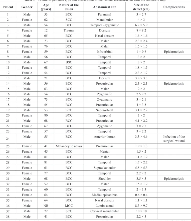
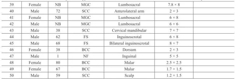
The patients were assessed by age, etiology, location, and size of the defect (according to measurements obtained du ring the anatomicalpathological examination, in cases of neoplasia), as well as the presence of postoperative compli cations. The proiles of the studied patients are described in Table 1.
Surgical Technique
The laps were prepared with consideration of the size and location of the original defect, as well as the force lines and elasticity of the adjacent tissues.
Flap preparation begins with drawing a diamond, with internal angles of 60 and 120 degrees, around the defect re sulting from the resection (Figure 1A). This drawing should be prepared, with two equilateral triangles with 60degree angles lined up from base to base so that all sides of the de fect have the same length (which, in practice, is equal to the shortest diagonal). The irst side of the lap is an exten sion outward of the defect, of the shortest diagonal in its own length; the second side of the lap is marked with a line the
same length as the irst, to the adjacent side of the defect in the diamond, producing an angle of 60 degrees at the lap apex2,9. The inal coniguration of the lap’s scar is foreseeable
at all times, as may be seen in Figure 1B.
For every defect, four rhomboid laps can be potentially produced. Then, according to the tension lines and thickness
of the skin, and the orientation and location of the defect,
the lap that best suits the defect is chosen. As evidenced in Figures 2 to 4, the measurements and production of the lap are performed prior to the resection of the defect’s edges, because of the posterior enlargement of the lesions. All edges of the lesion and the lap’s edges and base are thus detached, providing suitable approximation of tissues without tension
in the closure.
RESULTS
The average age of the patients was 59.6 years and ranged from 2 days to 81 years. Neoplastic lesions accounted for most of the cases (84%). The other cases involved defects de riving from meningomyelocele (8%), Fournier syndrome (4%), necrotizing fasciitis (2%), and trauma (2%). Among the neoplastic lesions, basal cell carcinoma was most frequent (81%), followed by squamous cell carcinoma (14.4%), basal squamous carcinoma (2.4%), and melanocytic nevus (2.4%).
With regard to the affected areas, the face was the most commonly affected, with 36 (72%) cases, followed by the lumbosacral region (8%), and by the dorsal and inguinos crotal regions (6%). The anterolateral arm, thorax, shoulder, and supraclavicular region each corresponded to 2% of the cases. On the face, the most commonly affected area was the temporal region (25%), followed by the malar region (19.4%). The number of laps used in facial reconstructions ranged from 1 to 9 (Table 2).
The defects came in different sizes. The largest defect was in the lumbosacral region, due to surgical correction of meningomyelocele, at 8.3 cm × 9.7 cm (Figure 4). In the fa cial region, the largest defect was 6.2 cm × 5.9 cm and affected the temporalzygomatic region.
Complications were observed in 4 (8%) patients: epider molysis in 3 (6%) cases and 1 (2%) case of surgical wound infection. All complications were treated with conservative treatment, and the outcomes were good.
DISCUSSION
This study demonstrates the usefulness of the rhomboid
lap, initially proposed by Limberg, for reconstruction of the most varied cutaneous defects, as has been observed by
several authors35. The lap provides the transference of adja
cent tissue to the defect with the same texture and skin color. The position of the scars resulting from the lap transposition is highly foreseeable and can always be taken into account in
planning the lap, with the aim of achieving reduced distortion of the underlying tissues and a less apparent scar (Figure 2).
Table 1 – Proile of patients: type and distribution of the lesions reconstructed with the rhomboid lap.
Patient Gender Age
(years)
Nature of the
lesion Anatomical site
Size of the
defect (cm) Complications
1 Male 53 BCC Paranasal 3 × 2.7
2 Female 62 SCC Mandibular 4 × 3
3 Male 54 BCC Temporalzygomatic 6.2 × 5.9
4 Female 12 Trauma Dorsum 8 × 8.2
5 Male 65 BCC Nasal dorsum 1.6 × 1.6
6 Male 73 BCC Malar 2.3 × 2.4
7 Female 76 BCC Malar 1.5 × 1.5
8 Female 59 BCC Infraorbital 1 × 0.8 Epidermolysis
9 Male 56 BCC Temporal 3 × 2
10 Male 67 BSC Temporal 3 × 2
11 Female 68 BCC Temporal 1.8 × 1.5
12 Female 54 BCC Temporal 2.3 × 1.7
13 Male 71 BCC Dorsum 3.8 × 3.3
14 Male 56 BCC Preauricular 2.3 × 2.1 Epidermolysis
15 Male 63 BCC Malar 2 × 2
16 Male 54 BCC Zygomatic 2.5 × 2
17 Male 73 BCC Zygomatic 3 × 2.1
18 Male 55 BCC Preauricular 4 × 3.5
19 Male 53 SCC Supraorbital 3.1 × 2.2
20 Female 80 BCC Temporal 3 × 2
21 Male 68 BCC Preauricular 4.1 × 2.2
22 Female 67 BCC Zygomatic 3 × 2.5
23 Female 57 BCC Temporal 3 × 2.2
24 Male 55 BCC Anterior thorax 3.3 × 4.6 Infection of the surgical wound
25 Female 41 Melanocytic nevus Preauricular 1.9 × 1.3
26 Female 45 BCC Mental 1.5 × 2
27 Male 81 BCC Malar 1.1 × 1.2
28 Female 81 BCC Temporal 1.7 × 2.2
29 Female 61 BCC Supraclavicular 5.5 × 5.3
30 Female 77 BCC Temporal 2.2 × 2
31 Male 68 BCC Shoulder 3.5 × 3 Epidermolysis
32 Female 52 BCC Malar 1.5 × 1.2
33 Female 60 BCC Temporal 2 × 1.3
34 Female 64 BCC Medial epicanthus 0.6 × 0.4
35 Female 64 BCC Nasal dorsum 1.1 × 1.1
36 Male NB MGC Lumbosacral 8.3 × 9.7
37 Male 72 SCC Cervical mandibular 10 × 10
38 Male 41 BCC Preauricular 2.2 × 3
BCC = Basal cell carcinoma; BSC = basal squamous carcinoma; FS = Fournier syndrome; MGC = meningomyelocele; NB = newborn; NF = necrotizing fasciitis; SCC = squamous cell carcinoma.
A B
Figure 1 – Rhomboid lap marking. In A, diamond with
internal angles of 60 and 120 degrees and marked lap. In B, inal coniguration of the scar.
A
B
C
Figure 2 – Patient with lesion secondary to dorsum trauma and
dificult scarring of the wound by secondary intention. Resection
and cutaneous coverage with Limberg’s fasciocutaneous lap.
In A, demarcation of the lap prior to resection.
In B, resected lesion. In C, at conclusion of operation. continuation
Table 1 – Proile of patients: type and distribution of the lesions reconstructed with the rhomboid lap.
39 Female NB MGC Lumbosacral 7.8 × 8
40 Male 72 SCC Anterolateral arm 2 × 3
41 Female NB MGC Lumbosacral 6 × 8
42 Male NB MGC Lumbosacral 6 × 6
43 Male 38 SCC Cervical mandibular 7 × 7
44 Male 62 FS Inguinoscrotal 6 × 8
45 Male 68 FS Bilateral inguinoscrotal 8 × 7
46 Female 38 BCC Dorsum 2 × 3
47 Male 1 NF Inguinal 5 × 5
48 Female 80 BCC Malar 2.5 × 2.5
49 Female 67 BCC Malar 1.7 × 1.5
50 Male 59 SCC Scalp 1.2 × 1.5
BCC = Basal cell carcinoma; BSC = basal squamous carcinoma; FS = Fournier syndrome; MGC = meningomyelocele; NB = newborn; NF = necrotizing fasciitis; SCC = squamous cell carcinoma.
trapping and hypertrophy, extremely low. This is, therefore, an attractive option for pediatric patients and/or those with a history of pathological scarring. The lap base, whenever possible, should be inferiorly positioned, in order to facilitate lymphatic drainage of the lap (Figures 2 and 3).
In facial reconstructions, even for small lesions, cuta
neous laps are preferable to primary closure and/or grafting, with the purpose of avoiding distortions of adjacent struc
tures and breaks in scar lines10. The great number of facial
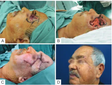
reconstructions in the temporalzygomatic and malar regions (44% of the facial defects) demonstrates the versatility of the rhomboid lap in this area, and that it is the preferable technique in these facial units.
A
C
B
D
Figure 3 – Patient with wide tumoral lesion in the left
temporal-zygomatic region. In A, preoperative appearance.
In B, the defect following resection. In C, at conclusion of the
operation. In D, appearance at 6 months postoperatively.
A B C
Figure 4 – Newborn with meningomyelocele.
In A, defect of 8.3 cm × 9.7 cm. In B, marked lap.
In C, sutured lap at the site of the defect.
Table 2 – Limberg’s lap application according to
aesthetic units of the face.
Facial unit Number of laps
Temporal 9
Malar 7
Preauricular 5
Zygomatic 3
Nasal dorsum 2
Cervical mandibular 2
Paranasal 1
Temporal zygomatic 1
Supraorbital 1
Mentum 1
Medial canthus of the eye 1
Scalp 1
Infraorbital 1
Mandibular 1
Total 36
are excisions of malignant tumors (84%), such resection pro vides a greater safety margin (extra tissue can be sent for
anatomicalpathological examination), without affecting the inal aestheticfunctional outcome11.
The largest defects, in this study, were those deriving from neurosurgical repairs for meningomyelocele correc tion. Although some authors suggest modiications with the association of two or more rhomboid laps for large defects
on the back3, we believe that the suitable planning of the
lap, as well as the maintenance of the paravertebral perfo rators as proposed by Muneuchi et al.8, enables the closure of
large areas with minimal complications. Moreover, the use of a single lap avoids suture lines over the previously repai red dural sac. The transfer of a fasciocutaneous lap of suitable thickness makes the use of muscular laps unnecessary for
clo sure of selected lesions.
Although it was not part of this case selection, the rhom boid lap has been considered the best option for treating sa crococcygeal pilonidal disease6.
There were few complications in this study, and they
did not affect the final result of the surgery; all of them re solved with conservative treatment (dressings and an ti bio tic the rapy), which demonstrates the safety of the rhom boid flap for the most varied reconstructions in the human body.
CONCLUSIONS
Limberg’s rhomboid lap provides for closure of small to large defects at a wide range of anatomical sites, with a high level of safety and predictability and a low index of com plications. The easy production of the lap design and the resulting strong scar, with no tension in the closure after lap rotation, make it the irst option in most reconstructions where the integrity of the skin has been broken.
REFERENCES
1. Tissiani LAL, Alonso N, Carneiro MH, Bazzi K, Rocco M. Versatilidade do retalho bilobado. Rev Bras Cir Plást. 2011;26(3):4117.
2. Limberg A. Mathematical principles of local plastic procedures on the surface of the human body. Leningrad: Medgis; 1946.
3. Turan T, Kuran I, Ozcan H, Baş L. Geometric limit of multiple local Limberg laps: a lap design. Plast Reconstr Surg. 1999;104(6):16758. 4. Chasmar LR. The versatile rhomboid (Limberg) lap. Can J Plast Surg.
2007;15(2):6771.
5. Ng SG, Inkster CF, Leatherbarrow B. The rhomboid lap in medial canthal reconstruction. Br J Ophthalmol. 2001;85(5):5569.
sacrococcygeal pilonidal disease? A metaanalysis of randomized con trolled trials. Colorectal Dis. 2012;14(2):14351.
7. Silva Neto MP, Adão O, Scandiuzzi D, Chaem LHT. Retalho romboide na reparação mamária imediata pósquadrantectomia e dissecção axilar. Rev Soc Bras Cir Plást. 2001;16(1):2934.
8. Muneuchi G, Matsumoto Y, Tamai M, Kogure T, Igawa HH, Nagao S. Rhomboid perforator lap for a large skin defect due to lumbosacral
Correspondence to: Gustavo Steffen Alvarez
Rua José Francisco Duarte Jr, 52 – ap. 601 – Menino Deus – Porto Alegre, RS, Brazil – CEP 90110-300 E-mail: gusalvarez@terra.com.br
meningocele: a simple and reliable modiication. Ann Plast Surg. 2005; 54(6):6702.
9. Limberg AA. Design of local laps. In: Gibson T, ed. Modern trends in plastic surgery. 2nd ed. London: ButterworthHeinemann; 1966. p. 3861.
10. Borges AF. The rhombic lap. Plast Reconstr Surg. 1981;67(4):45866. 11. Townend J. A template for the planning of rhombic skin laps. Plast


