Article
Printed in Brazil - ©2017 Sociedade Brasileira de Química0103 - 5053 $6.00+0.00*e-mail: [email protected]
Steroids from the Brazilian Zoanthids
Palythoa caribaeorum
and
Palythoa variabilis
Francisco C. L. Pinto,a José Gustavo L. Almeida,a Edilberto R. Silveira,a Arinice M.
Costa,b Larissa A. Guimarães,b Diego V. Wilke,b Letícia V. Costa-Lotufo,c Maria da
Conceição M. Torresd and Otília Deusdênia L. Pessoa*,a
aDepartamento de Química Orgânica e Inorgânica, Universidade Federal do Ceará,
60021-970 Fortaleza-CE, Brazil
bDepartamento de Fisiologia e Farmacologia, Universidade Federal do Ceará,
60430-270 Fortaleza-CE, Brazil
cDepartamento de Farmacologia, Universidade de São Paulo,
05508-900 São Paulo-SP, Brazil
dDepartamento de Química, Universidade Estadual da Paraíba,
58429-500 Campina Grande-PB, Brazil
Two unreported ergostane-type sterols 24(R)-7α-hydroperoxy-ergost-5-en-3β-ol and 6β-carboxyl-24(R)-(8→6)-abeo-ergostan-3β,5β-diol, along with seven known ones were isolated from the hexane and alcohol extracts from the zoanthids Palythoa caribaeorum and Palythoa variabilis. The structures of the new compounds were determined using spectroscopic techniques,
including 1D and 2D nuclear magnetic resonance (NMR), high-resolution electrospray ionization mass spectrometry (HRESIMS), and comparison with data previously published. 6β -Carboxyl-24(R)-(8→6)-abeo-ergostan-3β,5β-diol showed moderate cytotoxicity against colon cancer cells (HCT-116) as previously described for others abeo-sterol derivatives.
Keywords:Palythoa caribaeorum, Palythoa variabilis, ergostane sterols, cytotoxic activity
Introduction
Zoanthids are cnidarians found in most marine environments, from temperate to tropical areas and from the intertidal zone to the deep sea below 5000 m, but are particularly common in tropical and subtropical seas.1
The colonial zoanthid genus Palythoa (order Zoantharia)
is represented by ca. 92 species.2 It has been the subject
of constant taxonomy investigations due to an incomplete understanding of their diversity.3 Many chemical and
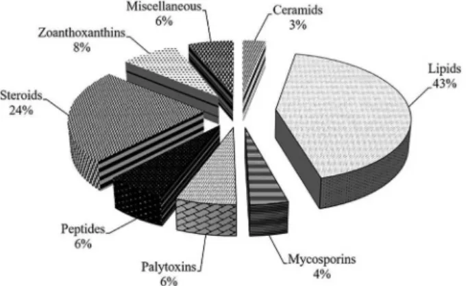
pharmacological studies have been conducted in order to investigate its biological potentialities. Several classes of compounds have been isolated from zoanthids, including ceramides,4 fatty acids,5 mycosporins,6 palytoxins,7
peptides,8 sterols,9,10 zoanthoxanthins,11 prostaglandins,12
lipidic α-amino acids,13 and pyrazines.14 Lipids and steroids
represent the commonest classes of compounds isolated from Palythoa spp. (Figure 1). The most potent toxic
non-protein known, palytoxin, has been isolated from Palythoa
species.15 The first natural lipid α-amino acids (C 30 and
C31) have been isolated from Protopalythoa variabilis,
and exhibited cytotoxic effects against human cancer cell lines.13 Protopalythoa taxonomy previously assigned4,13
is under revision based on mitochondrial DNA analysis, indicating that Protopalythoa and Palythoa are indeed
congeneric designations.16
The present investigation reports on the steroidal composition of P. caribaeorum and P. variabilis. Both
species are abundant in shallow waters along the Brazilian Northeast coastline.
Experimental
General
Melting points, not corrected, were determined on a digital MQAPF-302 melting point apparatus (Microquimica), with a heating rate of 2 °C min-1. 1H (500 or 300 MHz)
and 13C (125 or 75 MHz) nuclear magnetic resonance
(NMR) spectra were performed either on a Bruker Avance DRX-500 or Avance DPX-300 spectrometer. High resolution electrospray ionization mass spectrometry (HRESIMS) was acquired using a liquid chromatography-mass spectrometry ion trap and time-of-flight (LCMS-IT-TOF, Shimadzu) spectrometer, consisting of an UFLC (ultra fast liquid chromatography) system coupled to an IT-TOF mass spectrometer equipped with electrospray ionization (ESI) source operating either in positive or negative mode. The mass spectra were recorded in the range of m/z 100-1000 Da,
using a potential of 4.0 kV on the capillary, and nitrogen as the desolvation gas. The system was controlled by a LC-solution software, which was also used for data analysis. High-performance liquid chromatography (HPLC) analysis was carried out using an UFLC (Shimadzu) system equipped with a SPD-M20A diode array UV-Vis detector and a Phenomenex C18 column, 5 µm (4.6 × 250 mm). The mobile
phase consisted of H2O-CH3CN with a flow rate of 4.72 mL
min-1. For the column chromatography (CC) procedures,
silica gel 60 [70-230 mesh (Vetec) or 230-400 mesh (Merck)], or Strata C18-E cartridge (20 g/60 mL, 55 µm, 70 Å,
Phenomenex) were used. Thin layer chromatography (TLC) was performed on pre-coated 0.2 mm silica gel aluminum sheets (silica gel 60 F254, Merck), and the spots visualized
by heating (ca. 100 °C) the plates sprayed with a vanillin/ perchloric acid/EtOH solution.
Marine organisms
Both zoanthids, Palythoa caribaeorum and Palythoa variabilis,were collected during low tide at Paracuru beach
(3°24’0.22’’S and 39°0’48.60’’W), on the west coast of Ceará State, Brazil. Initially, the material was washed with
sea water and then stored under refrigeration. Specimens,
P. caribaeorum (voucher No. 000976) and P. variabilis
(voucher No. 000975), were authenticated by Dr Antonio Carlos Marques and deposited at the Museu de Zoologia, Universidade de São Paulo (MUZUSP-USP).
Extraction and isolation
The chemical investigation of P. caribaeorum and P. variabilis was conducted in two different ways. The first
one was performed using the usual method, i.e., extraction with EtOH at room temperature and gravitational column chromatography over silica gel, while the second one was performed using extraction by sonication with MeOH, liquid-liquid partition, SPE (solid phase extraction) cartridges and HPLC chromatography.
P. caribaeorum (4.7 kg) and P. variabilis (3.4 kg), were
cut in small pieces, washed with distilled water, dried at room temperature and extracted with n-hexane and then
followed by EtOH 96% (3 times 6 L for each solvent), for 24 h. The n-hexane and EtOH extracts were filtered
and evaporated under reduced pressure at 40 °C to yield, respectively, the crude extracts: 21.0 and 61.7 g from
P. caribaeorum, and 19.8 and 23.3 g from P. variabilis.
The P. caribaeorum n-hexane extract (21.0 g) was
initially fractionated over silica gel CC (108 g) using as the elution solvent n-hexane, n-hexane/EtOAc 4:1, 3:2, 2:3,
1:4, EtOAc and, finally, MeOH. The n-hexane/EtOAc 3:2
fraction (0.9 g) was subjected to silica gel CC by elution with n-hexane/EtOAc gradient (7:3 → 0:10) and then with MeOH to give 6 fractions (F1-F6), after TLC analysis. F6 (107.5 mg), a white amorphous powder, was washed with EtOAc to afford the pure compound 1 (31.5 mg).
The P. variabilis n-hexane extract (19.8 g) was
fractionated over a silica gel CC by elution with n-hexane, n-hexane/CH2Cl2 1:1, CH2Cl2, CH2Cl2/EtOAc 1:1, EtOAc,
and, finally, MeOH. The CH2Cl2/EtOAc 1:1 fraction (4.3 g)
was subjected to silica gel CC using a gradient of n-hexane/
EtOAc (7:3 → 0:10) followed by MeOH to yield 6 sub-fractions (F1-F6), after TLC analysis. F4 (197.2 mg) was subjected to silica gel CC by elution with an isocratic solvent system consisting of n-hexane/EtOAc 7:3 to afford
compound 2 (28.3 mg).
Additional samples of P. caribaeorum (2.0 kg)
and P. variabilis (1.8 kg) were extracted with MeOH
(3 × 1000 mL) under sonication for 15 minutes. The MeOH suspensions were filtered and concentrated under reduced pressure to approximately 1/3 of the total volume and then partitioned with n-hexane, CH2Cl2 and EtOAc
(3 × 200 mL of each solvent), from which were obtained the corresponding materials: 3.0, 0.9 and 0.5 g from
P. caribaeorum and 11.2, 0.1 and 0.2 g from P. variabilis,
after solvent evaporation under reduced pressure.
The EtOAc fraction (0.5 g), obtained from
P. caribaeorum, was fractionated through a C18 SPE
cartridge using a gradient of MeOH/H2O (2:8 → 10:0) as
eluent. The H2O/MeOH 6:4 (22.0 mg) and H2O/MeOH
4:6 (14.0 mg) fractions were further purified by HPLC using a semi-preparative C18 column, a solvent gradient
of H2O (0.1% TFA)/CH3CN(9.5:0.5 → 1:9.0), at a flow
rate of 4.7 mL min−1, affording peaks with retention times
(tR) corresponding to 20-hydroxyecdysone17 (4.6 mg,
tR = 13.0 min), 3-O-acetyl-20-hydroxyecdysone17 (2.6 mg,
tR = 14.9 min) and 2-O-acetyl-20-hydroxyecdysone17
(1.4 mg, tR = 15.6 min).
The EtOAc fraction (1.0 mg mL-1) from P. variabilis
was subjected to HPLC using the conditions above mentioned. Following this procedure, and by comparison with the retention time, UV and MS the sterols 20-hydroxyecdysone, 3-O-acetyl-20-hydroxyecdysone
and 2-O-acetyl-20-hydroxyecdysone were identified
(Figure S19, Supplementary Information (SI)).
24(R)-7α-Hydroperoxy-ergost-5-en-3β-ol (1)
White amorphous powder; m.p. 127.1 °C; [α]D20−34.0°
(c 0.04, CHCl3); IR (KBr) νmax 3425, 3297, 2936, 2875,
1461, 1376, 1058; 1H and 13C NMR data, see Table 1;
positive HRESIMS (m/z 433.3597 [M + H]+, calcd. for
C28H49O3, 433.3676).
6β-Carboxyl-24(R)-(8→6)-abeo-ergostan-3β,5β-diol (2)
White amorphous powder; m.p. 182.5 °C; [α]D20+28.0°
(c 0.06, CHCl3); IR (KBr) νmax 3492, 2948, 2928, 1711,
1152, 1081; 1H and 13C NMR data, see Table 1; negative
HRESIMS (m/z 447.3428 [M − H]−, calcd. for C
28H47O4,
447.3474).
Cytotoxic activity
The cytotoxic potential of 2 was assessed against a
human colorectal tumor cell line (HCT-116), obtained from the Banco de Células do Rio de Janeiro (RJ, Brazil), using the 3-(4,5-dimethyl-2-thiazolyl)-2,5-diphenyl-2H-tetrazolium (MTT) reduction assay as
described previously by Mosmann.18 Cells were seeded
at 5 × 10⁴ cells mL-1, treated with vehicle (0.4% DMSO),
or with 2 at concentrations ranging from 0.007 to 110 µM
and incubated for 24 or 72 h under 5% CO2 atmosphere
at 37 °C. Doxorubicin was used as positive control to confirm the proper sensitivity of the cell line. The inhibition concentration mean (IC50) values were calculated along
with their respective 95% confidence intervals, of at least
two experiments, by nonlinear regression using the software GraphPad Prism 6.0.
Cellular counting and viability
Cell viability was estimated on the basis of membrane integrity and evaluated by staining with propidium iodide (PI). HCT-116 cells were seeded in a 24-well plate (5 × 10⁴ cells mL-1) and treated with 2 at 20, 40 or 80 µM for
24 h along with vehicle (0.4% DMSO) or 3.5 µM doxorubicin, as negative and positive controls, respectively. Cells were harvested with trypsin-EDTA 0.1% (Gibco, New York, USA), centrifuged and resuspended in a 5 µg mL-1 PI solution
(Sigma-Aldrich, Saint Louis, USA). After 5 min incubation in the dark, ten thousand events were acquired using the Accuri C6 flow cytometer (BD Biosciences, Franklin Lakes, NJ, USA), on a gated region to exclude debris and doublets from the analysis. The experiments were repeated six times. The differences between negative control and experimental groups were determined by analysis of variance (ANOVA) followed by Dunnett’s test using GraphPad Software 6.0 (Intuitive Software for Science, La Jolla, CA, USA). The minimal significance level was set at p < 0.05.
Results and Discussion
The chemical investigation of the extracts from the Brazilian zoanthids P. caribaeorum and P. variabilis
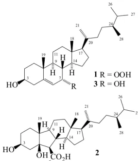
allowed the isolation and characterization of two new steroids 1 and 2 (Figure 2), in addition to seven known
sterols (Figure S1).
Compound 1, an amorphous white powder, m.p. 127.1 °C;
[α]D20 −34.0° (c 0.04, CHCl3) had its molecular formula
C28H48O3 deduced by HRESIMS analysis, by measurement of
the protonated molecular ion peak [M + H]+ at m/z 433.3597
(calcd. 433.3676), and by 13C NMR-CPD (composite pulse
decoupling) analysis. Comparing the 13C NMR data of 1
with those of 7α-hydroxycampesterol (3),19 also isolated in
this work (Table1), a good match was observed, but with a significant difference for the corresponding chemical shift of the C-7 oxymethine carbon, dC 78.6 for 1 instead
of 65.8 for 3 (∆dC 12.8 ppm). This is in agreement with
a carbon bearing a hydroperoxy moiety (−OOH),20 as
supported by the molecular formula C28H48O3. In fact,
the HSQC (heteronuclear single quantum correlation) spectrum exhibited correlations for the protons at dH 4.15
(br s, 1H, H-7) and 3.62 (m, 1H, H-3) with the carbons at
dC 78.6 (C-7) and 71.5 (C-3), respectively, in agreement
with the chemical shifts for oxymethines bearing a peroxyl and a hydroxyl groups, respectively.20 The deshielding of
H-7/C-7 (dH/dC 4.15/78.6), when compared with H-3/C-3
(dH/dC 3.62/71.5), is due to the β-effect of the additional
oxygen of the hydroperoxyl group when compared with that of the corresponding hydroxyl group of 3.19 According
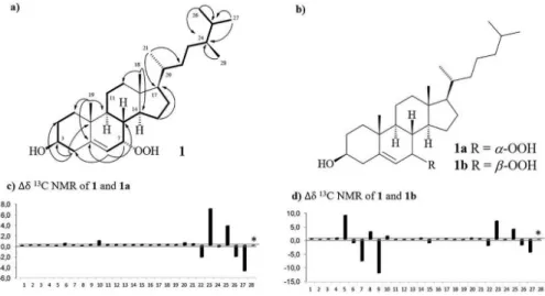
with the above data, and by analysis of the COSY (correlation spectroscopy) and HMBC (heteronuclear multiple bond correlation) spectra (Figure 3a), the structure of 1 was established. The NOESY (nuclear Overhauser effect
spectroscopy) spectrum did not show any key correlations to prove the β-position for HO-3 and the α-position for HOO-7. However, comparison of the carbon chemical shifts of 1 with those reported to 7α-hydroperoxycholesterol (1a)
and 7β-hydroperoxycholesterol (1b)20 (Figure 3b), showed
a very close similarity with the 1a, particularly for the
chemical shifts related to the carbocyclic chain (Figure 3c). Thus, the structure of compound 1 was defined as
7α-hydroperoxycampesterol.
Compound 2, an amorphous white powder, had its
C28H48O4 molecular formula determined by HRESIMS,
based on the deprotonated molecular ion peak [M − H]− at m/z 447.3498 (calcd. 447.3474), 13C CPD and DEPT 135
NMR spectra. Its IR spectrum (KBr) exhibited absorption bands at 3.492 (νOH) and 1.711 (νCO) cm-1 indicating a
carboxyl function. Comparison of 1H and 13C NMR spectra
of 2 with those of 1 (Table 1), revealed that these compounds
share the same ergostane backbone, but with significant differences in the chemical shifts related to the A and B rings. In the 13C CPD and DEPT NMR spectra, signals
corresponding to oxymethine carbons at dC 67.3 (C-3)
and 58.7 (C-6), and of a tertiary alcohol carbon at dC 82.9
(C-5), in addition to a carboxylic acid at dC 178.2 (C-7) were
observed. The unequivocal position for the two hydroxyl groups, including the carboxyl acid moiety, was supported by the HMBC spectrum. Correlations were observed for the diastereotopic methylene protons at dH 1.64/1.62 (m,
2H, H-2) and 2.13/1.68 (m, 2H, H-4) with the oxymethine carbon at dC 67.3 (C-3), as well as the methyl signal at
dH 0.96 (s, 3H, H-19) with the non-hydrogenated carbon
at dC 82.9 (C-5) supporting the presence of two hydroxyl
groups at C-3 and C-5, respectively. Likewise, correlations for the protons at dH 2.27 (H-6) and 2.09 (H-8) with the
carbon at dC 178.2 (C-7) warrant the carboxyl group at
C-6. These data, including COSY and HMBC analyses (Figure 4a) were in agreement with a rearranged five membered ring B of a nor-ergostane sterol.21 These kind
of C6-C5-C6-C5 tetracyclic ergostane steroids are rare in
nature. Few examples include similar sterols isolated from the sponges Stelletta hiwasaensis22 and Svenzea zeai.23
The β-positions for both hydroxyl groups at C-3 and C-5, for the carboxyl acid moiety at C-6, and the side chain at C-17, including the cis-configuration for the fused A/B
Figure 3. (a) Key 1H-1H COSY (H—H) and HMBC (H→C) correlations for 1; (b) structures of 7α-hydroperoxycholesterol (1a) and 7β-hydroperoxycholesterol
rings was inferred by direct comparison of the chemical shifts described to B-nor-ergostan-3β-5β-diol-6β-carboxyl acid (2a),24 which showed a good match from C-1 through
C-21 (Figure 4b). On the other hand, the observed NOE displayed for H-6 atdH 2.27 (d, J 9.7 Hz) with H-9 and
H-14 both at dH 1.16 (m), was an additional support for
a β-orientation of the carboxylic acid group.24 Thus, the
structure of 2 was established as 6β-carboxyl-24(R)-(8→
6)-abeo-ergostan-3β,5β-diol.
In addition to the new compounds 1 and 2, seven known
sterols have also been isolated: 7α-hydroxycampesterol (3),19
5α,8α-epi-dioxycampesterol (4),25 campesterol (5),2624(R
)-ergost-7-en-3β,5α,6β-triol (6),27 20-hydroxyecdysone (7),17
2-O-acetyl-20-hydroxyecdysone (8)17 and 3-O
-acetyl-20-hydroxyecdysone (9)17 (Figure S1). Additionally,
GC-MS analysis of the hexane fraction (see SI), allowed the identification of the following sterols: gorgosterol (m/z 426), 7α-hydroxycampesterol (m/z 416),campesterol
(m/z 400), crinosterol (m/z 398), cholest-4-en-3-one
(m/z 398), cholesta-3,5-dien-7-one (m/z 396), cholesterol
(m/z 386) and cholesta-5,22-dien-3-ol (m/z 384)10 (Table S2
and Figure S20).
Orostanal, an abeo-sterol isolated from a marine
sponge, and its derivatives, are known to have moderated cytotoxic effects.28 The scanty literature attributes the
cytotoxic activity of these class of compounds to apoptosis
Figure 4. (a) Key 1H-1H COSY (H—H) and HMBC (H→C) for compound
2; (b) structure of B-nor-ergostan-3β-5β-diol-6β-carboxyl acid (2a); (c) 13C NMR comparison between 2 and 2a.21 *Me-28 does not exist in
cholestane sterols.
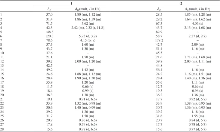
Table 1. 1H (500 MHz) and 13C (125 MHz) NMR data for 1 and 2 inCDCl 3
1 2
dC dH (mult, J in Hz) dC dH (mult, J in Hz)
1 37.0 1.80 (m), 1.12 (m) 28.5 1.85 (m), 1.28 (m)
2 31.4 1.86 (m), 1.59 (m) 28.2 1.64 (m), 1.62 (m)
3 71.5 3.62 (m) 67.3 4.06 (s)
4 42.3 2.42 (m), 2.32 (t, 11.8) 43.7 2.13 (m), 1.68 (m)
5 148.8 − 82.9 −
6 120.3 5.73 (d, 3.2) 58.7 2.27 (d, 9.7)
7 78.6 4.15 (br s) 178.2 −
8 37.3 1.60 (m) 42.7 2.09 (m)
9 43.7 1.30 (m) 51.4 1.16 (m)
10 37.6 − 45.5 −
11 21.1 1.50 (m) 21.6 1.71 (m), 1.68 (m)
12 39.2 2.00 (m), 1.20 (m) 39.8 2.03 (m), 1.11 (m)
13 42.5 − 44.8 −
14 49.2 1.42 (m) 56.4 1.16 (m)
15 24.6 1.88 (m), 1.12 (m) 24.2 1.16 (m), 1.51 (m)
16 28.4 1.90 (m), 1.30 (m) 28.4 1.40 (m), 1.36 (m)
17 55.9 1.20 (m) 55.6 1.11 (m)
18 11.5 0.66 (s) 12.7 0.69 (s)
19 18.4 0.99 (s) 19.3 0.96 (s)
20 36.3 1.38 (m) 36.2 1.36 (m)
21 19.1 0.91 (d, 6.6) 17.7 0.92 (d, 6.7)
22 33.9 1.32 (m), 0.98 (m) 33.9 1.38 (m), 0.95 (m)
23 30.6 1.40 (m), 0.99 (m) 30.7 1.36 (m), 0.95 (m)
24 39.2 1.20 (m) 39.2 1.18 (m)
25 31.7 1.58 (m) 31.6 1.55 (m)
26 20.7 0.86 (d, 6.6) 20.7 0.84 (d, 6.7)
27 17.8 0.79 (d, 6.6) 17.7 0.78 (d, 6.7)
induction.21,28 The cytotoxic activity of 2 was assessed by
the MTT assay against a human colorectal tumor cell line (HCT-116). Compound 2 showed inhibition concentration
mean (IC50) values of 50 and 4 µM after 24 and 72 h
treatment, respectively. Flow cytometry was performed after 24 h to assess the cellular concentration and viability as well (Figure 5). Compound 2 reduced cell number and
increased non-viable cells at the higher concentrations. Compound 1 was not tested because it decomposed
previously to the assay.
Conclusions
In this work, we described the chemical investigation of sterols from P. caribaeorum and P. variabilis. Nine
sterol compounds were isolated, two of which being new. A series of cholestane and ergostane sterols were identified by GC-MS analysis. Compound 2, an uncommon
C6-C5-C6-C5 tetracyclic abeo-ergostan sterol, showed
moderate cytotoxicity against the human colorectal tumor (HCT-116) cell line.
Supplementary Information
Experimental details of the isolation of the known compounds and supplementary information (Figures S1-S20 and Tables S1-S2) are available free of charge at http://jbcs.sbq.org.br as a PDF file.
Acknowledgments
The authors thank the Governmental Brazilian Agencies CNPq, FUNCAP and CAPES for financial support and fellowships.
References
1. Irei, Y.; Nozawa, Y.; Reime, J. D.; Zool. Stud.2011, 50, 426.
2. Reimer, J.; Palythoa lamouroux, 1816, 2014. Available at World Register of Marine Species at http://www.marinespecies.org/ aphia.php?p=taxdetails&id=205785, accessed in December 2016.
3. Hibino, Y.; Todd, P. A.; Yang, S.; Benayahu, Y.; Reimer, J. D.;
Hydrobiologia2013, 733, 31.
4. Almeida, J. G. L.; Maia, A. I. V.; Wilke, D. V.; Silveira, E. R.; Braz-Filho, R.; La Clair, J. J.; Costa-Lotufo, L. V.; Pessoa, O. D. L.; Mar. Drugs2012, 10, 2846.
5. Imbs, A. B.; Biochem. Syst. Ecol.2014, 54, 213; Carballeira,
N. M.; Reyes, M.; J. Nat. Prod. 1995, 58,1689; Miralles, J.; Diop, M.; Ferrel.; Kornprobst, J. M.; Comp. Biochem. Physiol. 1989, 94B, 91.
6. Hirata, Y.; Uemura, D.; Ueda, K.; Takano, S.; Pure Appl. Chem. 1979, 51, 1875; Ueda, K.; Takano, S.; Uemura, D.; Hirata, Y.; Tetrahedron Lett.1978b, 49, 4909; Ito, S.; Hirata,
Y.; Tetrahedron Lett.1977, 28, 2429.
7. Riobó, P.; Franco, J. M.; Toxicon2011, 57, 368.
8. Lazcano-Pérez, F.; Vivas, O.; Román-González, S. A.; Rodríguez-Bustamante, E.; Castro, H.; Arenas, I.; García, D. E.; Sánchez-Puig, N.; Arreguín-Espinosa, R.; Toxicon2014,
82, 112; Pettit, G.; Fujii, O.; Hasler, J. A.;Schmidt, J. A. M.;
Michel, C.; J. Nat. Prod. 1982, 45, 263; Pettit, G.; Fujii, O.; Hasler, J. A.;Schmidt, J. A. M.; J. Nat. Prod. 1982, 45, 272.
9. Elbagory, A. M.; Meyer, M.; Ali, A.-H. A. M.; Ameer, F.; Parker-Nance, S.; Benito, M. T.; Doyagüez, E. G.; Jimeno, M. L.; Hussein, A. A.; Steroids2015, 101, 110; Shigemori, H.; Sato, Y.; Kagata, T.; Kobayashi, J.; J. Nat. Prod. 1999, 62, 372;
Fedorov, S. N.; Stonik, V. A.; Elyakov, G. B.; Khim. Prirodn. Soedin. 1988, 4, 603.
10. Diop, M.; Leung-Tack, D.; Braekman, J. C.; Kornprobst, J. M.; Biochem. Syst. Ecol. 1986, 14, 151; Miralles, J.; Diop, M.;
Ferre, L.; Kornprobst, J. M.; Comp. Biochem. Physiol. 1988, 8, 209; Kelecom, A.; Salé-Cava, A. M.; Comp. Biochem. Physiol. 1982, 72, 677.
11. Cariello, L.; Crescenz, S.; Zanetti,L.; Comp. Biochem. Physiol. 1978, 63B, 77.
12. Han, C.; Qi, J.; Shi, X.; Sakagami, Y.; Shibata, T.; Uchida, K.; Ojika, M.; Biosci. Biotechnol. Biochem. 2006, 70, 706. 13. Wilke, D. V.; Jimenez, P. C.; Pessoa, C.; Moraes, M. O.; Araujo,
R. M.; Silva, W. M. B.; Silveira, E. R.; Pessoa, O. D. L.; Braz-Filho, R.; Lopes, N. P.; Costa-Lotufo, L. V.; J. Braz. Chem. Soc. 2009, 20, 1455; Wilke, D. V.; Jimenez, P. C.; Araújo, R. M.; Silva, W. M. B.; Pessoa, O. D. L.; Silveira, E. R.; Pessoa, C. O.; Moraes, M. O.; Skwarczynski, M.; Simerska, P.; Toth, I.; Costa-Lotufo, L. V.; Bioorg. Med. Chem. 2010, 18, 7997.
14. Uemura, D.; Toya, Y.; Watanabe, I.; Hirata, Y.; Chem. Soc. Jpn. 1979, 1481.
15. Moore, R. E.; Sheuer, P. J.; Science1971, 172, 495.
16. Reimer, J. D.; Ono, S.; Takishita, K.; Tsukahara, J.; Maruyama, T.; Zool. Sci.2006, 23, 87.
17. Cachet, N.; Genta-Jouve, G.; Ivanisevic, J.; Chevaldonne, P.; Sinniger, F.; Culioli, G.; Pérez, T.; Thomas, O. P.; Sci. Rep. 2015, 5, 1; Calcagno, M. P.; Camps, F.; Cool, J.; Melé, E.; Eur. J. Entomol. 1995, 92, 278.
18. Mosmann, T.; J. Immunol. Methods1983, 65, 55.
19. Kobayashi, M.; Krishna, M. M.; Haribabub, B. B.; Anjaneyulu, V.; Chem. Pharm. Bull. 1993, 41, 87.
20. Sung,P.-J.; Lin, M.-R.;Chen, J.-J.; Lin, S.-F.; Wu, Y.-C.; Hwang, T.-L.; Fang, L.-S.; Chem. Pharm. Bull.2007, 55, 666.
21. Lin, W.-H.; Fang, J.-M.; Cheng, Y.-S.; Phytochemistry1998,
48, 1392.
22. Miyamoto, T.; Kodama, K.; Aramaki, Y.; Higuchi, R.; Soest, R. W. M. V.; Tetrahedron Lett.2001, 42, 6349.
23. Wei, X.; Rodriguez, A. D.; Wang, Y.; Franzblau, S. G.;
Tetrahedron Lett.2007, 48, 8851.
24. Schenast, J. C.; Witter, D. P.; Grant, E.; Boldt, G. E.; Offer, J.; Wentworth, J. P.; Angew. Chem. Int. Ed. 2009, 48, 9469.
25. Ioannoua, E.; Abdel-Razika, A. F.; Zervouc, M.; Christfidisa, D.; Alexi, X.; Vagiasa, C.; Alxis, M. N.; Roussis, V.; Steroids 2009, 74, 73.
26. Zhang, X.; Cambrail, A.; Miesch, M.; Roussi, S.; Raul, F.; Marchioni, E.; J. Agric. Food Chem.2006, 54, 1196.
27. Madaio, A.; Piccialli, V.; Sica, D.; J. Nat. Prod. 1989, 52, 952.
28. Cui, J.; Qi, B.; Gan, C.; Liu, Z.; Huang, H.; Lin, Q.; Zhao, D.; Huang, Y.; Mar. Drugs2015, 13, 2488.
Submitted: August 26, 2016