Article
Synthesis and Characterization of Heptacoordinated Tin(IV) Complexes.
X-ray Crystal Structure of [
nBu
2Sn(dappt)]·(Me
2CO)
0.5[H
2dappt =
2,6-diacetylpyridine bis(4-phenylthiosemicarbazone)]
Gerimário F. de Sousaa*, Victor M. Deflona, Elke Niquetb and Anuar Abrasc
a
Instituto de Química, Universidade de Brasília, 70919-970, Brasília - DF, Brazil
b
Institut für Anorganische Chemie, Universität Tübingen, Auf der Morgenstelle 18, D-72076, Tübingen, Germany
c
Departamento de Física, Universidade Federal de Minas Gerais, 30123-970, Belo Horizonte - MG, Brazil
As reações do ligante 2,6-diacetilpiridina bis(4-feniltiossemicarbazona), H2dappt, com R4-mSnXm (m = 2, 3; R = Me, nBu, Ph e X = Cl) resultaram na formação de quatro novos complexos organoestânicos heptacoordenados, os quais foram caracterizados por análise elementar e pelas espectroscopias no IV e Mössbauer. O derivado n-butila, [nBu
2Sn(dappt)]·(Me2CO)0.5, foi também analisado por um estudo de difração de raios X em um monocristal. O complexo cristalizou-se no sistema monoclínico e grupo espacial C2/c, com a = 36,164(14), b = 9,7050(15), c = 26,194(11) Å,
β = 132,00(2)º, Z = 8. A determinação da estrutura revelou um complexo neutro de Sn(IV), numa geometria bipiramidal pentagonal (BPP), com o plano equatoral definido pelos átomos doadores SNNNS do ligante e dois grupos n-bultila nas posições axiais. Também é discutida uma correlação entre os dados de Mössbauer e de difração de raios X, baseada no modelo da carga-pontual.
The reactions of the 2,6-diacetylpyridine bis(4-phenylthiosemicarbazone) ligand, H2dappt, with R4-mSnXm (m = 2, 3; R = Me, nBu, Ph and X = Cl) led to the formation of four new heptacoordinated organotin(IV) complexes, which were characterized by microanalyses and by IR and Mössbauer spectroscopies. The n-butyl derivative [nBu
2Sn(dappt)]·(Me2CO)0.5 was also analyzed by a single crystal X-ray diffraction study. It crystallized in the monoclinic system with a space group C2/c, with a = 36.164(14), b = 9.7050(15), c = 26.194(11) Å, β = 132.00(2)º, Z = 8. The structure determination revealed a neutral complex of Sn(IV) in a distorted pentagonal bipyramidal (PBP) geometry, with the equatorial plane defined by the SNNNS donor system of the ligand and with the two n-butyl groups in the axial positions. Also, a correlation between Mössbauer and X-ray data based on the point-charge model is discussed.
Keywords: thiosemicarbazone complexes, heptacoordinated organotin(IV) complexes, X-ray diffraction analysis
Printed in Brazil 0103 - 5053 $6.00+0.00
Introduction
The chelating properties of 2,6-diacetylpyridine bis(thiosemicarbazones) have been investigated and six different coordination modes have been discovered so far. [Ph2Sn(daptsc)]·2DMF1 (H
2daptsc = 2,6-diacetylpyridine
bis(thiosemicarbazone), which was obtained from Ph2SnO in DMF (N,N-dimethylformamide), crystallizes in a regular PBP geometry with the dianion daptsc2- in the pentagonal
plane and the two phenyl groups in the axial positions. Similarly, in [nBu2Sn(H2daptsc)]Cl2·MeNO22 the Sn(IV) atom is heptacoordinated in a distorted PBP configuration, with the five SNNNS donor atoms of the H2daptsc in the pentagonal plane. In [Ph2Sn(Hdaptsc)]Cl3 the Sn(IV) atom
is heptacoordinated and also has a PBP geometry, where only
one of the H2daptsc arms has undergone deprotonation. A fourth coordination mode was reported for [Zn2(daptsc)2]·2MeOH·H2O4, where each of the two fully deprotonated ligands, daptsc2-, are coordinated to two Zn(II)
atom in a distorted octahedral geometry. The dinuclear complex [Zn2(daptsc)2]·MeOH·H2O4, crystallized from
Since both Sn(IV)6 and thiosemicarbazones7 have
significant pharmacological activity, some crystal structures of organotin(IV) complexes with 4-substituted thiosemicarbazones have already been discussed6. We report in this work the preparation and characterization of new heptacoordinated organotin(IV) complexes with the ligand 2,6-diacetylpyridine bis(4-phenylthiosemi-carbazone), whose structure is shown below.
Experimental
Materials
All solvents were purified and dried according to standard procedures. 2,6-diacetylpyridine (Aldrich) and the organotin halides (Aldrich) were used without further purification. IR spectra were recorded on a Nicolet 5ZDX-FT spectrophotometer in the 4000-400 cm-1 range using KBr
pellets. 119Sn Mössbauer spectra were recorded using a constant acceleration spectrometer moving a CaSnO3 source at room temperature. The samples were analyzed at 85K. The spectra were computer-fitted, assuming Lorentzian lines shapes. X-ray diffraction data were collected at room temperature using an Enraf-Nonius CAD-4 automatic diffractometer, with a graphite monocromated MoKα radiation obtained in a fine focus sealed tube (see Table 1).
Preparation of H2dappt and its Sn(IV) complexes
The pale yellow 2,6-diacetylpriridine bis(4-N
N C
N CH3
C
N H3C
N H
C C
N H
S S
N
H H
H2dappt
Table 1. Crystal data, data collection and structure refinement parameters for [nBu
2Sn(dappt)]·(Me2CO)0.5 (3).
Formula C32.5H42N7O0.5S2Sn
Formula weight (g mol-1) 721.54
Crystal system monoclinic
Crystal color orange
Formula units Z 8
Space group C2/c
Crystal dimensions (mm3) 0.40 x 0.40 x 0.10
Temperature (K) T 293(2)
Cell constants (Å, º) a 36.164(14)
b 9.7050(15)
c 26.194(11)
β 132.00(2)
Volume (Å3) V 6832(4)
Calculated density (g cm-3) ρ
x 1.403
Range of data collection θ 3-27º
h k l -1 → 46, 0 → 12, -33 → 25
F(000) 2976
Absorption coefficient (mm-1) µ 0.905
Radiation Mo-Kα
Wavelength (Å) λ 0.71073
Collected reflections 7813
Unique reflections/R(int) 7458/0.0255
Observed reflections {I > 2σ (I)} 5739
Absorption correction Ψ-Scan10
Min./max. Transmission 0.80698/0.95784
Structure solution heavy atom method11
Refinement method full-matrix least-squares on F2 12
Parameters refined 338
Goodness-of-fit S 1.016
Final structure factors {I > 2σ (I)} R1 0.0430
phenylthiosemicarbazone), H2dappt, was prepared by refluxing a 2:1 molar mixture of 4-phenylthiosemi-carbazide8 with 2,6-diacetylpyridine in absolute EtOH. The
Sn(IV) complexes were obtained by the following procedure: 0.097 g (0.21 mmol) of H2dappt were dissolved by refluxing a 1:1 molar mixture of MeOH/Me2CO (10 mL) for 10 min. To this solution was added 0.22 mmol of the appropriate organotin(IV) species, dissolved in 2 mL of MeOH, and the resulting mixture was refluxed for about 1 h. Cooling the solution and slow evaporation of the solvent led to the appearance of crystalline products with yields on the order of 60%. The microanalyses were performed with a Perkin Elmer 2400C analyzer, giving for C, H and N the following results.
H2dappt. Anal. Found: C, 59.05; H, 4.82; N, 21.24. Calcd. for C23H23N7S2: C, 59.87; H, 4.99; N, 21.26%.
1. MeSnCl(dappt)], m.p. 157 ºC (dec.). Anal. Found:
C, 47.22; H, 3.68, N, 15.51. Calcd. for C24H24ClN7S2Sn: C, 45.85, H, 3 85, N, 15.59%.
2. [Me2Sn(dappt)], m.p. 231-234 ºC. Anal. Found: C, 48.60; H, 4.39, N, 15.97. Calcd. for C25H27N7S2Sn: C, 49.36; H, 4.47; N, 16.12%.
3. [nBu
2Sn(dappt)]·(Me2CO)0.5, m.p. 238-241 ºC. Anal.
Found: C, 53.58; H, 5.64, N, 13.53. Calcd. for C32.5H42N7O0.5S2Sn: C, 52.77; H, 5.48; N, 14.13%.
4. [Ph2Sn(dappt)], m.p.134 ºC (dec.). Anal. Found: C, 48.03; H, 3.71; N, 9.24. Calcd. for C35H31N7S2Sn: C, 49.11; H, 3.74; N, 8.53%.
Results and Discussion
Crystal and molecular structure of [nBu
2Sn(dappt)]·
(Me2CO)0.5 (3).
The ORTEP drawing of the molecular structure of di-n-butyl[2,6-diacetylpyridine bis(4-phenylthiosemi-carbazone)]tin(IV) acetone solvate, [nBu2Sn(dappt)]· (Me2CO)0.5 (3), is shown in Figure 1. Crystal structure parameters and conditions of data collection are given in Table 1. Selected bond distances and angles are presented in Table 2.
All non hydrogen atoms were refined with anisotropic displacement parameters with the exception of those of the n-butyl groups and those of the solvate molecule. For the latter, greater freedom for thermal displacements was observed in accord with their low steric hindrance. The hydrogen atoms were fitted on idealized positions.
The n-butyl group bonded to Sn(IV) via C(31) shows considerably greater displacement parameters than the other one. This should be related to thermal movements. The second butyl group is proximate to the solvate
molecule, resulting in additional constraints on its freedom. The carbon atom C(33) was found close to C(32) and too far from C(34), with unrealistic interatomic distances for both C(32)-C(33) and C(33)-C(34) bonds that were then restrained to have similar values, as expected.
Table 2. Selected bond distances (Å) and angles (º) for
[nBu
2Sn(dappt)]·(Me2CO)0.5 (3). Standard deviation in parentheses.
Sn-C(21) 2.148(4) C(2)-N(3) 1.292(5)
Sn-C(31) 2.155(5) C(3)-N(4) 1.348(5)
Sn-N(3) 2.450(3) C(7)-N(4) 1.338(5)
Sn-N(4) 2.411(3) C(8)-N(5) 1.292(5)
Sn-N(5) 2.465(3) C(10)-N(2) 1.306(5)
Sn-S(1) 2.6210(17) C(17)-N(6) 1.309(5)
Sn-S(2) 2.6878(15) C(10)-N(1) 1.372(5)
N(2)-N(3) 1.374(4) C(17)-N(7) 1.366(5)
N(5)-N(6) 1.374(4) C(10)-S(1) 1.740(4)
N(1)-O(41) 2.937(4) C(17)-S(2) 1.738(4)
N(1)’-O(41) 2.937(4) N(7)-S(2)” 3.686(4)
C(21)-Sn-C(31) 168.12(18) N(4)-Sn-S(1) 138.30(8)
C(21)-Sn-N(4) 84.78(14) N(4)-Sn-S(2) 137.05(8)
C(31)-Sn-N(4) 83.56(16) N(3)-Sn-S(1) 72.05(9)
C(21)-Sn-N(3) 88.59(14) N(5)-Sn-S(2) 70.87(8)
C(31)-Sn-N(3) 88.82(16) N(5)-Sn-S(1) 155.25(8)
C(21)-Sn-N(5) 85.16(14) N(3)-Sn-S(2) 156.55(8)
C(31)-Sn-N(5) 88.07(16) S(1)-Sn-S(2) 84.62(4)
N(4)-Sn-N(5) 66.36(11) C(10)-S(1)-Sn 99.85(15)
N(4)-Sn-N(3) 66.26(11) C(17)-S(2)-Sn 99.75(14)
N(3)-Sn-N(5) 132.56(11) C(7)-N(4)-Sn 119.9(2)
C(21)-Sn-S(1) 94.06(12) C(3)-N(4)-Sn 120.1(3)
C(31)-Sn-S(1) 96.16(14) C(8)-N(5)-Sn 120.1(3)
C(21)-Sn-S(2) 95.49(12) N(2)-N(3)-Sn 123.9(2)
C(31)-Sn-S(2) 91.48(15) N(6)-N(5)-Sn 124.3(2)
N(1)-H(1)···O(41) 138.5 N(7)-H(7)···S(2)” 161.1 Symmetry transformations: – x + 1, y, – z + ½; – x + 1, – y + 2, – z + 1.
Figure 1. Ellipsoid representation9, with labeled atoms and 50%
probability, of the asymmetric unit of [nBu
2Sn(dappt)]·(Me2CO)0.5 (3).
The single crystal X-ray diffraction studies revealed the occurrence of a heptacoordinated neutral complex of approximately pentagonal bipyramidal (PBP) geometry, with the organic ligand lying in the equatorial plane. The approximate coplanarity between the phenyl rings and the bipyramidal equatorial plane suggests electron delocalization along the equatorial atoms. The mean deviation from planarity for the dappt2- atoms is 0.077 Å, except for the side methyl groups and the N-phenyl groups that were not fitted. The Sn(IV) atom has a deviation of 0.046(1) Å from this plane. The angle between the equatorial plane and the phenyl ring attached to the nitrogen atom N(1) is 6.8(1)°. The other phenyl group, bound to N(7), is somewhat more twisted, with a torsion angle of 36.5(1)°.
Due to geometrical requirements of the thiosemicar-bazone structure the pentagon is not regular. The S(1)-Sn-S(2) angle is significantly larger [84.62(4)º] than that of a regular pentagonal bipyramide (72º), while the other equatorial angles involving the Sn(IV) atom are in the 66.25(11)º - 72.05(9)º range. The axial n-butyl groups contribute to the distortion of the bipyramide structure since the C(21)-Sn-C(31) angle is 168.12(l8)º.
The Sn-S and Sn-N bond distances, which average 2.654 and 2.442 Å, respectively, are in good agreement with the values (Sn-S = 2.593 - 2.692; Sn-N = 2.417 - 2.436 Å) found in two other heptacoordinated pentagonal bipyramidal diorganotin(IV) complexes containing a pentadentate thiosemicarbazone in the equatorial plane, namely [nBu2Sn(Achexim)]2 and [Ph2Sn(daptsc)]·2DMF1 [H2Achexim = 2,6-diacetylpyridine bis(3-hexamethyl-eneiminylthiosemicarbazone) and H2daptsc = 2,6-diacetylpyridine bis(thiosemicarbazone)]. However, these distances are considerably longer than the corresponding bonds Sn-S = 2.5425(8) and Sn-N(3) = 2.199(2) Å in the trigonal bipyramidal (TBP) complex, [Me2Sn(L)]6 (H2L = salicylaldehydethiosemicarbazone), as would be expected from the structural differences between the ligands and the consequent different hybridizations of the Sn(IV) atom in each complex. The Sn-C distances in the three heptacoordinate complexes are quite similar and evidently insensitive to the different nature of the s-bonded organic groups.
Due to the deprotonation of the hydrazine nitrogen atoms, N(2) and N(6), a delocalization of the negative charges along the N(3)-N(2)-C(10)-S(1) and N(5)-N(6)-C(17)-S(2) atoms is in agreement with the shortening of the following bond distances N(2)-C(10) = 1.306(5) and N(6)-C(17) = 1.309(5) Å, thus confirming the N-C double bond character. As a result of the thiolate sulfur coordination, the C(10)-S(1) = 1.740(4) and C(17)-S(2) = l.738(4) Å bonds lose their π-character, becoming essential, single bonds.
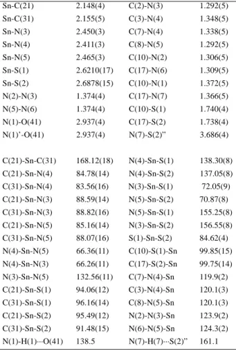
The solvate acetone carbonyl C(41) and O(41) atoms occupy special positions so that the C=O bond coincides with the two fold rotation axis. The oxygen O(41) atom participates through its free electron pairs in two hydrogen bonds with neighboring complex molecules via the nitrogen N(1) atom. The N(1)···O(41) distance [2.937(4) Å] is in good agreement with the normally observed distance for this type of bond (2.93 Å)13. The N(1)-H(1)···O(41) angle [138.5°] shows appreciable deviation from linearity. Hydrogen bonds are also observed between the N(7) and S(2) atoms, of two neighboring molecules, as shown in Figure 2. The N(7)···S(2)” distance [3.686(4) Å] is in this case 0.34 Å longer than the sum of the van der Waals radii of the two atoms (3.35 Å)13 and the N(7)-H(7)···S(2)” angle [161.1°] is compatible with the presence of a hydrogen bond.
Figure 2. Representation14 of the crystal structure of [nBu
2Sn
(dappt)]·(Me2CO)0.5 (3), showing three molecules of the tin complex and one of the acetone solvate. The dashed lines show the possible hydrogen bonds. Except for H(1) and H(7) all other hydrogen atoms are omitted for the sake of clarity.
Infrared spectroscopy
The main vibrational bands of H2dappt and of its complexes are shown in Table 3. The high frequency bands of the uncomplexed ligand, centered at 3354, 3321 and 3214, 3114 cm-1, are attributed to ν(N-H) stretching vibrations.
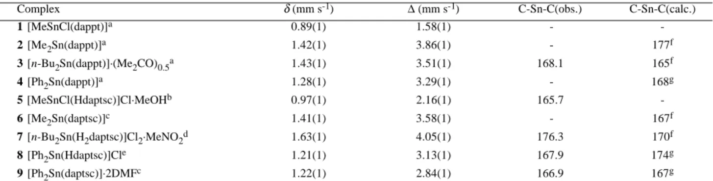
Table 4. Mössbauer data and C-Sn-C angles (º) for heptacoordinated bis(thiosemicarbazones) complexes.
Complex δ (mm s-1) ∆ (mm s-1) C-Sn-C(obs.) C-Sn-C(calc.)
1 [MeSnCl(dappt)]a 0.89(1) 1.58(1) -
-2 [Me2Sn(dappt)]a 1.42(1) 3.86(1) - 177f
3 [n-Bu2Sn(dappt)]·(Me2CO)0.5a 1.43(1) 3.51(1) 168.1 165f
4 [Ph2Sn(dappt)]a 1.28(1) 3.29(1) - 168g
5 [MeSnCl(Hdaptsc)]Cl·MeOHb 0.97(1) 2.16(1) 165.7
-6 [Me2Sn(daptsc)]c 1.41(1) 3.58(1) - 167f
7 [n-Bu2Sn(H2daptsc)]Cl2·MeNO2d 1.63(1) 4.05(1) 176.3 170f
8 [Ph2Sn(Hdaptsc)]Cle 1.21(1) 3.13(1) 167.9 174g
9 [Ph2Sn(daptsc)]·2DMFc 1.22(1) 2.84(1) 166.9 167g
H2dappt = 2,6-diacetylpyridine bis(4-phenylthiosemicarbazone), H2daptsc = 2,6-diacetylpyridine bis(thiosemicarbazone), H2daps = 2,6-diacetylpyridine bis(salicyloylhydrazone), aThis work, bRef. 15, cRef.1, dRef. 2, eRef. 3, fCalculated using [alkyl] = - 0.97 mm s-1, gCalculated
using [Ph] = - 0.77 mm s-1. Table 3. Main IR bands (cm-1) for H
2dappt and its Sn(IV) complexes.
Compound ν(N-H) ν(C=N, C=C) ν(C-S) + ν(C-N) ν(C-S)
H2dappt 3337, 3321, 3214, 3114 1597, 1546, 1511, 1448 1354 814
1 [MeSnCl(dappt)] 3375, 3320 - - 1594, 1561, 1521, 1493 1316 690
2 [Me2Sn(dappt)] 3375, 3326 - - 1594, 1553, 1532, 1497 1308 694
3 [nBu
2Sn(dappt)]·(Me2CO)0.5* - 3328 - - 1590, 1544, 1519, 1476 1310 691
4 [Ph2Sn(dappt)] - 3363 - - 1600, 1558, 1527, 1474 1317 693 *ν(C=O) =1695 cm-1
Mössbauer spectroscopy
The 119Sn Mössbauer data for the complexes are reported in Table 4, which includes parameters from the literature for comparison. All complexes present one heptacoordinated Sn(IV) central atom in a distorted pentagonal bipyramidal configuration (PBP), with the SNNNS donor atoms in the pentagonal plane and the two other groups (R, Cl) in the axial positions.
Isomer shifts
The isomer shifts (δ) of complexes 1 (0.89 mm s-1) and 5 (0.97 mm s-1) are lower than that of the parent acid
MeSnCl3 (1.20 mm s-1)15. The same can be seen for
complexes 2 (1.42 mm s-1) and 6 (1.41 mm s-1); 3 (1.43
mm s-1) and 7 (1.63 mm s-1); 4 (1.28 mm s-1), 8 (1.21 mm s-1) and 9 (1.22 mm s-1), compared to their parent acids
Me2SnCl2 (1.49 mm s-1), nBu2SnCl2 (1.75 mm s-1)16 and Ph2SnCl2 (1.32 mm s-1), respectively.
In general, isomer shifts decrease upon complexation as a result of rehybridization of the Sn atoms in complexes with a greater involvement of Sn(IV) d-orbitals, thus reducing the s-character in the overall hybridization of the metal17,18.
The complexes having the same Sn(IV) precursor with different ligands, i.e., 2 and 6, show very similar isomer shifts. This can be explained by considering that the only difference between the ligands consists in the thioamide
nitrogen atoms, N(1) and N(7) which are attached to a phenyl group in H2dappt and to a hydrogen atom in H2daptsc, respectively. As this difference is outside the coordination sphere of the Sn(IV) atom, no sensible change in the s-electron density of Sn(IV) nucleus is expected.
On the other hand, the considerable increase in the isomer shift of complex 7 (1.63 mm s-1) as compared to
complex 3 (1.43 mm s-1) may be related to the presence of a Me2CO molecule in 3 and two Cl- counter ions in 7,
which are less electronegative than the oxygen atom. This accounts for the higher δ of 7 as compared to 3 as a consequence of the inverse dependence of isomer shift with eletronegativity15.
On going now from 1 (0.89 mm s-1) to 4 (1.28 mm s-1), one sees that the ligand remains unchanged, but the Cl
-was replaced by an alkyl group and the isomer shift of 4 increases in relation to 1. This result is also consistent with the inverse dependence of δ with the electronegativity of the bonded atoms at the Sn(IV) center.
Quadrupole splitting
From the quadrupole splitting (∆) values, presented in Table 4, it is not possible to characterize Sn(IV) complexes as being tetra-, penta-, hexa-, or heptacoordinated15,19,20.
| ∆ | = 4[R](1-3/4sin2θ)1/2, where θ is the R-Sn-R angle
and [R] is the partial quadrupole splitting of the R group. Considering complexes 3 and 7, whose structures were determined by single crystal diffraction, the [nBu] can be estimated. Inserting the values ∆ = 3.51 mm s-1 and
θ = 168º for complex 3 and ∆ = 4.05 mm s-1 and θ = 176º for complex 7 in the above equation, the following values are obtained for [nBu] = -0.91 mm s-1 and -1.00 mm s-1, respectively (average value = -0.95 mm s-1), which are in
excellent agreement with the -0.97 mm s-1 value for [alkyl]19. Now, using [alkyl] = -0.97 mm s-1, R-Sn-R angles of 177º
and 167º, respectively, are predicted for complexes 2 and 6. From the [Ph] values of complexes 8 (-0. 80 mm s-1)3,
9 (-0.74 mm s-1) and [Ph2Sn(dapa)] (-0.78 mm s-1)19, where H2dapa = 2,6-diacethylpyridine bis(2-aminobenzoyl-hydrazone), an estimated value for [Ph] in similar heptacoodinated organotin(IV) complexes of -0.77 mm s-1
is obtained. By inserting ∆ (3.29 mm s-1) for 4 and [Ph] = -0.77 mm s-1 into the equation, the Ph-Sn-Ph angle
is calculated to be 168º.
Acknowledgments
The authors are grateful to Prof. Dr. J. Strähle for the possibility of data collection for the crystal structure determination, and to CNPq and FAPEMIG in Brazil for financial support.
Supplementary Information
Crystallographic data (excluding structural factors) for the structure in this paper have been deposited with the Cambridge Crystallographic Data Center as supplementary publication number CCDC 138942. Copies of the data can be obtained, free of charge, on application to CCDC, 12 Union Road, Cambridge CB2 1EZ, UK, (fax: +44 1223 336033 or e-mail: [email protected]).
References
1. Casas, J. S.; Castiñeiras, A.; Sánchez, A.; Sordo, J. Vásques-Lópes, A.; Rodriguez-Arqüelles, M. C.; Russo, U. Inorg. Chim. Acta 1994, 221, 61.
2. De Sousa, G. F.; Valdéz-Martínez, J.; Abras. A.; Filgueiras, C. A. L. Unpublished results.
3. Moreno, P. C.; Francisco, R. H. P.; Gambardella, M. T. do P.; De Sousa, G. F.; Abras, A. Acta Cryst. 1997, C53, 1411.
4. Bino, A.; Cohen, N. Inorg. Chim. Acta 1993, 210, 11. 5. Toukhy, A. Inorg. Chim. Acta 1991, 180, 85. 6. Casas, J. S.; Sánchez, A.; Sordo, J.; Vásques-Lópes,
A.; Castellano, E. E.; Zukerman-Schpector, J.; Rodriguez-Arqüelles, M. C.; Russo, U. Inorg. Chim. Acta 1994, 216, 169.
7. Iskander, M. F.; Labib, L.; Nour El-din, M. M. Z.; Tawfik, M. Polyhedron 1989, 23, 2755.
8. Offiong, O. E. Transition Met. Chem. 1997, 22, 263. 9. Farrugia, L. J. J. Appl. Cryst. 1997, 30, 565. 10. Spek, A. L.; PLATON, Program for the Treatment
of Crystallographic Data, University of Utrecht, Holland, 1996.
11. Sheldrick, G. M.; SHELXS97, Program for the Solution of Crystal Structures, University of Göttingen, Germany, 1997.
12. Sheldrick, G. M.; SHELXL97, Program for the Refinement of Crystal Structures, University of Göttingen, Germany, 1997.
13. Vinogradov, S. N.; Linnel, R. A. Hydrogen Bonding, Van Nostrand Reinhold, New York, 1971, 176. 14. Keller, E.; SCHAKAL92, Program for Graphical
Representation of Molecular Models, University of Freiburg, Germany, 1992.
15. De Sousa, G. F.; Filgueiras, C. A. L.; Abras, A.; Aljuaid, S. S.; Hitchcock, P. B.; Nixon, J. F. Inorg. Chim. Acta 1994, 218, 139.
16. Davies, A. G. Chem. Brit. 1968, 403.
17. Randall, R. S.; Wedd, R. W. J.; Sams, R. J. J. Organomet. Chem. 1971, 30, C19.
18. Cunningham, D.; Little, M.; Mcloughlin, K. J. Organomet. Chem. 1979, 165, 287.
19. Carini, C.; Pelizzi, G.; Tarasconi, P.; Pelizzi, C.; Molloy, K. C.; Waterfield, P. C. J. Chem. Soc., Dalton Trans. 1989, 289.
20. De Sousa, G. F.; Mangas, M. B. P.; Francisco, R. H. P.; Gambardella, M. T. do P.; Rodrigues, A. M. G. D.; A . Abras. J. Braz. Chem. Soc. 1999, 10, 222.
![Table 1. Crystal data, data collection and structure refinement parameters for [ n Bu 2 Sn(dappt)]·(Me 2 CO) 0.5 (3).](https://thumb-eu.123doks.com/thumbv2/123dok_br/18988869.459889/2.892.56.420.333.533/table-crystal-data-collection-structure-refinement-parameters-dappt.webp)
![Figure 2. Representation 14 of the crystal structure of [ n Bu 2 Sn (dappt)]·(Me 2 CO) 0.5 (3), showing three molecules of the tin complex and one of the acetone solvate](https://thumb-eu.123doks.com/thumbv2/123dok_br/18988869.459889/4.892.438.784.479.716/figure-representation-crystal-structure-showing-molecules-complex-acetone.webp)