New Heptacoordinated Organotin(IV) Complexes Derivatives of
2,6-diacetylpyridine
bis
(2-furanoylhydrazone), H
2dapf, and
2,6-diacetylpyridine
bis
(2-thenoylhydrazone), H
2dapt. Crystal and
Molecular Structure of [Me
2Sn(Hdapt)]Br.H
2O
Gerimário F. de Sousa
a,*, Maria B.P. Mangas
a, Regina H.P. Francisco
b,
Maria T. do P. Gambardella
b, Ana M.G.D. Rodrigues
b, Anuar Abras
caInstituto de Química,ICC, Universidade de Brasília,
70919.900 Brasília - DF, Brazil
bInstituto de Química de São Carlos, IQSC, Universidade de São Paulo,
13560.250 São Carlos - SP, Brazil
cDepartamento de Física, ICEx, Universidade Federal de Minas Gerais,
30123.970 Belo Horizonte - MG, Brazil
As reações dos ligantes H2dapf e H2dapt, com R4-mSnXm (m = 2, 3; R = Me, Ph e X = Cl, Br) resultaram na formação de oito novos complexos organoestânicos heptacoordenados, os quais foram estudados por análise elementar, espectroscopias no IV, RMN de 1H e Mössbauer, para investigar suas propriedades estruturais. O derivado metilado, [Me2Sn(Hdapt)]Br.H2O, foi também caracteri-zado por difração de raio X por monocristais. O complexo cristaliza no sistema monoclínico e grupo de espaço P21/c, com a = 21.920(3), b = 7.4470(5), c = 16.805(2) Å, β = 110.18(1)º e Z = 4. A determinação estrutural revelou um complexo monocatiônico de Sn(IV), [Me2Sn(Hdapf)]+, numa geometria bipiramidal trigonal, na qual o Br- está como contra-íon e uma molécula de água ajuda o empacotamento cristalino. Os parâmetros Mössbauer para o complexo [Me2Sn(Hdapf)]2 [Me2SnCl4] evidenciaram dois sítios de Sn(IV), como observado pela determinação da estrutura cristalina. Também uma correlação entre os dados de Mössbauer e de raio X, baseada no modelo carga-ponto, é discutida.
The reaction of the ligands H2dapf and H2dapt, with R4-mSnXm (m = 2, 3; R = Me, Ph and X = Cl, Br) led to the formation of eight new heptacoordinated organotin(IV) complexes, which were studied by microanalysis, IR, NMR and Mössbauer spectroscopy to investigate their structural properties. The methyl derivative [Me2Sn(Hdapt)]Br.H2O was also studied by single crystal X-ray diffraction. It crystallizes in the monoclinic system, space group P21/c, with a = 21.920(3), b = 7.4470(5), c = 16.805(2) Å, β = 110.18(1)º, Z = 4. The structure determination revealed a monocationic complex of Sn(IV) in a distorted bipyramidal geometry [Me2Sn(Hdapf)]+, with Br -as counter ion and one molecule of water helping the crystal packing. Mössbauer parameters of the complex [Me2Sn(Hdapf)]2[Me2SnCl4] have evidenced two Sn(IV) sites, as observed in the crystal structure determination. Also a correlation between Mössbauer and X-ray data based on the point-charge model is discussed.
Keywords: hydrazone complexes; heptacoordinated organotin(IV) complexes
Introduction
The chelating properties of 2,6-diacetylpyridinebis(hy-drazones) have been investigated towards several
organo-tin chlorides, R4-mSnClm (m = 2, 3). It has been found that these ligands act as pentadentate and coordinate to Sn (IV) through the two enolate oxygen atoms, the two azomethine Article
nitrogens and the pyridil nitrogen1,2,3. However, the nature of coordination depends on the metal ion, the pH of the medium, the reaction conditions, and also the nature of the hydrazone4. For instance, the pentadentate hydrazone can remain protonated as in the compound [MeSnCl(H2dapsc)]2+, H2dapsc = 2,6-diacetylpyridine-bis(semicarbazone)2, it can be deprotonated as in the com-plex [Et2Sn(dapt)], H2dapt = 2,6-diacetylpyridinebis(2-thenoylhydrazone)1, or partially protonated as in the monocation [Me2Sn(Hdapf)]+, H2dapf = 2,6-diacetylpyridinebis(2-furanoylhydrazone)3. Single crystal X-ray diffraction studies have shown that in all three modes of complexation, the Sn(IV) atom exhibits a nearly pentagonal bipyramidal coordination geometry with equa-torial plane defined by the ONNNO donor set of the hydra-zone ligand. The present work deals with the preparation and characterization of eight new complexes with the li-gands 2,6-diacetylpyridinebis(2-furanoylhydrazone), H2dapf, and 2,6-diacetylpyridinebis(2-thenoylhydrazone), H2dapt, whose structures are shown below. These two ligands were chosen since they have the potential to form pentacoordinated or higher coordinated Sn(IV) complexes. Furthermore, different possibilities of binding to the metal in the enolate or ketone forms make this study extremely interesting. The complexes formed were studied by mi-croanalysis, IR, 1H-NMR, Mössbauer spectroscopy and crystal X-ray diffraction.
Experimental
Synthesis
The ligands H2dapf and H2dapt were prepared accord-ing to the literature5, using EtOH as solvent. The Sn(IV) complexes were prepared by the following method. The methanol solution of the organotin(IV) derivative was added to the hot methanol solution of appropriate hydra-zone in a 1/1 mol ratio. The resulting mixture was refluxed for 1 h and filtered to form a clear solution and allowed to stand at room temperature. After cooling and slow evapo-ration of the solvent, crystalline products were formed with yields about 60%, which did not melt below 250 °C. Crystals, which were suitable for X-ray analysis, were
isolated only for the complexes 2 and 7. The structure of 2
was determined previously3. The microanalysis were per-formed with a Perkin Elmer 2400C analyser, giving for C, H and N the following results.
1. [MeSnCl(dapf)]. Anal. Calcd. for
(C20H18ClN5O2Sn): C, 43.90; H, 3.32; N, 12.81. Found: C, 43.40; H, 2.90; N, 12.72%.
2. [Me2Sn(Hdapf)]2[Me2SnCl4]. Anal. Calcd. for (C23H28Cl4N5O2Sn2): C, 39.31; H, 3.67; N, 10.41. Found: C, 38.72; H, 3.39; N, 10.62%.
3. [Me2Sn(Hdapf)]Br. Anal. Calcd. for (C21H22BrN5O2Sn): C, 41.53; H, 3.62; N, 11.54. Found: C, 40.25; H, 3.87; N, 11.40%.
4. [Ph2Sn(dapf)]. Anal. Calcd. for (C31H25N5O2Sn): C, 57.26; H, 3.11; N, 10.77. Found: C, 58.57; H, 2.93; N, 10.50%.
5. [MeSnCl(dapt)]. Anal. Calc.d for
(C20H18ClN5O2Sn): C, 41.52; H, 3.11; N, 12.10. Found: C, 40.73; H, 2.93; N,12.11%.
6. [Me2Sn(Hdapt)]2[Me2SnCl4]. Anal. Calcd. for (C23H28Cl4N5S2Sn2): C, 37.51; H, 3.58; N, 9.94%. Found: C, 37.07; H, 3.44; N, 9.77%.
7. [Me2Sn(Hdapt)]Br.H2O. Anal. Calcd. for (C21H22BrN5S2Sn): C, 38.25; H, 3.61; N, 10.62%. Found: C, 38.55; H, 3.41, N, 10.33%.
8. [Ph2Sn(dapt)]. Anal. Calcd. for (C31H25N5S2Sn): C, 54.55; H, 3.66; N, 10.26. Found: C, 53.81; H, 3.49; N, 9.87%.
Infrared spectra were recorded on a Nicolet 5ZDX-FT spectrophotometer in the 4000-400 cm-1 range using KBr pellets. Due to poor solubility of the complexes, it was possible to perform 1H NMR spectra only for the com-plexes 2, 3, 6 and 7 in CDCl3, using a 250 MHz Bruker spectrometer. Chemical shifts are relative to internal tetra-methylsilane. 119Sn Mössbauer spectra were measured us-ing a constant acceleration spectrometer movus-ing a CaSnO3 source at room temperature. The isomer shift values are given with respect to this source. The samples were meas-ured at liquid nitrogen temperature and all spectra were computer fitted assuming Lorentzian line shapes. X-ray diffraction data were collected at room temperature using an Enraf-Nonius CAD-4 automatic diffractometer, with a graphite monocromated MoKα radiation (λ = 0.71073 Å), obtained in a fine focus sealed tube.
X-ray structure determination of [Me2Sn(Hdapt)]Br.H2O
The unit cell of the complex 7 was determined by a
least-squares fit of settings for 25 strong reflections, with 10° ≤ θ ≤ 18°. Intensity measurements were carried out up to 27°, using ω/2θ scan mode. A yellow, prismatic, single-crystal with dimensions: 0.2 x 0.15 x 0.3 mm was used. During the data collection the intensity decay was checked through the measurement of 3 standard reflections every X = O, S
N N C C N N CH3
H3C
120 min and a correction factor was applied (average value = 0.9976). Absorption corrections were also taken into account according to an empirical method, ψ scan6. The maximum and minimum transmission factors were respec-tively 0.9964 and 0.7905. The crystal data and structure refinement parameters are given in Table 1.
The structure was solved by heavy-atom method. The refinement on (F(hkl))2 through iterative full-matrix least-squares calculations including all data was done, and weights ω = 1/[S2(F2
obs) + (0.0817P)2 + 10.9505P] where P = (F2
obs + 2 F2calc)/3 were assigned to them7. The observed criterion was used only for calculating R factor for ob-served reflections. The refinement process was applied until convergence (mean value of shift/esd = 0.000) and the maximum, minimum and average peaks of electronic den-sity residues were 2.80, -2.34 and 0.14 e Å-3, respectively. These peaks are high. The most positive is 0.44 Å from C132 and the negative is 0.50 Å from S135. Anisotropic displacement parameters were assigned to non-H atoms, with exception of one of the thiophene rings (C131, C132, C133, C134, S135). The rings have different behaviors during the refinement process, one of them is close to the bromide and the water molecule. The other one has a greater volume to occupy in the crystal structure. Due to this, the model was refined with isotropic temperature parameters for the atoms of this thiophene ring. Conse-quently, the highest peak and the deepest hole in the
resi-dual eletronic density are close to these atoms. Several attempts to refine them anisotropicaly and/or including disorder effects were done, without any success.
The H-atom positions were calculated and refined as riding atoms, except HN1, HO3A and HO3B, whose frac-tional atomic coordinates were determined in a difference Fourier map, calculated after convergence of the model with all other atoms and refined. An isotropic displacement parameter was applied to all H-atoms, using values 20% greater than the equivalent temperature factor of the corre-sponding C-atom.
Results and Discussion
Crystal structure of [Me2Sn(Hdapt)]Br.H2O
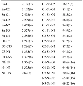
The structure determination revealed the presence of a complex cation of tin(IV), trans-dimethyl[2,6-diace-tylpyridinebis(2-thenoylhydrazone]tin(IV), with bromide as counter ion. A water molecule helps the packing mode. The metal is heptacoordinated, showing a distorted pentagonal bipyramidal geometry, with the pentahapto li-gand, Hdapt-, at the equatorial plane and two methyl groups at the axial positions. The major ligand is planar, except for the thiophene rings, which make dihedral angles of 27.4(1)° and 11.4(3)°, respectively, with the equatorial plane. Table 2 contains the fractional atomic coordinates and the equiva-lent isotropic displacement parameters. Table 3 contains
selected bond distances and angles. Figure 1 shows the molecule with labeled atoms.
During the reaction the proton from one of the azomethine groups leaves the molecule. The other, HN1, remains in the structure. As a consequence, differences between bond distances and angles around the metal can be observed, O1 atom is in a keto form and O2 is an enolate, which has greater basicity (Table 3).
This fact was also observed in the structures of other complexes, in which a similar ligand loses one proton, such as in [Ph2Sn(Hdaptsc)]Cl8 [H2daptsc = 2,6-diacetylpyrid-inebis(thiosemicarbazone)] with distances Sn-S: 2.592(1) and 2.703(1) Å; Sn-N: 2.348(4), 2.353(4) and 2.491(4) Å; [Me2Sn(Hdapf)]2[Me2SnCl4]3 [H2dapf = 2,6-diace-tylpyridinebis(furanoylhydrazone)] with distances Sn-O: 2.227(4) and 2.474(4) Å; Sn-N: 2.246(4), 2.263(5), and 2.314(4) Å; and [MeSnCl(Hdaptsc)]Cl.MeOH2 with dis-tances Sn-S: 2.527(2) and 2.633(2) Å, Sn-N: 2.288(6), 2.328(6), and 2.430(6) Å. On the other hand, when both protons, from the two arms of the molecule remain attached or leave it, the observed values of the homologue bond distances and angles are similar, reflecting a higher sym-metry for these complexes. Two examples of the first case are: [MeSnCl(H2dapsc)]Cl2.2H2O2, with distances Sn-O: 2.177(6) and 2.180(6) Å; Sn-N: 2.252(7), 2.262(6) and 2.284(7) Å; [ClSnCl(H2dapsc)]Cl2.2H2O9 with distances Sn-O: 2.127(5) and 2.123(6) Å; Sn-N: 2.259(6), 2.260(7), and 2.272(7) Å. Examples of the second case are: [PhSnPh(daptsc)].2DMF10, with distances Sn-S: 2.593(1) and 2.603(1) Å, Sn-N: 2.368(3), 2.421(4) and 2.427(4) Å; [EtSnEt(dapt)]1 with distances: Sn-O: 2.251(13) and 2.28 5(14 ) Å, Sn-N: 2.30 0(16), 2.306(17) and 2.356(16) Å.
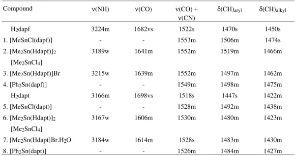
In the crystal packing, pairs of [Me2Sn(Hdapt)] Br.H2O are observed. These pairs are built by H-interac-tions through the proton from the azomethine groups, the water molecules and the bromide. This can be seen in
Fig. 2. The H-bond distances and angles are N1-O3: 2.889(8), HN1-O3: 2.24(7), N1-HN1-O3: 166(9)°, O3-Br: 3.294(7), O3-HO3A: 0.58(10), O3-HO3B: 0.78(9), Br-HO3A: 2.86(12) Å, HO3-O3-HO3B: 109(9)°, O3-HO3A-Br: 134(14)°.
Figure 2. View of one pair of [Me2Sn(Hdapt)]Br.H2O, to show Br- and water molecule roles in the crystal packing.
Table 1. Crystal data and structure refinement parameters of
[Me2Sn(Hdapt)]Br.H2O.
Molecular Formula C21H24N5O3S2SnBr Molecular Weight 657.18
Crystal System monoclinic Space group P21/c
a (Å) 21.920(3)
b (Å) 7.4470(5)
c (Å) 16.805(2)
β (º) 110.178(10)
V (Å)3 2574.8(5)
Z 4
Dcalc (g.cm-3) 1.696
µ (mm-1) 2.738
T (K) 298
Range of h, k, l -25 → 27, -9→0, -20→0 No. measured reflections 5831
No. of observed reflections (F > 4σ (F))
4218
F(000) 1304
No. of refined parameters 289
Robs 0.0472
Rall 0.0659
Rω 0.1351
Infrared spectroscopy
The main vibrational bands of the ligands H2dapf and H2dapt, and their complexes are shown in Table 4. The disappearance of the ν(NH) and ν(CO) bands in complexes
1, 4, 5 and 8, derived from the most acidic organotin
precursors, namely MeSnCl3 and Ph2SnCl2, is a
conse-quence of the double deprotonation of the ligands, indicat-ing that these groups are involved in the formation of deprotonated complexes via enolisation. On the other hand, the IR spectra of complexes 2, 3, 6 and 7, obtained with the
same ligands and the less acidic precursors Me2SnCl2 and Me2SnBr2 in this region are different, showing that the ν(CO) band shifts to lower frequencies, which is indicative of bonding of the carbonyl oxygen to the metal atom.
The position of the ν(NH) absorption shifted to smaller frequencies in complexes 2 and 3, is probably due to the
hydrogen bond in which the N-H group is involved being weaker than in free ligands. The N5-HN5 distance3 of 0.82(6)° found in complex 2 is longer than the N1-HN1
distance of 0.67(7)° found here in complex 7. The IR results
suggest that deprotonation may not occur upon complexa-tion with Me2SnX2 (X = Cl, Br). However, the structure determined by X-ray diffraction of the complexes 2 and 7,
shows that partial deprotonation of the ligands indeed take place.
It is seen from Table 4 that [ν(CO) + ν(CN)], δ(CH)aryl and δ(CH)alkyl absorption bands have shifted to higher frequencies in all the complexes. This is due to a greater rigidity shown by these groups. Similar results can be found in the literature1,3,11.
1H-NMR spectroscopy
Table 5 shows the 2J(117,119Sn, 1H) coupling constants only for the ionic complexes 2, 3, 6 and 7, obtained from
the partial deprotonation of H2dapf and H2dapt. The neutral complexes 1, 4, 5 and 8, resulting from the double depro-Table 2. Fractional atomic coordinates and equivalent isotropic
displace-ment parameters (Å2) for [Me2Sn(Hdapt)]Br.H2O.
Atom x y z U
Sn 0.207648(17) -0.03068(5) 0.19223(2) 0.03021(14) Br 0.43929(4) 0.64998(11) 0.35530(4) 0.0567(2) C1 0.2356(3) -0.2973(9) 0.2287(4) 0.0469(15) C2 0.1867(4) 0.2452(9) 0.1891(4) 0.0490(15) O1 0.24297(19) 0.0069(7) 0.3490(3) 0.0439(11) C3 0.3017(3) 0.0305(8) 0.3888(3) 0.0336(12) C31 0.3296(3) 0.0118(8) 0.4807(4) 0.0335(12) C32 0.3910(3) -0.0279(10) 0.5304(4) 0.0482(15) C33 0.3971(4) -0.0434(12) 0.6162(4) 0.0592(19) C34 0.3413(3) -0.0142(11) 0.6296(4) 0.0541(18) S35 0.27854(7) 0.0287(3) 0.53857(10) 0.0444(4) N1 0.3444(2) 0.0678(8) 0.3490(3) 0.0378(12) HN1 0.374(4) 0.096(10) 0.372(5) 0.045 N2 0.3230(2) 0.0504(7) 0.2628(3) 0.0333(10) C4 0.3616(3) 0.0671(8) 0.2210(4) 0.0354(12) C5 0.4321(3) 0.1109(12) 0.2581(4) 0.0532(18) C6 0.3307(3) 0.0337(8) 0.1286(4) 0.0352(12) C7 0.3645(3) 0.0393(9) 0.0723(4) 0.0453(15) C8 0.3328(4) 0.0041(10) -0.0113(5) 0.0533(18) C9 0.2661(3) -0.0337(10) -0.0397(4) 0.0476(15) C10 0.2351(3) -0.0432(8) 0.0190(4) 0.0371(12) N3 0.2666(2) -0.0094(6) 0.1009(3) 0.0325(10) C11 0.1657(3) -0.0961(9) -0.0038(3) 0.0386(13) C12 0.1256(4) -0.1434(11) -0.0933(4) 0.0559(19) N4 0.1449(2) -0.1017(7) 0.0585(3) 0.0361(10) N5 0.0830(2) -0.1621(8) 0.0459(3) 0.0427(12) C13 0.0705(3) -0.1573(9) 0.1174(4) 0.0411(13) C131 0.0077(3) -0.2312(10) 0.1135(4) 0.0485(15) C132 -0.0455(2) -0.3020(7) 0.0296(3) 0.0230(9) C133 -0.0953(5) -0.3664(14) 0.0663(6) 0.079(3) C134 -0.0814(5) -0.3511(14) 0.1470(6) 0.080(3) S135 -0.01130(15) -0.2609(4) 0.1990(2) 0.0954(8) O2 0.1088(2) -0.0990(7) 0.1893(3) 0.0468(11) O3 0.4614(3) 0.2544(9) 0.4486(4) 0.0652(17) HO3A 0.474(6) 0.306(16) 0.433(7) 0.078 HO3B 0.477(5) 0.258(14) 0.498(6) 0.078
Table 3. Selected bond distances (Å) and angles (°) for [Me2Sn(Hdapt)] Br.H2O.
tonation showed poor solubility in CDCl3 and in other usual solvents. The observed Sn-H coupling constants are in the range characteristic of heptacoordinated organotin(IV) complexes11. 2J values are very close for the heptacoordi-nated complexes 2, 3, 6 and 7, suggesting similar structures
for these species. The same can be concluded for the anionic hexacoordinated counter ions in 2, 6 and 9.
Mössbauer spectroscopy
119Sn Mössbauer spectroscopy was performed on all eight complexes, giving the results in Table 5, which in-cludes parameters from the literature for comparison. Some of the spectra are shown in Fig. 3.
All the complexes present one heptacoordinated Sn(IV) site, except 2 and 6 which show the presence of two
different Sn(IV) sites in a 2:1 ratio. The most abundant site corresponds to a monocationic complex with heptacoordi-nated Sn(IV), in a distorted pentagonal bipyramidal geome-try, and the other site corresponds to the hexacoordinated Sn(IV) which is a dianionic complex acting as counter ion. This conclusion is in accordance with single crystal X-ray analysis3 of the complex 2.
The isomer shift (δ) of the complex 1 (0.68 mm/s) and 5 (0.65 mm/s) are lower than that of the parent acid
MeSnCl314 (1.20 mm/s). The same is true for the heptaco-ordinated site of 2 (1.29 mm/s) and 6 (1.25 mm/s), 3 (1.31
mm/s) and 7 (1.25 mm/s), and 4 (1.01 mm/s) and 8 (1.03
mm/s) compared to their parent acids Me2SnCl214 (1.49 mm/s), Me2SnBr215 (1.59 mm/s) and Ph2SnCl214 (1.32 mm/s), respectively.
Isomer shifts decrease on complexation, as a result of rehybridization to higher coordination for Sn atoms in the complexes, indicating lower s-electron density at the Sn nucleus in the complexes as compared to the parents acids. This can be attributed to a greater involvement of d-orbi-tals, which now take part in the Sn(IV) hybridization scheme, thus reducing the weight of s-orbitals in the overall hybridization of the metal16,17. This conclusion is consis-tent with that observed for the Sn-H coupling constants, i.e., an increase in the coordination number produces an in-crease in 2J values1,11 (Table 5). Similar results have been reported for a great variety of other Sn(IV) com-pounds1,18,19.
The complexes of the same Sn(IV) precursor with dif-ferent ligands, i.e., 1 and 5, 2 and 6, 3 and 7, and 4 and 8,
have very similar isomer shifts. This is in accordance to the fact that the only difference between the ligands is in the 5-member ring, which has oxygen in H2dapf and sulfur in H2dapt. Since this difference is outside of the coordination sphere of the Sn atom, no sensible change in the s-electron density of Sn(IV) nucleus is expected.
From Table 5 it can be seen that, except for the complex
7, the same conclusion is true for the quadrupole splittings.
The difference between complex 3 (∆ = 4.01 mm/s) and 7
(∆ = 3.80 mm/s) may be related to the presence of a H2O molecule in 7.
On the other hand, the complexes of the same ligand with different Sn(IV) precursors show that the inverse dependence of the isomer shift values with the electrone-gativity of the substituents is quite reasonable18-21. This behavior can be seen either for the complexes of H2dapf or H2dapt. Complexes 1, 2 (monocationic), 3 and 4 have the
same ligand H2dapf in the equatorial plane and different axial groups. Going from 1 to 2, chloride was replaced by
a less electronegative Me group, which accounts for higher δ of 2 compared to 1. Complexes 2 and 3 have the same
axial groups, therefore the same δ is observed. Now going from 3 to 4, Me is replaced by a more electronegative group
Ph, leading to a decrease in δ.
The Mössbauer parameters δ and ∆ obtained for the dianionic complexes 2 and 6 are in agreeement with similar
species, complexes 10, 11 and 12 (Table 5).
Quadrupole splitting (∆) values, presented in Table 5, are not sufficient to characterize a Sn(IV) complex as either tetra-, penta-, hexa-, or heptacoordinated1. However, a
successful correlation between quadrupole splitting and the structure of different coordinated Sn(IV) using, either sim-ple point-charge approach10,22-24 or a more elaborate hy-bridization treatment25, has been reported. According to these models, one assumes that L, a parameter [L], is assigned to each ligand, which makes a fixed and inde-pendent contribution to the quadrupole splitting.
For Sn(IV) complexes containing SnR2 moiety, such a quadrupole splitting is dominated by highly covalent Sn-C bonds, and one can show by ignoring the contribution of the other ligands, that ∆ is given by22,23,
∆ = 4 [R] (1 − 34 sin2θ)1⁄2
(1)
where [R] denotes the partial quadrupole splitting of the group R and θ is the R-Sn-R angle. Eq. (1) has been satisfactorily applied to four-, five- and six-coordinated Sn(IV) compounds, using appropriate values of [R] for each coordination number21,22,24. Although little Mössbauer and structural data are available in the literature for heptacoordinated Sn(IV) compounds, the existing data have shown a reasonable and consistent behavior for Eq. (1)1,9,23.
Considering the complexes 2 and 7 whose structure
were determined by single crystal X-ray diffraction, the [Me] can be estimated. Inserting the values ∆ = 3.96 mm/s and θ = 166° 3 for the heptacoordinate site of complex 2 and ∆ = 3.80 mm/s and θ = 164.9° of 7 into Eq. (1) we get
[Me] = -1.01 mm/s and [Me] = -0.98 mm/s, respectively. These values agree within 4% with [alkyl] = -0.97 mm/s reported for heptacoordinate Sn(IV) complexes embodyng pentacoordinate ligands1. Now using the value [Me] = 1.00 mm/s (average of -1.01 and -0.98 mm/s) and ∆ = 4.01 mm/s
of complex 3 and ∆ = 3.94 mm/s for the heptacoordinate
Sn(IV) of complex 6, Eq. (1) allows us to predicte for the Me-Sn-Me angle θ = 180º and θ = 169° for the complexes
3 and 6, respectively. The later value is in excellent
agree-ment with θ = 166° found for complex 2.
The Ph-Sn-Ph angle for the phenyl derivatives, com-plexes 4 and 8, can be also predicted through Eq. (1) and
appropriate values of [Ph].
Combining Mössbauer and structural data for the hep-tacoordinate complex [Ph2Sn(Hdaptsc)]Cl8 [H2dapstc = 2,6-acetylpyridinebis(thiosemicarbazone)], i.e., ∆ = 3.13 mm/s) and θ = 167.9°, we found [Ph] = -0.80 mm/s, which agrees very well with a previous reported1 value [Ph] = -0.78 mm/s. Inserting ∆ = 3.22 mm/s 4 and ∆ = 3.24 mm/s 8 and [Ph] = -0.80 mm/s into Eq. (1) we get θ = 180º for
both complexes.
Finally the predicted Me-Sn-Me angles for the hexaco-ordinate Sn(IV) sites of complexes 2 and 6, namely
[Me2SnCl4]2- can be estimated using [Me] = -1.03 mm/s for hexacoordinate species21,22. Inserting ∆ = 4.11 mm/s 2 and ∆ = 3.99 mm/s 6 together with [Me] = -1.03 mm/s into Eq.
(1) we obtained θ = 175° (X-ray diffraction: 180°) and θ = 163°, respectively for Me-Sn-Me angle of complexes 2 and 6.
The correlation between Mössbauer and X-ray struc-tural data, using a simple point-charge model, gave values for [Me] and [Ph] in excellent agreement with earlier reported values. We think that this correlation is important considering the poor number of published structural data for heptacoordinate complexes as compared to penta- and hexacoordinate species.
Table 4. Main IR bands (cm-1) of the ligands H2dapf and H2dapt, and their complexes.
Compound ν(NH) ν(CO) ν(CO) +
ν(CN)
δ(CH)aryl δ(CH)alkyl
H2dapf 3224m 1682vs 1522s 1470s 1450s
1. [MeSnCl(dapf)] - - 1553m 1506m 1474s
2. [Me2Sn(Hdapf)]2 3189w 1641m 1552m 1519m 1466m
[Me2SnCl4]
3. [Me2Sn(Hdapf)]Br 3215w 1639m 1552m 1497m 1462m
4. [Ph2Sn(dapf)] - - 1549m 1498m 1475m
H2dapt 3166m 1698vs 1518s 1447s 1422m
5. [MeSnCl(dapt)] - - 1528m 1492m 1438m
6. [Me2Sn(Hdapt)]2 3167w 1606m 1530m 1480m 1423m
[Me2SnCl4]
7. [Me2Sn(Hdapt]Br.H2O 3184w 1614m 1528s 1483m 1430m
Concluding, eight new heptacoordinated organotin(IV) complexes of the ligands H2dapf and H2dapt were obtained, which were investigated by means of microanalysis, IR, 1H-NMR, Mössbauer spectroscopy and X-ray diffraction.
The crystallographic data were deposited at the Cam-bridge Crystallographic Data Centre under the number CCDC114597.
Acknowledgments
The authors are greateful for finantial support from CNPq, CAPES, FINEP, FAPEMIG and FAPESP in Brazil, as well as for a CNPq scholarship to M.B.P.M.
References
1. Carini, C.; Pelizzi, G.; Tarasconi, P.; Pelizzi, C.; Mol-loy, K.C.; Waterfield, C. J. Chem. Soc. Dalton Trans.
1989, 289.
2. De Sousa, G.F.; Filgueiras, C.A.L.; Abras, A;. Al-Juaid, S.S.; Hitchcock, P.B.; Nixon, J.F. Inorg. Chim. Acta 1994, 218, 139.
3. Moreno, P.C; Francisco, R.H.P.; Gambardella, M.T. do P.; Mangas, M.B.P.; De Sousa, G.F.; Abras, A. Acta Cryst. 1998, C54, 1444.
4. Sharma, V.K.; Pandey, O.P.; Sengupta, S.K.; Hale-poto, D.M. Trans. Met. Chem. 1989, 14, 263.
5. Lorenzini, C.; Pelizzi, C.; Pelizzi, G.; Pidiere, G. J. Chem. Soc. Dalton Trans 1983, 2155.
6. North, A.C.T.; Phillips, D.C.; Mathews, F.S. Acta Cryst. 1968, A24, 351.
7. Sheldrick, G.M. SHELX97 A Program for the Refine-ment of X-ray Structures, University of Göttingen; Germany,1997.
8. Moreno, P.C.; Francisco, R.H.P.; Gambardella, M.T. do P.; De Sousa, G.F.; Abras, A. Acta Cryst. 1997,
C53, 1411.
9. Sommerer, S.O.; Palenik, G.J. Inorg. Chim. Acta
1991, 183 217.
10. Casas, J.S.; Castiñeiras, A.; Sanchez, A.; Sordo, J.; Vasquez-Lópes, A; Rodrigues-Argüelles, M.C.; Russo, U. Inorg. Chim. Acta 1994, 221, 61.
11. Careri, M.; Mangia, A.; Prediere, G.; Vignali, C. J. Organomet. Chem. 1989, 375, 39.
12. Valle, G.; Gonzáles, A.S.; Etore, R.J. J. Organomet. Chem. 1989, 348, 39.
13. Nasser, F.A.K.; Hossain, M.B.; van der Helm, D.; Zuckerman, J.J. Inorg. Chem. 1984, 23, 606.
14. De Sousa, G.F.; Abras, A.; Filgueiras, C.A.L. Pro-ceeding of the International Conference on the Appli-cations of the Mössbauer Effect, ICAME-95, Ortalli, I., ed.,SIF, Bologne, v. 50, p. 76, 1996.
15. Stöckler, H.A.; Sano, H. Trans. Faraday Soc. 1968,
64, 577.
16. Randall, R.S.; Wedd, R.W.J.; Sams, R.J. J. Or-ganomet. Chem. 1971, 30, C19.
17. Cunningham, D.; Little, M.; Mcloughlin, K. J. Or-ganomet. Chem. 1979, 165, 287.
Table 5.1H-NMR and Mössbauer spectroscopy data for the complexes.
Complex C.N 2J(117Sn,1H)
(Hz)
2J(119Sn,1H)
(Hz) δ (mm/s) ∆ (mm/s)
1. [MeSnCl(dapf)]a 7 - - 0.68 1.89
2. [Me2Sn(Hdapf)]2 7 107 110 1.29 3.96
[Me2SnCl4]a 6 70 81 1.56 4.11
3. [Me2Sn(Hdapf)]Bra 7 99 109 1.31 4.01
4. [Ph2Sn(dapf)]a 7 - - 1.01 3.22
5. [MeSnCl(dapt)]a 7 - - 0.65 1.83
6. [Me2Sn(Hdapt)]2 7 106 113 1.25 3.94
[Me2SnCl4]a 6 78 81 1.49 3.99
7. [Me2Sn(Hdapt]Br.H2Oa 7 106 111 1.25 3.80
8. [Ph2Sn(dapt)]a 7 - - 1.03 3.24
9. [C5H7N2]2[Me2SnCl4]b 6 79 83 -
-10. (Cs)2[Me2SnCl4]c 6 - - 1.63 4.32
11.[2-H3NC6H4C(O)NH2]2 [Me2SnCl4]c 6 92 1.19 4.09
12.(C5H5NH)2[Me2SnCl4]c 6 - - 1.59 4.32
18. Barbieri, R.S.; Beraldo, H.O.; Filgueiras, C.A.L.; Abras, A.; Nixon, J.F.; Hitchcock, P.B. Inorg. Chim. Acta 1993, 206, 169.
19. Telles, W.A.; Allain, L.R.; Filgueiras, C.A.L.; Abras, A. Hyp. Int.1994, 83, 175.
20. Marques, E.V.; Ribeiro, W.F.; Filgueiras, C.A.L.; Abras, A. Hyp, Int. 1995, 96, 259.
21. Moura, E.M.; Rodrigues, C.M.; Santos, D.C.; Siebald, H.G.L.; Abras, A. Hyp. Int. 1997, C2, 116.
22. Sham, T.K.; Bancroft, G.M. Inorg. Chem. 1975, 14,
2281.
23. Parish, R.V. Structure and Bonding in Tin Compounds in “Mössbauer Spectroscopy Applied to Inorganic Chemistry”, Long, G.J., ed.; Plenum Press, New York-London, v. 1, p. 527, 1984.
24. Abras, A.; De Sousa, G.F.; Filgueiras, C.A.L. Hyp. Int.
1994, 90, 459.
25. Clark, M.G.; Maddock, A.G.; Platt, R.H. J. Chem. Soc. Dalton Trans 1972, 281.
![Figure 1. View of the asymmetric unit of [Me 2 Sn(Hdapt)]Br.H 2 O, with labelled atoms and 50% probability ellipsoids.](https://thumb-eu.123doks.com/thumbv2/123dok_br/18988461.459746/3.918.149.745.638.1046/figure-view-asymmetric-hdapt-labelled-atoms-probability-ellipsoids.webp)
![Figure 2. View of one pair of [Me 2 Sn(Hdapt)]Br.H 2 O, to show Br - and water molecule roles in the crystal packing.](https://thumb-eu.123doks.com/thumbv2/123dok_br/18988461.459746/4.918.475.821.456.968/figure-view-hdapt-water-molecule-roles-crystal-packing.webp)