Article
3D-WHIM Pattern Recognition Study for Bisamidines. A Structure-Property Relationship Study.
Fabiano A. S. Menezesa, Carlos A. Montanaria* and Roy E. Brunsb
a
Departamento de Química, Universidade Federal de Minas Gerais, Campus da Pampulha, 31270-901, Belo Horizonte - MG, Brazil
b
Instituto de Química, Universidade Estadual de Campinas, CP 6154, 13083-970, Campinas - SP, Brazil
Um modelo para a interação de bisamidinas aromáticas com o B-DNA foi estabelecido através de estudos de relações estruturas-propriedades derivadas dos cálculos de descritores tridimensionais WHIM-3D. Uma análise de componentes principais, PCA, dos descritores revelou três componentes principais significativas e agrupou as bisamidinas em diferentes conformações: estendida, semi-estendida e semifechada com interações tipo π-π e também por ligações de hidrogênio. O método SIMCA classificou as conformações de acordo com essas características. A interação das 29 bisamidinas estudadas com o B-DNA dá-se através de suas propriedades de forma, distribuição e dimensão.
A proposed model for the interaction of bisamidine analogues with the B-DNA receptor is established by structure-property relationship studies derived from 3D-WHIM descriptor calcula-tions. Three classes, each with relevant information about structural relationships, were determined by PCA and SIMCA analyses for molecular conformations described by 3D-WHIM descriptors for a set of 29 bisamidines with antileishmaniasis and anti-PCP activities. Shape, distribution and dimension properties mostly govern the interaction of bisamidines with B-DNA through the minor groove AT rich regions.
Keywords: 3D-QSAR, principal component analysis, pattern recognition
Printed in Brazil 0103 - 5053 $6.00+0.00
Introduction
AIDS is a fatal disorder for which no successful che-motherapy has yet been developed. Patients who suffer
from this disease are also susceptible to Pneumocystis
carinii pneumonia, PCP, which leads to a 100% death
rate1. Another causa mortis that is seriously widespread
in tropical countries, is visceral leishmaniasis, caused by
the protozoa Leishmania amazonensis2. It has been
esti-mated by WHO that over 17 million deaths were due to
infectious or parasitic diseases3.
Various compounds have been reported to treat both diseases. Pentamidine, a highly flexible bisamidine
ana-logue, has been found to be useful as anti-PCP4 and
antileishmaniasis agents5-7. Although it prolongs the life
of AIDS8-13 and Leishmania patients10,14, it does exhibit
some side effects. For this reason, several other drugs15
have been tried for treating these patients, but their usefulnesses have not been established, yet.
Since most of the drugs tried so far belong to the same structural class, one common mechanism of action has been accepted for these cationic drugs, that is, interaction with
B-DNA through the minor groove AT rich sequences16,17,19a.
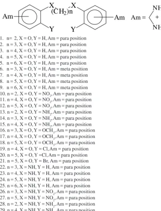
This provides good reason to believe that any drug of this class which encompasses DNA isohelicity could have re-stricted side effects. Figure 1 shows the drug pentamidine: 1A shows the structure obtained from CCDC (Cambridge Crystallographic Data Centre), and 1B represents the pre-ferred isohelical conformation adopted upon binding to B-DNA. Therefore, attempts have been made to find out the pharmacophoric conformation16 within this chemical fam-ily that may be more sequence specific and less toxic. As a result, 29 members of this family, Figure 2, were studied in order to disclose the structural features that would lead to the development of a rationale for synthesising new isohelical drugs to B-DNA. Thus, the present paper discusses a struc-ture-property relationship (SPR) investigation undertaken and aimed at obtaining better understanding of the mode of action with the goal of rationalising substituent selection.
Methods
Initial structures for molecules were built and the con-formational analysis of each pentamidine analogue, showed
in Figure 2, was carried out using HyperChem® program.
mechanics, MM-MD-MM, routine in the AMBER18 force
field was used, according to published procedure19. The
simulation temperature was set to 900 K, but the final tem-perature was 300 K. The simulation temtem-perature was adopted to allow molecules to span a better range of pos-sible conformations. The solvent effect was simulated by the use of a distance-dependent dielectric constant of the
form e = 4rij19. All atomic charges are derived from AM1
calculations20. Atom-centred charges for each molecule
were calculated and fitted to the entire molecules.
Ninety-eight WHIM21 descriptors were calculated and
subjected to PCA analysis using the TSAR22a, SIMCA in the
Sybyl-QSAR22b and ARTHUR22c packages. WHIM23,24
provide 3D molecular descriptors that are invariant to rota-tional and translarota-tional transformations, thus avoiding mol-ecule alignment problems. WHIM descriptors are able to dis-tinguish different conformations of the same molecule, and thus it seems appropriate for carrying out this study.
Priories to PCA analysis, the original 3D WHIM data were subjected to a scaling procedure according to aver-age/standard deviation calculations. The averaged values were used for pre-classification using PCA and also for SIMCA calculations.
Results and Discussion
WHIM (Weighted Holistic Invariant Molecular) descrip-tors are 3D molecular indices that represent different sources of chemical information. They contain information on 3D molecular structure in terms of size, shape, symmetry and atom distribution. The indices are calculated from x, y, z-coordinates of a 3D structure of the molecule, i.e. from a
spatial conformation of minimum energy23,24.
The representative conformations for all studied com-pounds can be found in Figure 3.
A
B
A
B
Figure 1. a: X-Ray structure for pentamidine; b: “Bounded” isohelical conformation to B-DNA.
X (CH2)n X
Y Y
Am
Am Am =
NH2
NH2
+
1. n= 2, X = O, Y = H, Am = para position 2. n = 3, X = O, Y = H, Am = para position 3. n = 4, X = O, Y = H, Am = para position 4. n = 5, X = O, Y = H, Am = para position 5. n = 6, X = O, Y = H, Am = para position 6. n = 3, X = O, Y = H, Am = meta position 7. n = 4, X = O, Y = H, Am = meta position 8. n = 5, X = O, Y = H, Am = meta position 9. n = 6, X = O, Y = H, Am = meta position 10. n = 2, X = O, Y = NO2, Am = para position
11. n = 4, X = O, Y = NO2, Am = para position 12. n = 5, X = O, Y = NO2, Am = para position 13. n = 2, X = O, Y = NH2, Am = para position 14. n = 3, X = O, Y = NH2, Am = para position 15. n = 4, X = O, Y = NH2, Am = para position
16. n = 3, X = O, Y = OCH3, Am = para position 17. n = 4, X = O, Y = OCH3, Am = para position 18. n = 5, X = O, Y = OCH3, Am = para position 19. n = 4, X = O, Y = Cl, Am = para position 20. n = 5, X = O, Y =Cl, Am = para position 21. n = 5, X = O, Y = Br, Am = para position 22. n = 3, X = NH, Y = H, Am = para position 23. n = 4, X = NH, Y = H, Am = para position 24. n = 5, X = NH, Y = H, Am = para position 25. n = 6, X = NH, Y = H, Am = para position 26. n = 3, X = NH, Y = NO2, Am = para position 27. n = 5, X = NH, Y = NO2, Am = para position 28. n = 2, X = NH, Y = NH2, Am = para position 29. n = 4, X = NH, Y = NH2, Am = para position
Figure 2. Structures for compounds investigated in this work.
Figure 3. Conformational families identified by WHIM descriptors through PCA and SIMCA. a: π-stacking family 1: analogues 1, 10-12, 17, 26-28. b: semi-extended family 2: 2, 6, 13-16, 20-23. c: extended family 3: 3-5, 7-9, 18, 19, 24, 25 29.
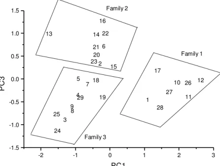
Figures 4 and 5 show the PCA score and loading plots, respectively. Supplementary information on calculated descriptors and their magnitude values can be found ei-ther from the authors or the Journal.
a
b
H2N NH2
N H2 H2N
O O
O H2N
NH2
N H2 NH2
O
H2N
NH
NH2 NH2
H2N
NH
A
B
C
a
b
The first three principal components explain 46.0, 22.2 and 11.2% of the total data variance. However, the first and third components show the larger discriminat-ing powers in defindiscriminat-ing three characteristic groups of com-pounds, which are detailed in Figure 3. The score and loading graphs in Figures 4 and 5, respectively, account for 57.2% of the total variance in the data. The three clusters of molecules in Figure 4 differ owing to the
differing number of carbon atoms in the bridge between the two aromatic amidines as well as to the type of
sub-stituent. Thus, they can adopt π-stacking intramolecular
and also H-bonding (family 1 in Figure 3), semi-extended (family 2) and extended (family 3) conformations. The SIMCA analysis confirmed this clustering pattern, clas-sifying all molecules in the three groups shown in the PCA score graph within 100%.
-2 -1 0 1 2 3
-1.5 -1.0 -0.5 0.0 0.5 1.0 1.5 1 2 3 4 5 6 7 8 9 10 11 12 13 14 15 16 17 18 19 20 21 22 23 24 25 26 27 28 29 Family 3 Family 1 Family 2 PC3 PC1
Figure 4. Score plot of PC1 versus PC3. Compounds are numbered according to Figure 2. The contoured lines were manoeuvred to show the so-called families, which were identified by PC1 and PC3 scores.
-0.15 -0.10 -0.05 0.00 0.05 0.10 0.15 -0.2 -0.1 0.0 0.1 0.2 1 2 3 4 5 6 7 8 9 10 11 12 13 14 15 16 17 18
19 20 21
22 23 24 25 26 27 28 29 30 31 32 33 34 35 36 37 38 39 40 41 42 43 44 45 46 47 48 49 50 51 52 53 54 55 56 57 58 59 60 61 62 63 64 65 66 67 68 6970 71 72 73 74 7576 77 78 79 80 81 82 8384 8586 87 88 89 90 91 92 93 94 959697 98 E G, V L P K, P 15 48 59 81 84 85 3 36 83 47 25 82 86 58 38 95 97 27 96 93 46 35 PC3 PC1
Accordingly, it has to be pointed out that the classifica-tion showed in Figure 4 is only dealt with 3D WHIM descrip-tors discriminating power and is, implicitly, in accordance with each conformation, though there may exist some appar-ent discrepancies in the classification. However, it has no meaning in terms of their conformational energies.
Neverthe-less, the energy ranges (kcal.mol-1) for all three families are as
following: family 1: 28.6-40.8, family 2: 21.4-35.5 and fam-ily 3, 27.6-49.9. Thus, it seems, of course, that these values are not capable of separating the three groups. Moreover, the apparent discrepancy, let us say, between compounds 27 and 28 might be better explained taking into consideration the conformations such compounds could adopt. In this case,
there is a hydrogen bond between moieties (NH2 and NO2) of
1.72 A, which explains their classifications. After all, mol-ecules with different energies may assume different confor-mations. In the process of drug-receptor interactions, the re-ceptor may recognise only one out of many. Hence, it is worth-while mentioning that such conformations may play an im-portant role in the nature of the binding processes. It could also be reasoned that families 1 and 2 are fairly similar. Com-pound 1, for instance, is classified into family 1 instead of
family 2 due to the fact that its conformation is in closer π
-contact (3-7 A), which resembles this family. A comparison to its nearest neighbour, compound 15 classified in family 2,
where the π-contacts are in longer range (6-10 A), sheds some
light into their classifications.
It should be mentioned that although the score graph of the first two principal components explains more vari-ance (68.3%) than the one in Figure 4, it is not capable of discriminating between the three groups of compounds. Only two clusters are evident in this graph (not shown), family 1 and combined families 2 and 3.The separation owes to the discriminating power of the first principal com-ponent, PC1, given by Equation 1.
The terms in Equation 1 are the most important ones for
PC1, having loadings with the larger absolute magnitudes
as can be seen upon inspection of the loading graph in Figure 5. This principal component is better understood in
terms of molecular size and shape. The P1m and K (that can
assume one of the following weights: unit, mass, van der Waals volume, electronegativity, polarizability and electrotopological as holistic measurement) descriptors have the largest absolute magnitude of loadings in Equation 1
and Figure 5. P1m and P2m are related to molecular shape
according to atomic masses22,23. They are directional
de-scriptors that search for the principal axes (spread along orthogonal axes) with respect to the atomic mass properties.
PC1 ~ -0.146(Ks) -0.147(P1v) + 0.145(P2m) -0.147(P1m) (1)
Nevertheless, K represents shape within any axis
direction. Thus, these descriptors comprise the eingevalue proportions for all studied conformations.
Other descriptors showed so far in Figure 5 play a
simi-lar trend. G describes the symmetry of molecules according
to Van der Waals volumes and electronegativity, V accounts
for all dimensions (unit, mass, Van der Waals,
electronega-tivity, electrotopological and polarizability). L stands also
for dimension, but as directional WHIM descriptors, as G
-not V, that are non-directional descriptors. E is distribution
embedded along axes, and it is also directional.
Hence, each “straight molecule” that in this study is repre-sented by isohelicity to B-DNA, as pentamidine itself, Figure 1, should be the choice ones for best binding selectivity.
Equation 2 shows the major descriptors depicted in Figure 5.
PC3 ~ -0.146(E2u) + 0.205(L3u) + 0.189(Vu) (2)
Finally, it has to be pointed out that there is a correlation between variables depicted in Figure 5. For instance, K and P
are correlated in the range of 0.7-0.9 (r2); G’s are in the range
of 0.7. However, this might not be the case for individuals. This means that in the case of selecting variables, those highly correlated may be represented by just one member.
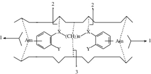
Based upon the above, a simple model can be proposed for the binding of bisamidine derivatives with the receptor, Figure 6. There are three distinct characteristics that appear to be of relevance: (i) the isohelicity to DNA through the “bridge” between the two bisamidine moieties. In this case, compounds that belong to families 2, and certainly 1, would not fit properly into the proposed B-DNA minor groove
mode of action for such compounds17,19a; (ii) hydrogen
bonding via the amidines, due to the size of molecules. If molecules can adopt family 1 conformations, an
intramo-lecular H-bonding and/or π-stacking interaction would
pre-vent the capabilities of H-bonding formation with minor groove base pairs of B-DNA; and (iii) the alkyl linker lipophilicity. This might be due to the fact that linker’s size would suffice for different shape molecules can adopt, and perhaps to any lipophilic interaction into the DNA wells.
X
Y Y
(CH2)n
X
Am Am
2 2
3
1 1
Conclusions
It seems quite reasonable, therefore, that the above char-acteristics could induce selective binding by promoting rigidity of analogues. This rationale could lead to less side effects if selectivity is achieved. Nonetheless, the cal-culation of holistic 3D WHIM descriptors is capable of dealing with conformations that can be envisaged through pattern recognition using PCA and SIMCA. It is, never-theless, noteworthy that the most prominent descriptors that account for classifying all conformations come from shape, distribution and dimension.
Overall, conformations were explored and classified based on physicochemical descriptors that encompass such 3D information content. This seems to be a very powerful way of dealing with molecules where the search for pharmacophoric conformations may play an important role in the drug design field.
Acknowledgements
We wish to thank CNPq for the grants and Oxford Molecular for providing us with TSAR. Thanks are also due to Prof. Roberto Todeschini, for 3D WHIM.
Electronic supplementary information
Includes definitions and values for the calculated descriptors. Available at: http://www.sbq.org.br/jbcs/2000/ vol11_n4/indice.
References
1. Gazzard, B. G. J. Antimicrob. Chemother. 1989,23, 67.
2. (a) Bell, C. A.; Hall, J. E.; Kyle, D. E.; Grogl, M.;
Ohemeng, K. A. E; Tidwell, R. R. Antimicrob. Agents
Chemother. 1990,34, 1381. (b) Jeronimo, S. M. B.; Teixeira, M. J.; Sousa, A. D.; Thielking, P.; Pearson,
R. D.; Evans, T. G. Clin. Infect. Dis. 2000, 30, 608.
3. Word Health Report 1997: http://www.who.ch/whr/
1997/factse.htm.
4. Graves, D. C.; CharyReddy, S.; BeckerHapak, M. Mol.
Cell. Probes1997, 11, 1.
5. Basselin, M.; BadetDenisot, M.A.; Lawrence, F.;
RobertGero, M. Exp. Parasitol. 1997, 85, 274.
6. Basselin, M.; Lawrence, F.; RobertGero, M. Biochem.
J. 1996, 315, 631.
7. Kandpal, M.; Tekwani, B. L.; Chauhan, P. M. S.;
Bhaduri, A. P. Life Sci.1996, 59, 75.
8. Montgomery, A. B.; Luce, J. M.; Turner, J.; Lin, E. T.; Debs, R. J.; Corkery, K. J.; Brunette, E. N.; Hopewell,
P. C. Lancet, 1987, 2, 480.
9. Bielawski, K.; Galicka, A.; Bielawska, A.; Sredzinska, K. Acta Biochim. Pol.2000, 47, 113.
10. Wispelwey, B.; Pearson, R. D. Infect. Control. Hosp.
Epidemiol.1991, 12, 375.
11. Donkor, I. O.; Queener, S. F.; Dalton, J. T. Bioorg.
Med. Chem. Lett. 1996, 6, 1967.
12. Santamauro, J. T.; Storer, D. E. Med. Clin. North Am.
1997, 81, 299.
13. Miller, R. F.; Lenoury, J.; Corbett, E. L.; Felton, J.
M.; Decock, K. M. J. Antimicrob. Chemother.1996,
37, 33.
14. Sands, M.; Kron, M. A.; Brown, R. B. Rev. Infect. Dis.
1985, 7, 625.
15. Lee, M. B.; Gilbert, H. M. Infect. Med.1999, 16, 37.
16. Greenidge, P. A.; Jenkins, T. C.; Neidle, S. Mol.
Pharmacol.1993, 43, 982.
17. Montanari, C. A.; Tute, M. S.; Beezer, A. E.; Mitchell,
J. C. J. Comput.-Aided Mol. Des. 1996, 10, 67.
18. Weiner, S. J.; Kollman, P. A.; Case, D. A.; Singh, U. C.;
Ghio, C.; Alagano, G.; Profeta, S.; Weiner, P. J. Am.
Chem. Soc.1984, 106, 765.
19. (a) Montanari, C. A.; Trent, J. O. and Jenkins, T. C. J. Braz. Chem. Soc. 1998, 9, 175. (b) Orozco, M.;
Laughton, C. A.; Herzyk, P.; Neidle, S. J. Biomol.
Struct. Dyn.1990, 8, 359.
20. Guadagnini, P. H. E.; Bruns, R. E. Quim. Nova1996,
19, 148.
21. WHIM-3D/QSAR version 3.2, Talete srl, Milano (Italy), 1996.
22. (a) TSAR, Version 3.21, Oxford Molecular, Ltd.. (b) Sybyl/QSAR, Version 6.3, Tripos, Inc... (c)
Scarminio, I. S.; Bruns, R. E. Trends Anal. Chem.
1989, 8, 326.
23. Todeschini, R.; Gramatica, P. Quant. Struct.-Act.
Relat.1997,16, 113.
24. Todeschini, R.; Gramatica, P. Quant. Struct.-Act.
Relat.1997,16, 120.
25. Bostock-Smith, C. E.; Searle, M. S. Nucleic Acids
Res.1999, 27, 1619.
26. Clark, G. R.; Boykin, D. W.; Czarny, A.; Neidle, S. Nucleic Acids Res.1997, 25, 1510.
27. Neidle, S.; Nunn, C. M. Nat. Prod. Rep.1998, 15, 1.
Received: May 12, 1999