Marginal microleakage of class II composite resin
restorations due to restorative techniques
Microiniltração marginal de restaurações classe II de resina
composta devido às técnicas restauradoras
Andreia A. Carvalho a Francine C. L. Moreira b Larissa M. Cunha c Samara M. de Moura c João Batista de Souza d Carlos Estrela e
Lawrence G. Lopes e
a Post-graduate Program, School of Dentistry,
Federal University of Goiás, Goiânia, GO, Brazil
b Department of Prevention and Oral Rehabilitation,
School of Dentistry, Federal University of Goiás, Goiânia, GO, Brazil
c School of Dentistry, Federal University of Goiás,
Goiânia, GO, Brazil
d Department of Prevention and Oral Rehabilitation,
School of Dentistry, Federal University of Goiás, Goiânia, GO, Brazil
e Department of Prevention and Oral Rehabilitation,
School of Dentistry, Federal University of Goiás, Goiânia, GO, Brazil
Correspondence:
Lawrence Gonzaga Lopes
Department of Prevention and Oral Rehabilitation School of Dentistry – Federal University of Goias Praça Universitária esq. c/ 1ª Avenida s/n – Setor Universitário
Goiânia, GO – Brazil 74605-220
E-mail: [email protected]
Received: December 30, 2009 Accepted: March 31, 2010
Conflict of Interest Statement: The authors state that there are no financial and personal conflicts of interest that could have inappropriately influenced their work. The materials were donated by Dentsply.
Copyright: © 2010 Carvalho et al.; licensee EDIPUCRS. This is an Open Access article distributed under the terms of the Creative Commons Attribution-Noncommercial-No Derivative Works 3.0 Unported License.
Abstract
Purpose: To evaluate the marginal microleakage of class II composite resin (CR) restorations due to restorative techniques.
Methods: Forty human extracted premolars were assigned to 4 groups (n=10). Class II cavities were prepared (4-mm wide, 2-mm axially, with the gingival margin located 1 mm beyond the cementum-enamel-junction), and the restorative adhesive system Prime & Bond 2.1/TPH3
(Dentsply) was used. CR was inserted by the oblique incremental technique (OIT) and cured in continuous exposure. The restoratives techniques were: group 1 (control): OIT; group 2: flowable resin (1 mm) applied in the gingival wall + OIT; group 3: OIT + three pre-cured spheres inserted in the first increment of CR; and, group 4: OIT + strip of fiberglass inserted in the first increment of CR. The specimens were subjected to a thermocycling regimen of 500 cycles (1 min at 5º-37º-55ºC), coated with two layers of nail varnish up to 1 mm from the restoration margins, and immersed in 0.5% basic fuchsine solution for 24 h. The extension of dye penetration at the cervical wall (µm) was evaluated using an optic microscope at x40. Data were analyzed using analysis of variance (ANOVA) (α=0.05).
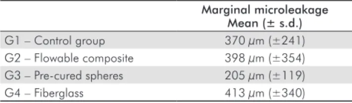
Results: The microleakage values were: G1: 370 µm ± 241; G2: 398 µm ± 354; G3: 205 µm ± 119; and G4: 413 µm ± 340. No statistically significant differences were found among the restorative techniques (P=0.081).
Conclusion: Marginal microleakage values were not influenced by the different restorative techniques tested.
Key words: Composite resin; marginal microleakage; restorative technique
Resumo
Objetivo: Avaliar a microinfiltração marginal de restaurações classe II de resina composta (RC) em função de técnicas restauradoras.
Metodologia: Quarenta pré-molares humanos extraídos foram divididos em 4 grupos (n=10). Cavidades classe II foram preparadas (4 mm de largura, 2 mm de altura e margem gengival localizada a 1 mm além da junção amelo-cementária, e foi usado o sistema adesivo Prime & Bond 2.1/TPH3 (Dentsply). A RC foi inserida pela técnica incremental
oblíqua (OIT) e polimerizada em exposição contínua. As técnicas restauradoras foram: grupo 1 (controle): OIT; grupo 2: resina fluida (1 mm) aplicada na parede gengival + OIT; grupo 3: OIT + três esferas pré-polimerizadas no primeiro incremento de RC; e grupo 4: OIT + tira de fibra de vidro inserida no primeiro incremento de RC. Os espécimes foram submetidos à termociclagem por 500 ciclos (1 min a 5º-37º-55ºC), cobertos com duas camadas de esmalte até 1 mm das margens da restauração e imersos em solução de fucsina básica a 0,5% por 24 h. A extensão da penetração do corante na parede cervical (µm) foi avaliada com microscópio ótico (x40). Os dados foram analisados por ANOVA (α=0,05).
Resultados: Os valores de microinfiltração foram: G1: 370 µm ± 241; G2: 398 µm ± 354; G3: 205 µm ± 119 e G4: 413 µm ± 340. Não houve diferença estatisticamente significativa entre as técnicas restauradoras (P=0,081).
Conclusão: Os valores de microinfiltração marginal não foram influenciados pelas diferentes técnicas restauradoras neste estudo.
Introduction
The demand for esthetic restorative materials has greatly increased in recent years. However, for class II cavities, the factors primarily responsible for microleakage problems are related to the initial shrinkage stress of the composite
resin (CR), the difference between the coeficient of
thermal expansion of materials with hard dental tissue, the inaccessibility of the cervical area and, in particular, problems of bonding to the cervical substrate (1).
Microleakage may be deined as the passage of bacteria, luids, molecules or ions between the cavity wall and the
restorative material applied to it (2). These events may affect the property of the materials, resulting in recurrent caries, or causing post operative sensibility (3). Obviously, the longevity of the restoration may also be compromised. Polymerization shrinkage is a complex process depending on several factors. The volumetric contraction causes debonding forces at the material/tooth interface (3). The stress resulting
from this shrinkage has been associated with open margins between the restoration and the tooth, overt tooth straining or fracture, and post operative sensitivity (3,4). Adhesive failure can also occur at the internal interface, leading to the formation of an internal gap between the material and the dentin surface (2). Such openings or gaps are considered
deleterious since they allow the transit of luid between
the dentin-pulp complex and the oral environment (3,5,6). Thus, different restorative techniques have been proposed to improve the adaptation marginal, such as pre-cured CR spheres inserted with the composite increments (7), a thin
layer of lowable resin applied at the gingival wall and a strip
of iberglass inserted with the increments of CR (8,9).
The aim of this study was to evaluate the marginal micro- leakage of CR restorations as a function of different restorative techniques. The null hypothesis was that there are no differences in marginal microleakage of CR with respect to restorative techniques.
Methodology
The present study was approved by the Research Ethics Committee of the Federal University of Goias – COEP/UFG (Protocol number 076/06).
Forty sound, non-carious human premolar teeth extracted due to orthodontic reasons, were used in this study. The teeth were immersed in a 0.05% thymol solution for no longer than 6 months. Class II cavities were done using a drill #245 (KG Sorensen Ind. Com. Ltda, Barueri, SP, Brazil), in high-speed turbine under water coolant (Kavo do Brasil Ind. Com. Ltda, Joinville, SC, Brazil). The mesial surface of each tooth was prepared, and the cavities (vertical slot) were standardized in the following dimensions: 4-mm wide, 2-mm axially, with the gingival margin located 1 mm beyond the cementum-enamel-junction (CEJ). Prior to the restorative procedures, prophylaxis with pumice stone and water was performed. The 40 premolars were randomly assigned to 4 groups of ten teeth each according to the different experimental conditions.
Dentin and enamel were etched with 35% phosphoric acid gel (Denstply Ind. Com. Ltda, Petrópolis, RJ, Brazil) for 30 s in enamel and 15 s in cementum and dentin, followed by water spray for 30 s and drying with absorbent paper, leaving the surface visibly moist. The Prime & Bond 2.1 (Denstply Ind. Com. Ltda) adhesive system was applied with a microbrush for 20 s and gently air dried. A slight drying using a triple air syringe was performed at 10-cm distance for 5 s before the application of a new adhesive layer. A visible light-curing quartz-tungsten-halogen unit (Ultralux; Dabi Atlante, Ribeirão Preto, SP, Brazil) was used at continuous intensity of 500 mW/cm2. The adhesive system
was cured for 40 s.
The polyester matrix was adapted and ixed with adhesive tape in the cervical region. All cavities were illed with a
microhybrid composite resin (TPH3; Denstply Ind. Com.
Ltda). The 40 teeth had approximately the same size. In order to standardize the volume of each increment to be
used (5 increments per tooth), the cavity was illed entirely
with CR. Then, the resin was removed from the preparation
and divided into ive equal parts. A matrix of acrylic resin
was made (1/5 of total volume of the prepared cavity) to standardize the CR increment before its application into the cavity.
Group 1 – Control group
The teeth were restored with the oblique incremental
technique (OIT), where the irst increment was placed
horizontally, i.e., the resin composite was applied directly on the gingival wall of the cavity without any combination of materials. The other increments were inserted so that the wall did not join the vestibular and palatal/lingual surfaces. Each increment was cured for 20 s followed by 40 s on the occlusal, buccal and lingual sides.
Group 2 – Flowable composite
The irst increment was a layer of 1 mm resin low (Fill Magic
Flow; Vigodent SA Ind. Com., Rio de Janeiro, RJ, Brazil) applied directly on the gingival wall of the cavity using the
ine tip of a syringe that accompanies the material. Then,
curing was performed for 20 s, and four more increments of CR were inserted into the cavity by OIT. Each increment
was cured for 20 s, and the inal restoration was cured for
40 s on each side similarly to the control group.
Group 3 – Pre-cured spheres
The pre-cured spheres with diameter of 1 mm was confectioned to be used in restorative technique. The teeth were restored with OIT, with three pre-cured spheres in the
irst increment of CR that were applied on the gingival wall.
The increment was cured for 20 s. The other increments were done without pre-cured spheres, and were cured as in the control group.
Group 4 – Fiberglass
The teeth were restored with a 3x2 mm strip of glass iber
was positioned in the irst increment of CR cured for 20 s.
The remaining four increments of CR were inserted by using OIT and cured as in the other groups.
After the restorations were inished, all groups were stored in distilled water at 37ºC for 48 h. The polishing/inishing
procedures were performed using a sequential series of aluminum oxide disks (Sof-Lex; 3M/ESPE, Sumaré, SP, Brazil). Afterwards each tooth was dried with absorbent paper, and two layers of cyanocrylate resin Super Bonder (Henkel Ltda, Itapevi, SP, Brazil) were placed in the root apex to seal it. The specimens were immersed in physiological solution at 37ºC for 24 h.
The four groups were subjected to a thermocycling regimen of 500 cycles with 1-min immersions in distilled water at 37ºC, 5ºC, 37ºC and 55ºC. The teeth were coated with two layers of nail polish to leaving 1 mm from the resin/ tooth interface margins and were immersed in a 0.5% basic fuchsine solution for 24 h. All teeth were included in
orthoftalic transparent resin (T 208; Redeibra, São Paulo,
SP, Brazil) to be sectioned longitudinally to a mesiodistal direction. In order to analyze the extent of leakage in the cervical wall (µm), the slice that showed the highest degree of dye penetration per group was selected for observation
under alternate microscopic high-power ields (x40) using an integration graticule (4740680000000-Netzmik-rometer
12.5, Carl Zeiss, 131 Göttingen, Germany). The extension of dye penetration were measured using this standard scale (µm) located in the microscopy lenses by two calibrated examiners. The microleakage data were analyzed by analysis
of variance (ANOVA) at the signiicance level of 0.05.
Results
Table 1 displays the values of marginal microleakage according to the restorative technique. The analysis of variance showed that the restoration techniques did not
inluence the values of microleakage (P=0.081). Although
not statistically different, the group 3 (pre-cured spheres) had the lowest extent of penetration of the dye when compared with the other groups.
The CR is widely used in clinical practice, because of the
aesthetic beneits, ease of use and bonding with the tooth
structure (6). When applied in a cavity with the adhesive system, it establishes a proper interaction with the enamel and/or dentin, and the longitudinal stability is directly related to the steps resulting from cavity preparation, intrinsic characteristics of the adhesive and restorative materials, operational procedures and manipulative weather of the oral environment (5,10).
Another important factor for CR refers to its mechanism of polymerization shrinkage (11). The marginal gap caused by the polymerization shrinkage may lead to pain on biting and failure of adhesion by repeated occlusal loading (6). In addition, cavity-wall gap formation may cause marginal staining, postoperative sensitivity and secondary caries (4,6), although for Lima et al. (12) microleakage
and surface roughness did not inluenced caries lesion
formation.
The adaptation of the material to the cavity depends essentially on their thermal expansion and dimensional changes during the process of polymerization (2,13,14).
Differences in the coeficient of linear thermal expansion
between the dental restorative materials and structures are
largely responsible for the leakage (2), which justiies the
use of thermal cycling in this work (11,15). Theoretically,
the greater the difference between the coeficient of thermal
expansion of the restorative material and the tooth, the greater the marginal leakage that occurs during temperature changes (15). On the other hand, Cenci et al. (16) evaluated
the inluence of thermal stress on the marginal integrity
of restorative material with different adhesive and thermal properties. They concluded that CR was only affected in extreme situations, which are not present in normal oral conditions. In present study the specimens were leaved for 1 min in each temperature.
The class II cavities were confectioned only in one part of the teeth, since the photo-curing of the resin restoration
could be to inluence the shrinkage stress generation
on the restoration of the other side. In class II cavities,
another factor that signiicantly inluences the marginal it
is the location of cervical margin (5,17). If the margins are localized around 1.0 mm coronal to the CEJ a good marginal integrity may result following a conventional enamel acid
etching (17). Soares et al. (18) evaluated the marginal
integrity and microleakage of direct and indirect composite
inlays in cavities with cervical inishing line prepared in dentine or enamel. There were no signiicant differences
between the direct and indirect techniques for the cervical
inishing line in enamel, but for the inishing line in dentin,
the indirect technique allowed less microleakage than the direct technique. Thus, the authors decided to assemble the end cervical cavities with 1 mm below the CEJ, as are those most likely to microleakage and therefore there is greater
need for studies to obtain satisfactory restorations (18). The
dimensions of the cavity used in the present study were chosen because they represent the modern cavities that are indicated in the clinical practice.
Table 1. Means (± standard deviation – s.d.) of marginal microleakage of CR according to the restorative technique.
Marginal microleakage Mean (± s.d.)
G1 – Control group 370 µm (±241)
G2 – Flowable composite 398 µm (±354)
G3 – Pre-cured spheres 205 µm (±119)
G4 – Fiberglass 413 µm (±340)
Discussion
Dye penetration test are known to be valid tools for the determination of marginal gaps in vitro studies (19). It is an established method for the determination of marginal leakage in vitro, mostly performed after cutting the teeth
in a longitudinal direction (19). However, the longer the
penetration time is, the higher might be a risk of dye diffusion into the adhesive resulting in stained adhesive
layers might be interpretated as gaps (19). The immersion in basic fuchsine solution for 24 h appears to be suficient time
for assessment of leakage with no impairment of adhesive interface, which is in agreement with the work of Cenci et al. (16) and Pazinatto et al. (20).
Knowing that the CR suffers shrinkage, it is important the clinical use of technical resources to try to minimize this problem, with the aim of obtaining a proper marginal sealing (3). For this, some factors involving the material and operative technique should be considered, such as the volume of CR inserted in the cavity and association of CR to other materials, whose properties are more suitable for application in the cervical area (10).
Li et al. (21), on work done in class V cavities with gingival margins in dentin/cementum, concluded that low viscosity CR can improve the marginal adaptation in dentine restorations in this type of cavity. Chuang et al. (22) investigated the
inluence of lowable composite in different thicknesses as
marginal and internal porosity of class II restorations with CR. They concluded that the group with a thin layer of
lowable composite (0.5 a 1mm) exhibited superior marginal
quality in interface microleakage evaluation compared to the other groups (22). Alonso et al. (23) evaluated the effect of an adhesive applied in layers of different thickness or in
association with a illed adhesive or with a low viscosity
composite on the microleakage of composite restorations. The group in which was used application of one layer of adhesive
followed by application of lowable composite showed less
leakage than group in which there was only the application of one layer of adhesive (23). The authors concluded that
the use of resin liners with lowable composites can reduce
the microleakage of composite restorations (23).
These results differ from those obtained in the present study, in which the values of the group with the low viscosity resin were higher than the control group, although there was no
statistically signiicant difference. This can be explained
by the high percentage of organic matrix of lowable
composites that increase the shrinkage stress (3,10). So this could encourage the creation of an elastic layer in the cervical margins, just having high polymerization shrinkage, generating high stress at the interface of bonding (10). This is in accordance to Castañeda-Espinosa et al. (24),
who described that the use of a lowable composite as an
intermediate layer (0.5 or 1mm) promoted an increase in shrinkage stress force values.
The G3 (pre-cured spheres) had the lowest extent of penetration of the dye when compared with other groups,
although this was not statistically signiicant. These data are
in agreement with those obtained by Conceição et al. (7), in an in vitro study, which examined four techniques restored with CR in Class II cavities. In the group that was used in pre-polymerized portions of CR, there was increased ability to limit leakage of dye (7). The CR spheres probably decreased the shrinkage of volume and linear polymerization of CR and, if allowed to exert pressure against the walls of the material of the cavity, facilitating its adaptation as well as obtaining a point of contact (7). In this context, this technique can be an alternative to be used in complex clinical cases, especially when gingival wall is located in dentin or cement substrate.
The null hypothesis was not rejected because there were not differences among marginal microleakage of CR with respect to restorative techniques. Although some studies
used a smaller number of specimens per group (9,21) or the same amount (17,19) than the present study, our data showed
high standard deviation values. It is possible that a larger
sample size per group could lead to signiicant differences,
especially in the group 3.
Conclusions
Under the conditions of this in vitro study, it can be concluded
that the marginal microleakage values were not inluenced
by the different restorative techniques tested.
Acknowledgments
The authors would like to acknowledge Núbia Miranda
Ferreira (Dentsply) for supplying the material.
References
1. Dietrich TH, Lösche AC, Lösche GM, Roulet JF. Marginal adaptation of direct composite and sandwich restorations in class II cavities with cervical margins in dentine. J Dent 1999;27:119-28.
2. Kidd EA. Microleakage: a review. J Dent 1976;4:199-206. 3. Lopes LG, Franco EB, Mondelli RF, Souza Jr MH, Lauris JR. Evaluation
of the effect of the cavity configuration factor on the marginal microleakage of the aesthetic restorative materials. Am J Dent 2003;6:211-4.
4. Santos GO, Silva AH, Guimarães JGA, Barcellos AAL, Sampaio EM, Silva EM. Analysis of gap formation at tooth-composite resin interface: effect of C-factor and light-curing protocol. J Appl Oral Sci 2007;15:270-4.
6. Yoshikawa T, Burrow MF, Tagami J. A light curing method for improving marginal sealing and cavity wall adaptation of resin composite restorations. Dent Mater 2001;17:359-66.
7. Conceição EM, Vidor MM, Pacheco JFM, Manfredi DAB. Capacidade de selamento marginal de diferentes técnicas restauradoras com resina composta em dentes posteriores. Rev Fac Odontol Porto Alegre 1997;38:20-2.
8. Portero OO, Grullón PG, Ditterich RG, Gomes OMM, Gomes JC. A utilização das fibras de reforço na Odontologia. Publ UEPG Ci Biol Saúde 2005;11:47-52.
9. Xu HHK, Schumacher GE, Eichmiller FC, Peterson RC, Antonucci JM, Mueller HJ. Continuous-fiber perform reinforcement of dental resin composite restorations. Dent Mater 2003;19:523-30.
10. Franco EB, Lopes LG. Conceitos atuais na polimerização de sistemas restauradores resinosos. Biodonto 2003;1:1-59.
11. Kenshima S, Grande RHM, Singer JM, Ballester RY. Effect of thermal cycling and filling technique on leakage of composite resin restoration. J Appl Oral Sci 2004;12:307-11.
12. Lima FG, Romano AR, Correa MB, Demarco FF. Influence of microleakage, surface roughness and biofilm control on secondary caries formation around composite resin restorations: an in situ evaluation. J Appl Oral Sci 2009;17:61-5.
13. Cunha LG, Alonso RC, Souza-Júnior EJC, Neves AC, Correr-Sobrinho L, Sinhoreti MA. Influence of the curing method on the post-polymerization shrinkage stress of a composite resin. J Appl Oral Sci 2008;16:266-70.
14. Braga RR, Ballester RY, Ferracane JL. Factors involved in the development of polymerization shrinkage stress in resin-composites: a systematic review. Dent Mater 2005;21:962-70.
15. Going RE. Microleakage around dental restorations: a summarizing review. J Am Dent Assoc 1972;84:1349-57.
16. Cenci MS, Pereira-Cenci T, Donassollo TA, Sommer L, Strapassom A, Demarco FF. Influence of thermal stress on marginal integrity of restorative materials. J Appl Oral Sci 2008;16:106-10.
17. Schuckar M, Geurtsen W. Proximo-cervical adaptation of class II – composite restorations after termocycling: a quantitative and qualitative study. J Oral Rehab 1997;24:766-75.
18. Soares CJ, Celiberto L, Dechichi P, Fonseca RB, Martins LR. Marginal integrity and microleakage of direct and indirect composite inlays – SEM and stereomicroscopic evaluation. Braz Oral Res 2005;19: 295-301.
19. Ernst CP, Galler P, Willershausen B, Haller B. Marginal integrity of class V restorations: SEM versus dye penetration. Dent Mater 2008;24:319-27.
20. Pazinatto FB, Campos BB, Costa LC, Atta MT. Effect of the number of thermocycles on microleakage of resin composite restoration. Pesqui Odontol Bras 2003;17:337-41.
21. Li Q, Jepsen S, Albers H-K, Eberhard J. Flowable materials as an intermediate layer could improve the marginal and internal adaptation of composite restorations in class-V-cavities. Dent Mater 2006;22:250-7.
22. Chuang SF, Jin Y-T, Liu J-K, Chang C-H, Shieh D-B. Influence of flowable composite lining thickness on class II composite restorations. Oper Dent 2004;29:301-8.
23. Alonso RCB, Sinhoreti MAC, Sobrinho LC, Consani S, Goes MF. Effect of resin liners on the microleakage of class V dental composite restorations. J Appl Oral Sci 2004;12:56-61.