Article
J. Braz. Chem. Soc., Vol. 27, No. 1, 10-14, 2016. Printed in Brazil - ©2016 Sociedade Brasileira de Química 0103 - 5053 $6.00+0.00
A
*e-mail: [email protected]
New Biphenyls from
Garcinia multiflora
Xue-Mei Gao,a,b Bing-Kun Ji,a,b Yin-Ke Li,a,c Yan-Qing Ye,a Zhi-Yong Jiang,a Hai-Ying
Yang, a Gang Du,a Min Zhou,a Xiao-Xia Pan,a Wen-Xing Liua and Qiu-Fen Hu*,a
aKey Laboratory of Chemistry in Ethnic Medicinal Resources, State Ethnic Affairs
Commission & Ministry of Education and bJoint Research Centre for International
Cross-Border Ethnic Regions Biomass Clean Utilization in Yunnan, Yunnan Minzu University, 650031 Kunming, P. R. China
cCollege of Resource and Environment, Yuxi Normal University,
653100 Yuxi, P. R. China
Three new biphenyls were isolated from Garcinia multiflora. The structures of these biphenyls were elucidated by spectroscopic methods, and their rotavirus activity was evaluated.
Keywords:Garcinia multiflora, biphenyls, anti-rotavirus activity
Introduction
Garcinia multiflora (clusiaceae) is a dioecious evergreen tree that grows to a height between 3-10 m in Southern China. This is a widely adaptable species growing in various habitats and at various elevations. The seeds yield up to 50% oil (seed pulp up to 55% oil), which is used for manufacturing soap or as a lubricant for machines.1 The bark is used as an external
medicine to reduce inflammation, whereas the timber is used for making furniture, boats, and woodcuts.1
Previous phytochemical studies of Garcinia multiflora
revealed the presence of xanthones,2,3 benzophenone
derivatives,4-6 and biflavonoids,2,7-9 and a wide range of
biological activities such as anti-hepatocellular carcinoma activity,4 anti-inflammatory,5 anti-HIV,9 antioxidant,3,10
and anti-tuberculosis activities have been reported for this species.8 Motivated by the search for new bioactive
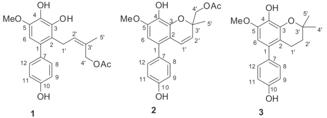
metabolites from local plants, we investigated the chemical constituents of the stems of Garcinia multiflora growing in Xishuangbanna Prefecture, leading to the characterization of three new biphenyls (1-3) (Figure 1), along with maclurin(4),3 2,4,6,3’-tetrahydroxybenzophenone(5),3
4-methoxybenzoic acid(6),10 luteolin(7),10 and apigenin
(8).10 The anti-rotavirus activity of 1-3 has also been
evaluated.
Experimental
General experimental procedures
UV (ultraviolet) spectra were obtained using a Shimadzu UV-2401A spectrophotometer. A Tenor 27 spectrophotometer was used for scanning infrared (IR) with KBr pellets. 1D and 2D nuclear magnetic resonance (NMR) spectra were recorded on a DRX-500 NMR spectrometer with tetramethylsilane (TMS) as internal standard (IS). Chemical shifts (d) are expressed in ppm with reference to the solvent signals. Electrospray ionization mass spectra (ESIMS), electron ionization mass spectra (EIMS) and high resolution electron ionization mass spectra (HREIMS) were performed on a VG Autospec-3000 spectrometer. Semi-preparative high performance liquid chromatography (HPLC) was performed on a Shimadzu LC-8A preparative liquid chromatograph with Zorbax PrepHT GF (21.2 mm × 25 cm) or Venusil MP C18
(20 mm × 25 cm) columns. Column chromatography (CC) was performed using silica gel (200-300 mesh, Qing-dao Marine Chemical, Inc.), Lichroprep RP-18 gel (40-63 µm, Merck), 3-[4,5-dimethylthiozol-2-yl]-2,5-diphenyltetrazolium bromide (MTT, Sigma) and middle chromatogram isolated (MCI) gel (75-150 µm, Mitsubishi Chemical Corporation). Fractions were monitored by thin layer chromatography (TLC), and spots were visualized by heating silica gel plates sprayed with 5% H2SO4 in
Plant material
Garcinia multiflora was collected in Xishuangbanna Prefecture, Yunnan Province, People’s Republic of China, in September 2012. The identification of the plant material was verified by PhD Huang Jian-Ping. A voucher specimen (YNNU 2012-09-16) has been deposited in our laboratory.
Extraction and isolation
The air-dried and powdered stems and leaves of
Garcinia multiflora (5.5 kg) were extracted four times with 80% aqueous ethanol (4 × 50 L) at room temperature and filtered. The filtrate was evaporated under reduced pressure, and the crude extract (260 g) was extracted by ethyl acetate and decolorized by MCI gel. The portion of the extract soluble in 90% methanol (85 g) was chromatographed on a silica gel column eluting with a CHCl3-acetone gradient
system (20:1, 9:1, 8:2, 7:3, 6:4, 5:5), to give six fractions (A-F). Fractionation of fraction B (9:1, 4.6 g) by silica gel CC, eluted with petroleum ether-acetone (9:1-1:2), yielded fractions B1-B7. Fraction B2 (8:2, 0.86 g) was subjected to silica gel CC using petroleum ether-acetone and semi-preparative HPLC (70% MeOH-H2O) to give 4 (4.8 mg),
5 (6.2 mg), and 6 (3.9 mg). Fraction C (8:2, 1.23 g) was subjected to silica gel CC using petroleum ether-acetone and semi-preparative HPLC (50% MeOH-H2O) to give
1 (5.7 mg), 2 (5.8 mg), and 3 (4.9 mg). Fractionation of fraction D (7:3, 2.5 g) by silica gel CC, eluted with petroleum ether-acetone (9:1-1:2), yielded fractions D1-D7. Fraction D3 (7:3, 0.85 g) was subjected to silica gel CC using petroleum ether-acetone and semi-preparative HPLC (45% MeOH-H2O) to give 7 (4.2 mg), 8 (4.5 mg).
Multibiphenyl A (1)
Pale yellow gum; [α]D23.1 –11.0 (c 0.07, MeOH); UV
(MeOH) λ
max / nm (log ε) 570 (2.16), 205 (4.71); IR (KBr)
ν / cm-1 3422, 2939, 1721, 1611, 1589, 1498, 1443, 1357,
1266, 1172, 1102, 1045, 1023, 838; 1H and 13C NMR
data (400 and 100 MHz, CD3OD), see Table 1; ESI-MS
(positive mode) m/z 381 [M + Na]+; EI-HRMS (M+)
calcd.: 358.1416; found: 358.1408 (C20H22O6).
Multibiphenyl B (2) Pale yellow gum; [α]
D23.1 –7.2 (c 0.05, MeOH); UV
(MeOH) λmax / nm (log ε) 570 (2.32), 205 (4.36); IR (KBr) ν / cm-1 3430, 2926, 2930, 1720, 1609, 1578, 1495, 1442,
1351, 1263, 1178, 1109, 1049, 1020, 830; 1H and 13C NMR
data (400 and 100 MHz, CD3OD), see Table 1; ESIMS
(positive mode) m/z 379.1163 [M + Na]+; EI-HRMS (M+)
calcd.: 356.1260; found: 356.1274 (C20H20O6).
Multibiphenyl C (3)
Pale orange gums; [α]D22.8 –11.3 (c 0.02, MeOH); UV
(MeOH) λ
max / nm (log ε) 570 (1.76), 266 (3.70), 226
(3.80), 204 (3.98); IR (KBr) ν / cm-13423, 2973, 2931,
1612, 1494, 1446, 1418, 1369, 1346, 1319, 1252, 1220, 1169, 1150, 1113; 1H NMR and 13C NMR data (400 and
100 MHz, CDCl3), Table 3; ESI-MS (positive mode) m/z
323 [M + Na]+; EI-HRMS (M+) calcd.: 300.1362; found:
300.1360 (C18H20O4).
Rotavirus bioassay
The human rotavirus Wa group was used to infect the cell culture MA104 in vitro, the 50% cytotoxicity concentration (CC50) and half maximal effective concentration (EC50)
were evaluated.11 Ribavirin was used as positive control.
MA-104 cells (1 × 10⁵ cells per well) were grown in 96-well
plates for 48 h. The media were removed and replaced by new media containing serial dilutions of compounds under test. After incubation for 72 h, the media were discarded, and 5 µL of MTT solution was added to each well. Plates were then incubated at 37 oC for 4 h. The solution was
removed, and 100 µL of 0.04 mol L-1 HCl-isopropanol
were added to each well to dissolve formazan crystals. Using a microplate reader, the absorbance of each well was measured at 540 nm. After subtracting the background absorbance at 655 nm, the 50% CC50 of each compound
was estimated by regression analysis.
In the mixed treatment assay, each compound was mixed with a 0.01 multiplicity of infection (MOI) of the rotaviruses at various concentrations (1-160 µg mL-1) and
incubated at 4 oC for 1 h. The mixtures were inoculated in
triplicates onto near confluent MA-104 cell monolayers (1 × 10⁵ cells per well) for 1 h with occasional rocking. The
solution was removed and the cells replaced with eagles minimum essential medium (EMEM) containing 1 µg mL-1
trypsin. The cells were incubated for 72 h at 37 oC under
5% CO2 atmosphere until the cells in the control showed
complete viral cytopathic effect (CPE) by light microscopy. EC50 was estimated by regression analysis.
Results and Discussion
Compound 1 was obtained as a pale yellow gum. The molecular formula was determined to be C20H22O6
from the molecular ion peak [M]+ at m/z 358.1408 in the
EI-HRMS. The IR spectrum indicated that 1 possesses hydroxy (3422 cm-1), phenyl (2939, 1498 cm-1), and carbonyl
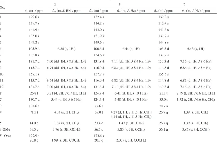
(1721 cm-1) functional groups. The 1H and 13C NMR spectra
119.7 (C-2), 144.9 (C-3), 135.0 (C-4), 147.2 (C-5), 105.9 (C-6)], one p-substituted benzene ring [dH 7.00 (2H, dd,
J 8.8, 2.4 Hz, H-8, H-12), 6.74 (2H, dd, J 8.8, 2.4 Hz, H-9, H-11); dC 133.8 (C-7), 131.7 (C-8, C-12), 115.7 (C-9, C-11), 157.1 (C-10)], one acetoxyprenyl group [dH 3.21 (2H, d,
J 6.7 Hz, H-1'), 5.44 (1H, d, J 6.7 Hz, H-2'), 4.33 (2H, s, H-4'), 1.39 (3H, s, H-5'), and 1.99 (3H, s, H-OAc); dC 26.8 (C-1'), 130.7 (C-2'), 134.6 (C-3'), 71.5 (C-4'), 14.0 (C-5'), 172.9, 20.8 (OAc)], and one methoxy group [dH 3.76 (3H, s, OMe-5);dC 56.5 (OMe)], which implied that compound 1 was a biphenyl derivative. This conclusion was confirmed by the heteronuclear multiple bond correlation (HMBC) correlations of H-6 with C-7, and of H-8 and H-12 with C-1 (Figure 2). HMBC correlations of H-1' with C-1, C-2, and C-3, and of H-2' with C-1 suggested the acetoxyprenyl group at C-2. The methoxy group was located at C-5 from the HMBC correlations of dH 3.76 (OMe) with C-5. Considering the signal for quarternary C-3, C-4, C-10 and the molecular formula of 1, three hydroxy groups were located at C-3, C-4, C-10, respectively. Thus, the structure of 1 was determined as shown (Figure 1), and named multibiphenyl A.
Compound 2 was obtained as a pale yellow gum and had the molecular formula C20H20O6, as inferred from the
EI-HRMS showing the molecular-ion peak at m/z 356.1274, indicating eleven degrees of unsaturation. The IR spectrum of 2 showed absorption bands at 3430 cm-1 for free OH
group. The 1H NMR spectrum of compound 2 showed
characteristic signals for an acetoxychromene ring, i.e., olefinic H-atoms at dH 6.41 (1H, d, J 10.1 Hz, H-1') and
dH 5.48 (1H, d, J 10.1 Hz, H-2'), a Me group at dH 1.47 (s, Me-5'), an oxygenated methylene at dH 4.27, 4.14 (2H, d,
J 11.5 Hz, H-4'), and an acetoxy group dH 2.00 (s, OAc). A set of signals at dC 124.7 (C-1'), 124.4 (C-2'), 77.6 (C-3'), 69.0 (C-4'), 23.4 (C-5'), and 172.6/20.7 (OAc) in the 13C NMR spectrum provided further support for the
presence of an acetoxy chromene system. The remaining unsaturation degrees suggested the presence of a typical biphenyl unit which was supported by the remaining 12 aromatic C-atom signals dC 132.4 (C-1), 114.2 (C-2), 142.0 (C-3), 131.9 (C-4), 149.6 (C-5), 106.6 (C-6), 134.6 (C-7), 131.8 (C-8, C-12), 116.0 (C-9, C-11), 157.7 (C-10). Further support for the determination of the structure was provided by the signal of H-6 showing a three-bond connectivity with C-7 (dC 134.6), and H-8 and H-12 showing three-bond connectivities with C-1 in the HMBC plot. The HMBC experiment allowed to position the substituents at the chromene benzene ring. The H-1' signal at dH 6.41 showed a two-bond connectivity with C-2 (dC 114.2), and a three-bond connectivity with C-1 (dC 132.4) and C-3 (dC 142.0), whereas H-2' (dH 5.48) correlated to C-2 in HMBC spectrum. Thus, the chromeme ring proved to be fused to the benzene ring at C-2 and C-3. The methoxy group was located at C-5 according to the HMBC correlation of OMe (dH 3.85) with C-5 (dC 149.6). A single H-atom at dH 6.44 (s, H-6) belonging to a 1,2,3,4,5-pentasubstituted benzene moiety showed a two-bond connectivity with C-1 (dC 132.4) and C-5, and a three-bond connectivity with C-2 (dC 114.2), and C-4 (dC 131.9), which suggested that one OH group was attached to C-4. The existence of two phenolic OH groups was confirmed by the signals of two oxygenated quarternary aromatic C-atoms in the 13C NMR spectrum at dC 131.9 (C-4) and 157.7 (C-10), and the molecular formula of compound 2. The presence of the second benzene ring was deduced from the 1H NMR spectrum, which showed a
set of ds of orto-coupled H-atoms at dH 7.11 (dd, J 8.4 Hz, 1.9, H-8, 12) and 6.82 (dd, J 8.4 Hz, 1.9, H-9,11), typical OH OH MeO OH O O 10 9 8 11 12 7 1 2 3 4 5 6 1' 2' 3' 4' 5'
Figure 2. Selected HMBC (H→C) and 1H-1H correlation spectroscopy
(COSY) (–) correlations of 1.
1 OH OH MeO OH OAc 5' 4' 3' 2' 1' 2 5 4 3 1 7 10 8 12 2 OH O OH MeO OAc 1 2 3 4 5 6 1' 2 ' 3' 4' 5' 1 0 7 OH MeO OH O 3 1 7 10 8 2 1' 2' 3' 4' 5' 5 3 8 9 9 9
11 11 11
12
12 4
6 6
for a para-substituted aryl moiety. Therefore, the other OH group was assigned to C-10. The configuration of C-3' was not assigned. Accordingly, the structure of compound 2 was determined as shown (Figure 1), and named multibiphenyl B.
Compound 3 was obtained as a pale orange gum, and its molecular formula was established to be C18H20O4 as
deduced by the EI-HRMS molecular ion peak (M+) at m/z
300.1360, indicating nine degree of unsaturation. The IR spectrum revealed absorption bands of hydroxyl (3423 cm-1)
and methyl (1446 cm-1) groups. The connectivity of the
protons and C-atoms was established by the heteronuclear single-quantum correlation (HSQC) spectrum. There were eighteen resolved peaks observed in the 13C NMR spectrum
(Table 1), including signals for twelve aromatic carbons, one methoxy group, two methylenes, and two methyl carbons. Its 1H NMR spectrum indicated the presence of
twenty protons including five aromatic protons at dH 6.43 (1H, s, C-6), 7.16 (2H, d, J 8.6 Hz, C-8), 6.86 (2H, d,
J 8.6 Hz, C-9), and two methyl protons at dH 1.39 (6H, s, C-3' and C-4') and 3.86 (3H, s, OMe). Analysis of the NMR data, indicated that the spectra of 3 were similar to those of 2 except for the absence of an acetoxy group and
one double bond and the appearance of two methylenes, thus suggesting that 3 and 2 possess the same substitution pattern of the benzene rings. The 13C NMR spectrum of 3,
when compared to that of 2, displayed an upfield shifted signal for C-4' (dC26.7), the absence of signals for OAc, and upfieldshifted signals for C-1' (dC21.1) and C-2' (dC33.0). These data indicated the presence of two methyl groups placed at C-4, and the absence of double bond between 1' and 2'. The structure of 3 was then established as shown and named multibiphenyl C.
The new biphenyls 1-3 were tested for their ability to prevent the cytopathic effects of rotavirus in MA104 cells, and their effects were measured in parallel with the determination of antiviral activity using ribavirin as positive control, showing potent anti-rotavirus activity with therapeutic index (TI) above 10 (Table 2).
Conclusions
Three new biphenyls (1-3) were isolated from
Garcinia multiflora, whose structures were elucidated by spectroscopic methods. The three compounds presented a promising anti-rotavirus activity.
Table 1. 1H and 13C NMR data for compounds 1-3 (d in ppm, 1 and 2 in CD
3OD, 3 in CDC13, 100 and 400 MHz)
No. 1 2 3
dC (m) / ppm dH (m, J, Hz) / ppm dC (m) / ppm dH (m, J, Hz) / ppm dC (m) / ppm dH (m, J, Hz) / ppm
1 129.6 s 132.4 s 132.3 s
2 119.7 s 114.2 s 112.4 s
3 144.9 s 142.0 s 141.5 s
4 135.0 s 131.9 s 132.7 s
5 147.2 s 149.6 s 144.8 s
6 105.9 d 6.26 (s, 1H ) 106.6 d 6.44 (s, 1H) 105.5 d 6.43 (s, 1H)
7 133.8 s 134.6 s 132.7 s
8 131.7 d 7.00 (dd, 1H, J 8.8 Hz, 2.4) 131.8 d 7.11 (dd, 1H, J 8.4 Hz, 1.9) 130.3 d 7.16 (d, 1H, J 8.6 Hz)
9 115.7 d 6.74 (dd, 1H, J 8.8 Hz, 2.4) 116.0 d 6.82 (dd, 1H, J 8.4 Hz, 1.9) 114.8 d 6.86 (d, 1H, J 8.6 Hz)
10 157.1 s 157.7 s 155.5 s
11 115.7 d 6.74 (dd, 1H, J 8.8 Hz, 2.4) 116.0 d 6.82 (dd, 1H, J 8.4 Hz, 1.9) 114.8 d 6.86 (d, 1H, J 8.6 Hz)
12 131.7 d 7.00 (dd, 1H, J 8.8 Hz, 2.4) 131.8 d 7.11 (dd, 1H, J 8.4 Hz, 1.9) 130.3 d 7.16 (d, 1H, J 8.6 Hz)
1' 26.8 t 3.21 (d, 2H, J 6.7 Hz, CH2) 124.7 d 6.41 (d, 1H, J 10.1 Hz) 21.1 t 2.59 (t, 2H, J 6.6 Hz, CH2)
2' 130.7 d 5.44 (t, 1H, J 6.7 Hz) 124.4 d 5.48 (d, 1H, J 10.1 Hz) 33.0 t 1.72 (t, 2H, J 6.6 Hz, CH2)
3' 134.6 s 77.6 s 74.7 s
4' 71.5 t 4.33 (s, 3H, CH3) 69.0 t 4.27 (d, 1H, J 11.5 Hz, CH2)
4.14 (d, 1H, J 11.5 Hz, CH2)
26.7 q 1.39 (s, 3H, CH3)
5' 14.0 q 1.39 (s, 3H, CH3) 23.4 q 1.47 (s, 3H, CH3) 26.7 q 1.39 (s, 3H, CH3)
3-OMe 56.5 q 3.76 (s, 3H, OCH3) 56.5 q 3.85 (s, 3H, OCH3) 56.1 q 3.86 (s, 3H, OCH3)
5'- OAc 172.9 s
20.8 q 1.99 (s, 3H, COCH3)
172.6 s
Supplementary Information
Supplementary data (1H, 13C, HSQC, HMBC, 1H-1H
correlation spectroscopy (COSY) NMR and mass spectrometry (MS) spectra of 1-3) are available free of charge at http://jbcs.sbq.org.br as PDF file.
Acknowledgements
This research was supported by the National Natural Science Foundation of China (21002085 and 21362044), and start-up funds of Yunnan University of Nationalities.
References
1. Li, X. W.; Li, J.; Stevens, P. F.; Flora of China2007, 13, 40.
2. Chen, F. C.; Lin, Y. M.; Hung, J. C.; Phytochemistry1975, 14, 300.
3. Chiang, Y. M.; Kuo, Y. H.; Oota, S.; Fukuyama, Y.; J. Nat. Prod.
2003, 66, 1070.
4. Lee, L. T.; Tsai, H. P.; Wang, C. C.; Chang, C. N.; Liu, W. C.; Hsu, H. C.; Hsieh, C. T.; Chen, Y. C.; Tseng, H. W.; Gau, R. J.; Liu, S. H.; Chen, I. S.; Iinuma, M.; Biomed. Prev. Nutr.2013, 3, 247.
5. Chen, J. J.; Ting, C. W.; Hwang, T. L.; Chen, I. S.; J. Nat. Prod.
2009, 72, 253.
6. Chien, S. C.; Chyu, C. F.; Chang, I. S.; Chiu, H. L.; Kuo, Y. H.; Tetrahedron Lett. 2008, 49, 5276.
7. Chen, F. C.; Lin, Y. M.; Hung, J. C.; Phytochemistry1975, 14, 818.
8. Lin, Y. M.; Flavin, M. T.; Cassidy, C. S.; Mar, A.; Chen, F. C.; Bioorg. Med. Chem. Lett. 2001, 11, 2101.
9. Lin, Y. M.; Anderson, H.; Flavin, M. T.; Pai, Y. H. S.; Greenwood, E. M.; Pengsuparp, T.; Pezzuto, J. M.; Schinazi, R. F.; Hughes, S. H.; Chen, F. C.; J. Nat. Prod. 1997, 60, 884. 10. Wu, J. H.; Tung, Y. T.; Chyu, C. F.; Chien, S. C.; Wang, S. Y.;
Chang, S. T.; Kuo, Y. H.; J. Wood Sci. 2008, 54, 383. 11. Zhu, S. L.; Luo, J. B.; Tan, X. M.; Xing, X. F.; J. Chin. Med.
Mat. 2010, 33, 785.
Submitted: May 13, 2015
Published online: September 15, 2015
Table 2. Anti-rotavirus activity of compounds 1-3a
No. CC50b / (µg mL-1) EC
50c / (µg mL-1) TId
1 125.72 ± 6.41 11.56 ± 1.13 10.93 ± 1.27
2 134.65 ± 8.34 10.94 ± 1.65 12.35 ± 1.75
3 159.83 ± 7.46 12.73 ± 1.75 12.58 ± 1.68
Ribavirin 274.27 ± 11.07 13.61 ± 1.04 20.14 ± 1.16
aAll results are expressed as mean ± SD (n = 3); bCC
50: mean (50%)
value of cytotoxic concentration; cEC
50: mean (50%) value of effective