Complementarity of radioautographic
and immunohistochemical techniques
for localizing neuroreceptors at the
light and electron microscopy level
Montreal Neurological Institute, McGill University, Montreal, Quebec, Canada
A. Beaudet, P. Dournaud and H. Boudin
Abstract
To assess relationships between neuropeptide-binding sites and recep-tor proteins in rat brain, the distribution of radioautographically labeled somatostatin and neurotensin-binding sites was compared to that of immunolabeled sst2A and NTRH receptor subtypes, respec-tively. By light microscopy, immunoreactive sst2A receptors were either confined to neuronal perikarya and dendrites or diffusely dis-tributed in tissue. By electron microscopy, areas expressing somato-dendritic sst2A receptors displayed only low proportions of mem-brane-associated, as compared to intracellular, receptors. Conversely, regions displaying diffuse sst2A labeling exhibited higher proportions of membrane-associated than intracellular receptors. Furthermore, the former showed only low levels of radioautographically labeled soma-tostatin-binding sites whereas the latter contained high densities of somatostatin-binding suggesting that membrane-associated receptors are preferentially recognized by the radioligand. In the case of NTRH receptors, there was a close correspondence between the light micro-scopic distribution of NTRH immunoreactivity and that of labeled neurotensin-binding sites. Within the substantia nigra, the bulk of immuno- and autoradiographically labeled receptors were associated with the cell bodies and dendrites of presumptive DA neurons. By electron microscopy, both markers were detected inside as well as on the surface of labeled neurons. At the level of the plasma membrane, their distribution was highly correlated and characterized by a lack of enrichment at the level of synaptic junctions and by a homogeneous distribution along the remaining neuronal surface, in conformity with the hypothesis of an extra-synaptic action of this neuropeptide. Inside labeled dendrites, there was a proportionally higher content of immu-noreactive than radiolabeled receptors. Some of the immunolabeled receptors not recognized by the radioligand were found in endosome-like organelles suggesting that, as in the case of sst2A receptors, they may have undergone endocytosis subsequent to binding to the endog-enous peptide.
Correspondence
A. Beaudet
Montreal Neurological Institute 3801 University Street Montreal, Quebec H3A 2B4 Canada
Presented at the 5th International Symposium on Radioautography, São Paulo, SP, Brasil,
August 24-26, 1997.
The present address of P. Dournaud is INSERM U.159, 2ter, rue d’Alésia, 75014 Paris, France.
Received November 19, 1997 Accepted November 24, 1997
Key words
•Neurotensin
•Somatostatin
•Internalization
•Neuropeptides
•Autoradiography
Introduction
For more than twenty years, radioautog-raphy has been the method of choice for visualizing neurotransmitter receptors or, more specifically, neurotransmitter-binding sites in mammalian brain. Original labeling
methods relied on in vivo administration of
radioligands by the parenteral (1) or intrace-rebroventricular (2) route followed by radio-autographic processing of frozen brain sec-tions using dry radioautographic techniques (3). Young and Kuhar (4) were later to
de-vise an in vitro radioligand-binding
tech-nique which, combined with the use of dry emulsion-covered coverslips (4) or tritium sensitive film (5), was to be universally ap-plied to the study of the regional distribution of a variety of neurotransmitter receptors in the central nervous system (CNS). The intro-duction of photoaffinity probes and of radioligands that lent themselves to cross-linking by divalent agents made it possible to visualize covalently labeled receptors by means of standard wet autoradiographic tech-niques (6,7). This technical improvement significantly increased the resolution of re-ceptor detection allowing their visualization at both the cellular (e.g., 8-11) and subcellu-lar levels (7,12-15).
During the last decade, the cloning of a multiplicity of neuroreceptor subtypes as well as of various subunits of ligand-gated chan-nels made it possible to visualize neurotrans-mitter receptors in the brain by immunohis-tochemistry through the development of an-tibodies directed against specific amino acid sequences of the cloned proteins. This ap-proach has the advantage of permitting the selective detection of molecularly defined receptor subtypes. It also affords a consider-ably higher resolution than radioautography, particularly when conjugated to immunogold detection systems. This increased resolution permits better differentiation between soma-todendritic (e.g., post-synaptic) and axonal (i.e., pre-synaptic) receptors than was
previ-ously possible with radioautography at the light microscopic level, even using high reso-lution dipping techniques. At the electron microscopy level, it permits the determina-tion of both the nature of the receptor-bear-ing element (neuronal or glial; dendritic ver-sus axonal) and the identity of the subcellu-lar organelles with which the receptors are associated.
These advantages of immunohistochemi-cal over radioautographic loimmunohistochemi-calization tech-niques have led many to believe that radio-autography was no longer a method of choice for studying the distribution of neuroreceptors in the brain. However, this is clearly not the case since radioautography i) remains a vi-able approach for visualizing receptors that have not yet been cloned, ii) is a method of choice for the recognition of functional re-ceptors (i.e., capable of ligand binding), and iii) is an approach which readily lends itself to the study of the pharmacological proper-ties of neurotransmitter-binding sites as well as to the visualization of the sites of action of a variety of centrally acting drugs.
In our own work, we have taken advan-tage of the complementarity of radioauto-graphic and immunohistochemical tech-niques to gain insight into the functional significance of the cellular and subcellular distribution of neuropeptide receptors in the CNS. The examples that follow are taken from recent studies from our laboratory on the light and electron microscopy distribu-tion of somatostatin and neurotensin recep-tors in the rat brain.
Light and electron microscopy localization of somatostatin sst2A receptors in rat brain
rat brain. sst2A receptors were labeled using tyramide-amplified peroxidase (light micros-copy) or immunogold (electron microsmicros-copy) techniques with an antibody directed against a specific amino acid sequence located in the C-terminus of the receptor, developed and characterized by Dr. Agnes Schonbrunn, Houston, TX. sst2-binding sites were
la-beled in vitro by incubation of frozen
sec-tions with an iodinated somatostatin
ana-logue (125I-D-Trp8-somatostatin), as
de-scribed by Dournaud et al. (17). Immunohis-tochemical results were also compared to radioautographic data generated with sst2-preferring radioligands, as reported in the literature (reviewed in 16).
At low light microscope magnification, sst2A-immunoreacted sections exhibited se-lective and intense sst2A receptor labeling in a number of brain areas, and most conspicu-ously in deep layers of cerebral cortex, CA1 and CA2 subfields of the hippocampus, me-dial habenula, bed nucleus of the stria termi-nalis, endopiriform nucleus, claustrum and amygdaloid complex. Dense sst2A immu-noreactivity was also observed within sev-eral brainstem nuclei, including the locus coeruleus, pontine nuclei, and nucleus
trac-Figure 1 - Comparative distribu-tion of somatostatin immunore-activity (a), sst2A receptor im-munoreactivity (b) and autoradi-ographically labeled somatosta-tin-binding sites (c) in the pari-etal cortex. a, Numerous soma-tostatin immunoreactive nerve cell bodies are distributed throughout layer II to layer VI. b, Numerous sst2A receptor-ex-pressing neurons are evident in layers II-III, whereas diffuse sst2A labeling pervades the outer part of layer V and the deeper part of layer VI. c, The distribution pattern of 125
I-D-Trp8-somatostatin-binding sites is superimposable over that of sst2A diffuse labeling in layers V-VI. ec: External capsule. Scale bars: 45 µm.
tus solitarius (18).
At higher light microscope magnifica-tion, sst2A immunolabeling was either con-fined to neuronal perikarya and dendrites or diffusely distributed in tissue. Comparison of this labeling pattern with that of sst2-binding sites labeled by radioautography,
using either 125I-D-Trp8-somatostatin or
sst2-preferring ligands, revealed that in regions in which sst2A immunoreactivity was present over nerve cell bodies and dendrites concen-trations of radiolabeled somatostatin-bind-ing sites were highly variable, rangsomatostatin-bind-ing from high in regions such as the medial habenula to very low in regions such as the upper layers of the cerebral cortex (Figure 1b,c) or the central nucleus of the amygdala. Quanti-tative analysis confirmed the total lack of correlation between the distribution of sst2A somatodendritic immunolabeling and that of somatostatin-binding sites radioautographi-cally labeled using sst2-preferring ligands (r = 0.24; P<0.3). By contrast, in areas in which it appeared diffusely distributed in tissue (e.g., in deep layers of the cerebral cortex, Figure 1b), sst2A immunolabeling showed a striking correspondence with the distribu-tion pattern of radioautographically labeled
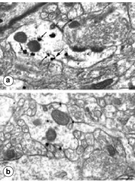
mainly concentrated inside neuronal peri-karya and dendrites rather than associated with their plasma membrane. In fact, quanti-tative analysis demonstrated that over 78% of labeled receptors were intracellular whereas only 22% were membrane bound. Inside labeled neurons, immunoreactive re-ceptors were evident over structures involved in protein synthesis and glycosylation (i.e., endoplasmic reticulum and Golgi apparatus) as well as over vesiculotubular organelles characteristic of endosomal compartments (Figure 3a).
In contrast, in areas in which immunola-beled sst2A receptors appeared diffusely dis-tributed by light microscopy, electron mi-croscopy revealed a predominant associa-tion of the label with neuronal plasma mem-branes (70%) as opposed to the interior of the cells (30%). As can be seen in Figure 3b, membrane-associated receptors were hap-hazardly distributed along the plasma mem-brane without obvious concentration over sites of synaptic specialization or opposite abutting axon terminals.
These results indicate that membrane-bound sst2A receptors preferentially recog-nize radiolabeled ligands. In contrast, intra-cellular receptors recognize radioactive so-matostatin analogs poorly, presumably be-cause of a different molecular conformation and/or functional state. What is particularly intriguing here is the finding that regions in which sst2A somatostatin receptors are pre-dominantly intracellular are the ones that receive a dense somatostatin innervation, whereas in regions in which sst2A receptors are predominantly membrane bound, soma-tostatin axon terminals are only sparse. One possible interpretation for these findings is that in regions of dense somatostatin inner-vation, membrane-associated sst2A recep-tors are chronically down-regulated because of a massive release of endogenous soma-tostatin. Such a down-regulation would ob-viously be much less pronounced in regions of sparse somatostatin innervation, hence
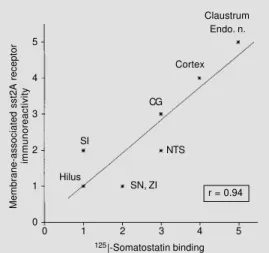
Figure 2 - Correlation between the density of membrane-asso-ciated immunoreactive sst2A re-ceptors and that of somatosta-tin-binding sites documented by quantitative autoradiography us-ing sst2-preferrus-ing ligands. Us-ing Spearman’s coefficient, a highly significant correlation is found between immunological and autoradiographic signals (r = 0.94, P<0.0001). Endo. n., Endopiriform nucleus; NTS, nucleus tractus solitarius; CG, central gray; SI, substantia innominata; SN, substantia nigra; ZI, zona incerta.
Membrane-associated sst2A receptor
immunoreactivity
5
4
3
2
1
0
0 1 2 3 4 5
125|-Somatostatin binding
Claustrum Endo. n.
Cortex
CG
SI
Hilus
SN, ZI NTS
r = 0.94
sst2-binding sites (Figure 1b,c). Furthermore, a high correlation was observed between the density of diffuse sst2A immunolabeling and that of radioautographically labeled sst2A-binding sites (Figure 2; r = 0.94; P<0.0001). An inverse relationship was observed when the distribution of sst2A-immunoreac-tive receptors was compared to that of soma-tostatin axon terminals immunohistochemi-cally detected in the same or adjacent sec-tions using a specific antibody directed against somatostatin (16). Specifically, there was a strong correlation between sst2A im-munolabeling and somatostatin terminal den-sities in regions in which sst2A immunola-beling was clearly somatodendritic (e.g., in the central nucleus of the amygdala). By contrast, in regions exhibiting diffuse sst2A immunolabeling (and dense radioautographic labeling of somatostatin-binding sites), so-matostatin-immunoreactive axons were only sparse (e.g., in deep layers of cerebral cor-tex, Figure 1a-c).
the large proportion of membrane-associ-ated receptors found in these areas. The frequency at which intracellular receptors were found in association with endosome-like organelles suggests that this down-regulation may be predicated on somatosta-tin-induced internalization of receptor-ligand complexes. Such an interpretation is consistent with the earlier demonstration that the sst2A receptor subtype may undergo ligand-induced internalization in transfected cells as well as in natural cell lines (19-21).
Comparative distribution of
high-affinity neurotensin receptors and specific neurotensin-binding sites in rat substantia nigra
In a separate set of experiments (Boudin H, Pélaprat D, Rostène W, Pickel VM and Beaudet A, unpublished results), we com-pared the light and electron microscopy dis-tribution of immunocytochemically labeled neurotensin receptor proteins with that of radioautographically labeled high-affinity neurotensin-binding sites. In contrast to so-matostatin, only two receptors for the tri-decapeptide neurotensin have been cloned to date: a high-affinity site (Kd = 0.1 nM) expressed in restricted areas of the rodent brain (9,22,23) and a low-affinity site (Kd = 1 µM) the expression of which is consider-ably more widespread (24; Sarret P, Beaudet A, Vincent JP and Mazella J, unpublished results). Of these two receptors, the high-affinity receptor (NTRH) is the one that appears to be involved in most of the docu-mented effects of neurotensin. In particular, it has been shown to be responsible for the potent excitatory effects of neurotensin on mesencephalic dopaminergic cells (25-27). This finding is consistent with the high con-centration of NTRH detected in association with dopaminergic cells in the substantia nigra and adjacent ventral tegmental area (28-30).
NTRH receptor proteins were immuno-labeled using either tyramide-amplified per-oxidase (light microscopy) or immunogold (electron microscopy) techniques with a spe-cific antibody directed against an amino acid sequence of the third intracellular loop of the receptor. This antibody was developed and characterized in Dr. William Rostène’s labo-ratory in Paris (23,31). High-affinity neuro-tensin-binding sites were detected in
sec-tions of the rat brain labeled in vitro, using
concentrations of iodinated neurotensin (0.1 nM) that ensure selective labeling of the high-affinity neurotensin receptor subtype (for a detailed description of the methodol-ogy for radioautographic labeling of NTRH at light and electron microscopic levels, see references 9,14).
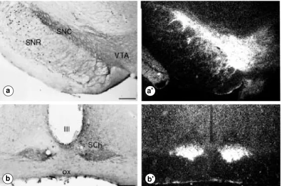
At the light microscopy level, there was a close correlation between the topographic distributions of immunolabeled and radio-autographically labeled NTRH receptors. This is illustrated in Figure 4 for two areas of particularly dense NTRH enrichment: the substantia nigra and suprachiasmatic nucleus of the hypothalamus. As previously reported (23,30,32), in both of these areas
neuro-Figure 3 - Electron microscopy localization of sst2A receptors using silver-enhanced immu-nogold in the bed nucleus of the stria terminalis (a) and claustrum (b). a, Immunolabeled receptors are detected inside a large den-dritic shaft. Note the frequent association of gold particles with endosome-like organelles (small arrows). Only one of the gold particles is associated with the plasma membrane (large arrow). b, Gold particles are predomi-nantly associated with the plasma membrane of a dendrite. Note the lack of receptor enrich-ment opposite abutting axon ter-minals. Scale bars: 0.3 µm.
tensin immunoreactive receptor proteins were mainly associated with neuronal perikarya and dendrites (Figure 4).
Electron microscopy examination of the rat substantia nigra confirmed the overall similarity between the fine structural distri-butions of radioautographically and immu-nolabeled NTRH receptors. Thus, as pre-dicted from light microscopy results (see Figure 4), both markers were almost exclu-sively associated with neuronal perikarya and dendrites which, on the basis of earlier double-labeling (30) and lesion studies (28), may be surmised to be mainly dopaminergic. However, quantitative analyses of immuno-gold and radioautographic silver grain distri-bution (for a description of the methodology for statistical analysis of radioautographs, see reference 33) revealed subtle differences between the distributions of the two mark-ers. Thus, whereas in electron microscopy radioautographs a slightly larger proportion of labeled-binding sites was associated with plasma membranes (55%) than with intra-cellular compartments (45%), in immunore-acted sections intracellular receptors were proportionally more numerous than plasma
membrane-associated ones (73% vs 27%).
In spite of these quantitative differences, both markers showed a remarkably similar distribution along the length of neuronal plasma membranes (Figures 5 and 6). A striking feature of this distribution was the lack of receptor enrichment at the level of synaptic junctions. Rather, both markers were more or less evenly distributed along soma-todendritic plasma membranes with no ap-parent predilection for any of the opposite abutting elements. Such a homogeneous dis-tribution is consistent with the earlier dem-onstration that, within the substantia nigra, neurotensin-containing axon terminals only rarely contact neurotensin receptor-bearing (i.e., dopaminergic) cells (34). It is also con-gruent with the previously proposed hypoth-esis that throughout several regions of the brain, including the substantia nigra (34), ventral tegmental area (14) and basal fore-brain (35), neurotensin acts in a parasynaptic manner by diffusion for short distances into the extracellular space.
Inside nerve cell bodies, both NTRH re-ceptor proteins and binding sites were evi-dent over the endoplasmic reticulum and
Figure 4 - Comparative light mi-croscopy distribution of NTRH immunoreactivity (left) and 125
I-NT-binding sites (right) in the substantia nigra and ventral teg-mental area (a,a’) and in the su-prachiasmatic nucleus (b,b’). In both regions, the distribution of immunostaining matches that of autoradiographic labeling. a,a’, The labeling is prominent in the substantia nigra, pars compacta (SNC) and in the ventral tegmen-tal area (VTA) where it is pre-dominantly associated with nerve cell bodies and dendrites. The dendritic meshwork is espe-cially dense in the SNC where-from it extends into the substan-tia nigra, pars reticulata (SNR). b,b’, The labeling is concentrated in the ventral part of the supra-chiasmatic nucleus (SCh) in which it also predominates over somatodendritic elements. III, Third ventricle; ox, optic chiasm. Scale bars: 300 µm.
b
a a'
Golgi apparatus, suggesting that newly syn-thesized NTRH receptors already possess the molecular conformation necessary for recognition of the ligand. At the level of labeled dendrites, however, there were marked discrepancies between the intracel-lular distributions of radioautographic and immunocytochemical markers. First, immu-noreactive NTRH receptor proteins were rela-tively more abundant than radiolabeled neu-rotensin-binding sites, which probably ac-counts for the larger proportion of intracellu-lar receptors found in immunocytochemical preparations. Second, a higher proportion of immunogold particles than of radioauto-graphic silver grains was found in associa-tion with vesicular and/or tubulovesicular endosome-like elements. Our interpretation for these discrepancies is that, as in the case of sst2A receptors, a proportion of intra-dendritic NTRH receptor proteins is either unrecognized by or unaccessible to the ra-dioligand. This unaccessibility may be due to the trapping of the receptors in endosomes subsequent to their agonist-induced inter-nalization into target cells. This interpreta-tion is supported by the earlier demonstra-tion of receptor-mediated internalizademonstra-tion of fluorescent neurotensin in nigrostriatal neu-rons (36) as well as by the demonstration that this internalization is mediated by the high-affinity neurotensin receptor subtype (37).
Conclusions
In summary, the present data illustrate the complementarity of radioautographic and immunohistochemical techniques for local-izing and assessing the functionality of neu-ropeptide receptors in the CNS at the light and electron microscopy levels. The correla-tive data obtained here by conjugating these two techniques for visualizing sst2A soma-tostatin and NTRH neurotensin receptors in the rat brain have made it possible to draw the following conclusions: 1) whereas
im-munocytochemical markers recognize both intracellular and membrane-associated forms of the receptors, radiolabeled ligands may or may not recognize intracellular receptors depending on the accessibility and/or molec-ular conformation of the latter. These results imply that radioautography, even when
car-ried out after in vitro labeling, may be a
better predictor of the sites of action of exog-enously administered than of endogexog-enously released neuropeptides. 2) At the level of neuronal plasma membranes, our studies showed a remarkable concordance between the distribution of neuropeptide receptor pro-teins and that of specific transmitter-binding
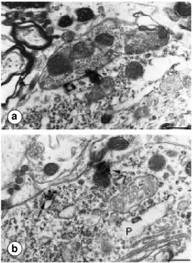
Figure 5 - Electron microscopy localization of immunogold-la-beled NTRH receptors in the rat substantia nigra, pars com-pacta. Gold particles are de-tected along the plasma mem-branes of two adjacent den-dritic profiles (D1 and D2). Note the presence of a labeled re-ceptor at the level of an asym-metrical synaptic junction (ar-row). Scale bar: 0.5 µm.
Figure 6 - Electron microscopy detection of radioautographically labeled 125I-NT-binding sites in
the rat substantia nigra. a) Two radioautographic silver grains are detected over an axosomatic ap-position. The abutting axon ter-minal contains densely packed clear synaptic vesicles. b, Two silver grains are associated with the plasma membrane of a neu-ronal perikaryon (P). One of the silver grains is detected oppo-site an astroglial sheath (arrow) and the other at the level of an incoming axon terminal contain-ing only sparse clear synaptic vesicles (arrowhead). Scale bars: 0.6 µm.
sites. Both markers were found to be uni-formly distributed along the membranes, with no evidence of enrichment at the level of synaptic specializations. These results sug-gest that receptor proteins present on the surface of the neurons, and not only those found opposite abutting axon terminals or at the level of synaptic junctions, are func-tional (i.e., are in a conformation that recog-nizes endogenous as well as exogenous ligands). This proposal is consistent with the parasynaptic mode of neurotransmission pre-viously proposed for a number of neuropep-tides in the CNS. 3) Our results suggest that a proportion of intracellular receptors do not recognize radioactive ligands because they are trapped in endosome-like compartments. This interpretation implies that cell surface
receptors may undergo down-regulation upon agonist-induced internalization into their tar-get cells. This interpretation is in keeping with the recent proposal that there might be widespread receptor-mediated internalization of neuropeptides through receptor-mediated mechanisms in the CNS (e.g., 38-40). Inter-nalization is a process that has been linked to a variety of functions, including ligand se-questration and degradation (41), receptor desensitization/resensitization and recycling (42) and intracellular neuropeptide signal-ling (43). The extent of this internalization, however, appears to vary from one receptor to another and may be related in part to the density of innervation of receptor-bearing cells by axon terminals that store and release the receptor’s endogenous ligand.
References
1. Pert CB, Kuhar MJ & Snyder SH (1976). Opiate receptor: autoradiographic localiza-tion in rat brain. Proceedings of the Nalocaliza-tional Academy of Sciences, USA, 73: 3729-3733. 2. Hunt SP & Schmidt J (1978). The electron microscopic autoradiographic localization of α-bungarotoxin-binding sites within the central nervous system of the rat. Brain Research, 142: 152-159.
3. Stumpf WE & Roth LG (1966). High resolu-tion autoradiography with dry mounted, freeze-dried frozen sections. Comparative study of six methods using two diffusable compounds 3H-estradiol and 3
H-mesobi-lirubinogen. Journal of Histochemistry and Cytochemistry, 14: 274-287.
4. Young WS & Kuhar MJ (1979). A new method for receptor autoradiography: 3H
opioid receptor labeling in mounted tissue sections. Brain Research, 179: 255-270. 5. Palacios JM, Niehoff DL & Kuhar MJ
(1981). Receptor autoradiography with tri-tium-sensitive film: Potential for compu-terized densitometry. Neuroscience Let-ters, 24: 111-116.
6. Hamel E & Beaudet A (1984). Localization of opioid-binding sites in rat brain by elec-tron microscopic radioautography. Journal of Electron Microscopy Technique, 1: 317-329.
7. Pasquini F, Bochet P, Garbay-Jaureguiberry C, Roques BP, Rossier J & Beaudet A (1992). Electron microscopic localization of photoaffinity-labelled delta opiod receptors in the neostriatum of the rat. Journal of Comparative Neurology, 326: 229-244.
8. Herkenham M & Pert CB (1982). Light microscopic localization of brain opiate re-ceptors: a general autoradiographic method which preserves tissue quality. Journal of Neuroscience, 2: 1129-1149. 9. Moyse E, Rostène W, Vial M, Leonard K,
Mazella P, Kitabgi P, Vincent JP & Beaudet A (1987). Distribution of neuro-tensin-binding sites in rat brain: A light microscopic radioautographic study using monoiodo 125I-Tyr
3-neurotensin.
Neuro-science, 22: 526-536.
10. Epelbaum J, Moyse E, Tannenbaum GS, Kordon C & Beaudet A (1989). Combined autoradiographic and immunohistochemi-cal evidence for an association of soma-tostatin-binding sites with growth hor-mone-releasing factor-containing nerve cell bodies in the rat arcuate nucleus. Journal of Neuroendocrinology, 1: 109-115.
11. Moyse E, Beaudet A, Bertherat J & Epelbaum J (1992). Light microscopic ra-dioautographic localization of somatosta-tin-binding sites in the brainstem of the rat. Journal of Chemical Neuroanatomy, 5: 75-84.
12. Beauvilain JC, Moyse E, Dutriez I, Mitchell V, Poulain P & Mazzuca M (1992). Local-ization of mu opioid receptors on the mem-branes of nerve endings and tanycytes in the guinea pig median eminence by elec-tron microscopy. Neuroscience, 49: 925-936.
13. Hamel E & Beaudet A (1984). Electron microscopic autoradiographic localization of opioid receptors in rat neostriatum. Na-ture, 31: 155-157.
14. Dana C, Vial M, Leonard K, Beauregard A, Kitabgi P, Vincent JP, Rostène W & Beaudet A (1989). Electron microscopic localization of neurotensin-binding sites in the midbrain tegmentum of the rat. I. Ven-tral tegmental area and interfascicular nucleus. Journal of Neuroscience, 9: 2247-2257.
16. Dournaud P, Boudin H, Shonbrunn A, Tannenbaum G & Beaudet A (1998). Rela-tionships between somatostatin and the somatostatin sst2A receptor in rat brain: a light, confocal and electron microscopic double labeling study. Journal of Neuro-science (in press).
17. Dournaud P, Jazat-Poindessous F, Slama A, Lamour Y & Epelbaum J (1996). Corre-lations between water maze performance and cortical somatostatin mRNA and high-affinity-binding sites during aging in rats. European Journal of Neuroscience, 8: 476-485.
18. Dournaud P, Gu YZ, Shonbrunn A, Mazella J, Tannenbaum GS & Beaudet A (1996). Localization of the somatostatin receptor sst2a in rat brain using a specific anti-peptide antibody. Journal of Neurosci-ence, 16: 4468-4478.
19. Nouel D, Gaudriault G, Houle M, Reisine T, Vincent JP, Mazella J & Beaudet A (1997). Differential internalization of so-matostatin in COS-7 cells transfected with sst1 and sst2 receptor subtypes: a confo-cal microscopic study using novel fluores-cent somatostatin derivatives. Endocrinol-ogy, 138: 296-306.
20. Hukovic N, Panetta R, Kumar U & Patel YC (1996). Agonist-dependent regulation of cloned human somatostatin receptor types 1-5 (hSSTR1-5): subtype selective internalization or upregulation. Endocrinol-ogy, 137: 4046-4049.
21. Roth A, Kreienkamp HJ, Nehring RB, Roosterman D, Meyerhof W & Richter D (1997). Endocytosis of the rat somatosta-tin receptors: subtype discrimination, ligand specificity, and delineation of car-boxy-terminal positive and negative se-quence motifs. DNA Cell Biology, 16: 111-119.
22. Nicot A, Bérod A & Rostène W (1994). Neurotensin receptor expression in the rat forebrain and midbrain: A combined analysis by in situ hybridization and recep-tor aurecep-toradiography. Journal of Compara-tive Neurology, 341: 407-419.
23. Boudin H, Pélaprat D, Rostène W & Beaudet A (1996). Cellular distribution of neurotensin receptors in rat brain: immu-nohistochemical study using an antipep-tide antibody against the cloned high af-finity receptor. Journal of Comparative Neurology, 373: 76-89.
24. Kitabgi P, Rostène W, Dussaillant M, Schotte A, Laduron PM & Vincent JP (1987). Two populations of neurotensin-binding sites in murine brain: discrimina-tion by the antihistamine levocabastine reveals markedly different radioauto-graphic distribution. European Journal of Pharmacology, 140: 285-293.
25. Pinnock RD (1985). Neurotensin depolar-izes substantia nigra dopamine neurones. Brain Research, 338: 151-154.
26. Shi WX & Bunney BS (1992). Actions of neurotensin: a review of the electrophysi-ological studies. Annals of the New York Academy of Sciences, 668: 129-145. 27. Jiang ZG, Pessia M & North RA (1994).
Neurotensin excitation of rat ventral teg-mental neurons. Journal of Physiology, 474: 119-129.
28. Palacios JM & Kuhar MJ (1981). Neuro-tensin receptors are located on dopami-nergic-containing neurons in rat midbrain. Nature, 294: 587-589.
29. Quirion R, Chiueh CC, Everist HD & Pert A (1985). Comparative localization of neu-rotensin receptors on nigrostriatal and mesolimbic dopaminergic terminals. Brain Research, 327: 385-389.
30. Szigethy E & Beaudet A (1989). Corre-spondence between high affinity 125
I-neu-rotensin-binding sites and dopaminergic neurons in the rat substantia nigra and ventral tegmental area: a combined radio-autographic and immunohistochemical light microscopic study. Journal of Com-parative Neurology, 279: 128-137. 31. Boudin H, Gruaz-Guyon A, Faure MP,
Forgez P, Lhiaubet AM, Dennis M, Beaudet A, Rostène W & Pélaprat D (1995). Immunological recognition of dif-ferent forms of the neurotensin receptor in transfected cells and rat brain. Bio-chemical Journal, 305: 277-283. 32. François-Bellan AM, Bosler O, Tonon MC,
Tong WL & Beaudet A (1992). Associa-tion of neurotensin receptors with VIP-containing neurons and serotonin-contain-ing axons in the suprachiasmatic nucleus of the rat. Synapse, 10: 282-290. 33. Beaudet A (1993). Autoradiographic
local-ization of receptors at the electron micro-scopic level. In: Wharton J & Polak JM (Editors), Receptor Autoradiography. Prin-ciples and Practices. Oxford University Press, Oxford, 135-158.
34. Woulfe J & Beaudet A (1989). Neuro-tensin terminals form synapses primarily with neurons lacking detectable tyrosine hydroxylase immunoreactivity in the rat substantia nigra and ventral tegmental area. Journal of Comparative Neurology, 321: 163-176.
35. Szigethy E, Leonard K & Beaudet A (1990). Ultrastructural localization of [125I]
neurotensin-binding sites to cholinergic neurons of the rat nucleus basalis magno-cellularis. Neuroscience, 36: 377-391. 36. Faure MP, Nouel D & Beaudet A (1995).
Axonal and dendritic transport of internal-ized neurotensin in rat mesostriatal dopa-minergic neurons. Neuroscience, 68: 519-529.
37. Nouel D, Faure MP, St. Pierre JA, Alonso R, Quirion R & Beaudet A (1997). Differ-ential-binding profile and internalization process of neurotensin via neuronal and glial receptors. Journal of Neuroscience, 17: 1795-1803.
38. Faure MP, Alonso A, Nouel D, Gaudriault G, Dennis M, Vincent JP & Beaudet A (1995). Somatodendritic internalization and perinuclear targeting of neurotensin in mammalian brain. Journal of Neurosci-ence, 15: 4140-4147.
39. Mantyh PW, Allen CJ, Ghilardi JR, Rogers SD, Mantyh CR, Liu H, Basbaum AI, Vigna SR & Maggio JE (1995). Rapid endocyto-sis of a G protein-coupled receptor: sub-stance P-evoked internalization of its re-ceptor in the rat striatum in vivo. Proceed-ings of the National Academy of Sciences, USA, 92: 2622-2626.
40. Liu H, Mantyh PW & Basbaum AI (1997). NMDA-receptor regulation of substance P release from primary afferent nocicep-tors. Nature, 368: 721-724.
41. Beaudet A, Mazella J, Nouel D, Chabry J, Castel MN, Laduron P, Kitabgi P & Faure MP (1994). Internalization and intracellu-lar mobilization of neurotensin in neuronal cells. Biochemical Pharmacology, 47: 43-52.
42. Yu SS, Lefkowitz RJ & Hausdorff WP (1993). ß-adrenergic receptor sequestra-tion. A potential mechanism of receptor resensitization. Journal of Biological Chemistry, 268: 337-341.