Pedro Oliveira Quintas
Dissertation presented to obtain the Ph.D degree in Biochemistry
Instituto de Tecnologia Química e Biológica | Universidade Nova de LisboaOeiras,
June, 2013
Pedro Oliveira Quintas
Dissertation presented to obtain the Ph.D degree in Biochemistry
Instituto de Tecnologia Química e Biológica | Universidade Nova de LisboaOeiras, June, 2013
iii
A
CKNOWLEDGEMENTS
Like all scientific work, this would have been much harder to accomplish without the precious help from a number of people.
In the first place I must thank my supervisors Prof. David Turner and Prof. Teresa Catarino for all the help and support during these years. It is hard to imagine how much I have learned in this time, and most of it was owed to them.
I would also like to acknowledge Prof. Helena Santos and the rest of the group for listening and discussing a subject that was very different from what they were used to. Also, one can hardly think of a better group of people to be with every day. Thank you all for everything.
Most of the experimental work was a lonely affair, however, towards the end of the PhD I had the invaluable support of Márcia Oliveira and Andreia Cepeda, which was greatly appreciated.
I necessarily had to learn a few new techniques in order to perform my work and therefore I wish to express my gratitude to the following people: Dr. Nuno Borges for the lessons in molecular biology; Dr. Melinda Noronha for the instructions on the growth of E. coli and the expression of proteins; Prof. Carlos Salgueiro who
Additionally, I would like to express my appreciation to Prof. António Xavier for believing in my capacities and giving me the opportunity to join his group.
Finally, I would like to thank ITQB for providing all the conditions to per-form my work and FCT for funding.
v
S
UMMARY
Haem proteins are one of the most versatile groups of proteins in nature. They are able to perform several functions, such as transport and storage of oxygen, electron transfer, sensing of small molecules, and catalysis. The nature of the haem, the presence or absence and the nature of the axial ligands to the iron atom, and the effect of the polypeptide chain of the protein on the environment of the haem all contribute to this versatility. The work presented in this thesis focuses on the mechanisms of electron transfer and the discrimination of small ligands by cytochromes containing haem c with axial histidines.
Several haem proteins are known to bind and discriminate small ligands. Selec-tivity is thought to be achieved by regulating the relative binding affinities of the ligands through electrostatic interactions with the nearby aminoacids and/or steric hindrance. Some ligands also cause conformational changes on binding that change the active/inactive state of the protein. One of the proteins that is selective towards nitric oxide (NO), and has been implicated in the protection of cells against toxic levels of NO, is cytochrome c’, which is produced by several organisms, such as Alcaligenes xylosoxidans, Paracoccus denitrificans and Rhodobacter sphaeroides. A protein
featuring some similarities to cytochrome c’ has been isolated from Methylophilus methylotrophus and named cytochrome c”. Previous studies of this protein focused on
the characterization of electron transfer processes, as well as the determination of the three-dimensional structure. Because of its similarities to cytochrome c’, we sought to
characterize in detail the ligand binding and selectivity of cytochrome c”. We used
nitric oxide (NO), carbon monoxide (CO) and cyanide (CN
ligands and determine binding energies by extrapolation to normal conditions. Despite having a lower affinity and rate of binding than other NO-binding proteins, the selectivity of cytochrome c” for NO is bigger. The study of the kinetics of NO
binding by the stopped-flow technique revealed a slower and NO-independent process that follows the initial binding. Using resonance Raman spectroscopy to get information about the environment around the haem and understand the structural implications of NO binding, we concluded that the NO-bound form of cytochrome
c” is 5-coordinated. We attributed the second process to detachment of the proximal
axial histidine following the binding of NO.
Another goal of this work was the study of electron transfer in haem proteins. This topic has recently grown in importance following the discovery of organisms that are able to perform the extracellular reduction of metal oxides, such as those from the genera Geobacter and Shewanella. This characteristic has potential uses in
bioreme-diation of contaminated environments or in energy production by microbial fuel cells. Electrons are exported to the extracellular space by several multihaem proteins; therefore, the detailed thermodynamic and kinetic characterization of these proteins is of great importance. Haem reduction potentials, as well as the interactions between haems and those with ionisable centres, can be determined by following redox titrations using a combination of NMR and UV-visible spectroscopies. The kinetic properties of individual haems have been studied by the reduction of multihaem cytochromes with sodium dithionite in stopped-flow experiments, and then using the thermodynamic parameters for the individual haems with the Marcus theory of electron transfer to separate the contributions of each haem. It is assumed that variations in the rates of reduction of each haem depend only on the driving force, while any changes in electrostatic interactions were neglected, otherwise the model would fail. Using cytochrome c” as a model, we aimed to test this assumption. By studying the dependence of the rate of reduction of cytochrome c” by dithionite on
vii
eliminate electrostatic interactions. The change in these rates with pH can be compared with the change predicted by the Marcus theory of electron transfer, based solely on the driving force of the reaction. We concluded that most of the effect was caused by the change in the driving force; hence the assumption made in the model, that changing electrostatic interactions between the protein and dithionite may be neglected, now has an experimental basis.
The final part of the work concerned the electron transfer between two cyto-chromes c3 from Desulfovibrio africanus, identified as cytochrome c3 type I (TpIc3) and
cytochrome c3 type II (TpIIc3). It is believed that these cytochromes are physiological
partners and are involved in the transfer of electrons from the periplasmic hydrogenase to the transmembrane electron-transfer complex. Based on their thermodynamic properties, it is also possible that two electrons could be transferred in the same encounter between the two cytochromes. The study involved the determination of electron exchange rates by 1
H-NMR. Electron transfer between TpIc3 and TpIIc3 was
much faster than transfer between TpIc3 molecules, which is good evidence for the
two cytochromes being physiological partners. However, no evidence was found for two-electron transfers occurring in the lifetime of the TpIc3 encounter complexes.
In summary, the work presented in this thesis further advances the characteri-zation of cytochromes. The ligand selectivity by cytochrome c” is another example of
ligand discrimination by a haem protein, which extends the knowledge of these mechanisms. The verification of the main assumption of the kinetic model, that the changes in rates of reduction depend exclusively on the driving force, clears the way for the utilization of the model in the characterization of other multihaem cyto-chromes, such as those from Geobacter and Shewanella. The methodology developed to
determine the electron exchange rates between proteins confirmed that TpIc3 and
TpIIc3 from Desulfovibrio africanus are physiological partners and proved to be a useful
ix
R
ESUMO
As proteínas hémicas são um dos mais versáteis grupos de proteínas na nature-za. São capazes de executar funções tão distintas como transporte e armazenamento de oxigénio, transferência de eletrões, deteção de pequenas moléculas e catálise. Para esta versatilidade contribuem vários fatores, tais como a natureza do grupo hemo, a presença ou ausência e a natureza dos ligandos axiais ao átomo de ferro e o efeito da sequência polipeptídica da proteína na proximidade do hemo. O trabalho apresentado nesta tese foca-se nos mecanismos de transferência eletrónica e na discriminação de pequenos ligandos por citocromos que contêm hemos c com histidinas axiais.
Diversas proteínas hémicas são capazes de ligar e discriminar pequenos ligan-dos. Pensa-se que a seletividade é alcançada através da regulação das afinidades relativas dos ligandos por meio de interações eletrostáticas com os aminoácidos vizinhos e/ou limitações espaciais. Alguns ligandos causam ainda mudanças conforma-cionais que alteram o estado ativo/inativo da proteína. Uma das proteínas que é seletiva para o óxido nítrico (NO), e foi implicada na proteção das células contra níveis tóxicos do NO, é o citocromo c’, que é produzido por diversos organismos, tais
como Alcaligenes Xylosoxidans, Paracoccus Denitrificans e Rhodobacter Sphaeroides. Uma
proteína que possui algumas semelhanças com o citocromo c’ foi isolada de
Methylophilus Methylotrophus e denominada citocromo c”. Estudos anteriores nesta
proteína focalizaram-se na caracterização de processos de transferência eletrónica, e na determinação da sua estrutura tridimensional. Devido às similaridades com o citocro-mo c’, procurou caracterizar-se em detalhe a ligação de pequenas moléculas e a
seletividade do citocromo c”. Usou-se óxido nítrico (NO), monóxido de carbono
(CO) e cianeto (CN-) como ligandos e descobriu-se que apenas o NO é capaz de se
x
apesar de ter uma afinidade e uma velocidade de ligação mais baixas do que outras proteínas que ligam NO, a seletividade do citocromo c” para o NO é maior. O estudo da cinética de ligação do NO por stopped-flow revelou um processo mais lento e
independente do NO que ocorre após a ligação. Usando espectroscopia de ressonância de Raman, de modo obter informação sobre o ambiente em torno do hemo e compreender as implicações estruturais da ligação do NO, concluiu-se que o citocro-mo c” ligado ao NO apresenta o hemo na forma penta-coordenada. Atribuiu-se,
portanto, o segundo processo ao afastamento da histidina proximal, que ocorre na sequência da ligação do NO.
Outro objetivo deste trabalho era o estudo da transferência de eletrões em pro-teínas hémicas. Este tópico tem crescido recentemente em importância, na sequência da descoberta de organismos capazes de executar a redução extracelular de óxidos de metais, como, por exemplo, bactérias dos géneros Geobacter e Shewanella. Esta
característica tem aplicações potenciais na biorremediação de ambientes contaminados ou na produção de energia por pilhas de combustível microbianas. Os eletrões são exportados para o espaço extracelular por diversas proteínas multihémicas, logo, a caracterização detalhada das propriedades termodinâmicas e cinéticas destas proteínas é de grande importância. Os potenciais de redução dos hemos, assim como as interações entre hemos e destes com centros ionizáveis, podem ser determinados através de titulações redox usando uma combinação de espectroscopias de NMR e UV-visível. As propriedades cinéticas dos hemos individuais foram estudadas pela redução dos citocromos multihémicos com ditionito do sódio em experiências de stopped-flow, usando depois os parâmetros termodinâmicos dos hemos individuais e a teoria de Marcus para a transferência eletrónica de modo a separar as contribuições de cada hemo. Assume-se que as variações nas velocidades de redução de cada hemo depen-dem apenas da diferença de energia (driving force), ao passo que quaisquer mudanças em
xi
dependência da velocidade de redução do citocromo c” pelo ditionito com a força
iónica, foi possível determinar as velocidades a força iónica infinita, de modo a eliminar as interações eletrostáticas. A variação da velocidade com o pH pode ser comparada com a mudança prevista pela teoria de Marcus para a transferência eletrónica, baseada apenas na driving force da reação. Concluiu-se que a maior parte do
efeito é causada pela mudança na driving force; portanto, a suposição feita no modelo de
que variações nas interações eletrostáticas entre a proteína e o ditionito podem ser negligenciadas, tem agora um fundamento experimental.
A parte final do trabalho debruçou-se sobre a transferência de eletrões entre dois citocromos c3 de Desulfovibrio africanus, designados citocromo c3 tipo I (TpIc3) e
citocromo c3 tipo II (TpIIc3). Supõe-se que estes citocromos são parceiros fisiológicos e
estão envolvidos na transferência de eletrões da hidrogenase periplasmática para o complexo transmembranar de transferência eletrónica. Com base nas suas propriedades termodinâmicas, é também possível que dois eletrões possam ser transferidos no mesmo encontro entre os dois citocromos. O estudo envolveu a determinação das velocidades de permuta eletrónica por 1H-NMR. A transferência eletrónica entre o
TpIc3 e o TpIIc3 é muito mais rápida do que transferência entre as moléculas de TpIc3,
o que é um forte indício de que os dois citocromos são parceiros fisiológicos. Contudo, nenhuma evidência foi encontrada para a transferências de dois eletrões durante o tempo de vida dos complexos.
Em resumo, o trabalho apresentado nesta tese é mais um passo no avanço da caracterização de citocromos. A seletividade dos ligandos pelo citocromo c” é mais um
exemplo de discriminação de ligandos por uma proteína hémica, o que contribui para o aumento do conhecimento a respeito destes mecanismos. A verificação da hipótese restritiva do modelo cinético, de que as mudanças nas velocidades de redução dependem exclusivamente da driving force, permite a utilização do modelo na
xii
entre proteínas confirmou que o TpIc3 e o TpIIc3 de Desulfovibrio africanus são parceiros
xiii
A
BBREVIATIONS
2D – Two dimensional 5cc – five-coordinated 6cc – six-coordinated
ATP – Adenosine-5'-triphosphate ATPase – ATP synthase
Cyt – Cytochrome DTT – Dithiothreitol
EDTA – Ethylenediaminetetraacetic acid ET – Electron transfer
EXSY – Exchange spectroscopy Hase – Hydrogenase
Hb – Haemoglobin HS – High-spin
HSQC – Heteronuclear single quantum coherence IPTG – Isopropyl β-D-1-thiogalactopyranoside IUB – International union of biochemistry
IUPAC – International union of pure and applied chemistry LS – Low-spin
Mb – Myoglobin
MeV – Methyl viologen
NMR – Nuclear magnetic resonance
NOESY – Nuclear Overhauser enhancement spectroscopy OD – Optical density
PDB – Protein data bank
xiv
rB+ – Positive redox-Bohr effect
rB- – Negative redox-Bohr effect
Red5cc – Reduced five-coordinated Red6cc – Reduced six-coordinated RMSD – Root-mean-square deviation ROS – Reactive Oxygen Species RR – Resonance Raman
SDS-PAGE – Sodium dodecyl sulfate polyacrylamide gel electrophoresis sGC – Soluble Guanylate Cyclase
Tmc – Transmembrane electron-transfer complex TpIc3 – Type I cytochrome c3
TpIIc3 – Type II cytochrome c3
Tris – Tris(hydroxymethyl)aminomethane UV – Ultra-violet
xv
A
MINO ACID ABBREVIATIONS
xvi
C
ONTENTS
Acknowledgements ... iii
Summary ... v
Resumo ... ix
Abbreviations ... xiii
Amino acid abbreviations ... xv
Contents ... xvi
Figure index... xix
Table index ... xxi
CHAPTER 1 - INTRODUCTION ... 1
HAEM PROTEINS ... 3
CYTOCHROME C” ... 6
TETRAHAEM CYTOCHROMES C 3 ... 10
FURTHER SIGNIFICANCE OF MULTIHAEMPROTEINS ... 15
CHAPTER 2 - HIGHLY SELECTIVE LIGAND BINDING
BY
METHYLOPHILUS METHYLOTROPHUS
CYTOCHROME
C
” ... 17
ABSTRACT ... 20
INTRODUCTION ... 20
MATERIALS AND METHODS ... 23
Expression and purification of cytochrome c” ... 23
xvii
Effect of temperature ... 26
Effect of urea ... 27
Effect of DTT ... 28
Kinetic experiments ... 28
Resonance Raman Spectroscopy ... 29
RESULTS AND DISCUSSION ... 30
Binding of NO to cyt c” ... 30
Binding of CO and CN- to cyt c” ... 41
Effect of temperature in the reduced form ... 43
Effect of urea in the reduced form ... 45
CONCLUSIONS ... 48
CHAPTER 3 - RELATIVE IMPORTANCE OF DRIVING
FORCE AND ELECTROSTATIC INTERACTIONS IN THE
REDUCTION OF MULTIHAEM CYTOCHROMES BY
SMALL MOLECULES ... 53
ABSTRACT ... 55
INTRODUCTION ... 56
MATERIALS AND METHODS ... 63
Expression and purification ... 63
Sample preparation ... 64
Kinetic experiments ... 65
Redox titrations ... 66
RESULTS AND DISCUSSION ... 68
xviii
CHAPTER 4 - ELECTRON TRANSFER BETWEEN
MULTIHAEM CYTOCHROMES
C
3
FROM
DESULFOVIBRIO AFRICANUS
... 81
ABSTRACT ... 83
INTRODUCTION ... 84
MATERIALS AND METHODS ... 87
Expression and purification... 87
NMR sample preparation ... 91
NMR experiments ... 91
Data analysis ... 91
RESULTS AND DISCUSSION ... 92
CONCLUSIONS ... 99
CHAPTER 5 – CONCLUDING REMARKS ... 101
REFERENCES ... 109
xix
F
IGURE INDEX
Figure 1.1 Different types of haem groups ... 4
Figure 1.2 Reduction potential range of c-type cytochromes ... 5
Figure 1.3 Structure of oxidised cytochrome c” ... 7
Figure 1.4 UV-visible spectra of cytochrome c” oxidised, red5cc and red6cc ... 8
Figure 1.5 pH dependence of the reduction potential of cytochrome c” ... 8
Figure 1.6 Proposed hydrogen metabolism in Desulvovibrio africanus ...11
Figure 1.7 Superimposed structure of TpIc3 and TpIIc3 ...13
Figure 1.8 Surface charge at pH 7 of TpIc3 and TpIIc3 ...13
Figure 2.1 SDS-PAGE gel of the purification of cytochrome c” ...25
Figure 2.2 Deconvolution of the UV-visible spectrum ...27
Figure 2.3 UV-visible spectra of CN-, CO and NO adducts of cytochrome c” ...31
Figure 2.4 Binding curve of NO to reduced cytochrome c” ...31
Figure 2.5 Time-course traces for the reaction of cytochrome c” with NO ...33
Figure 2.6 Dependence of k1 and k2 on the concentration of NO ...34
Figure 2.7 Resonance Raman spectra of cytochrome c” in the oxidised, reduced and reduced NO-bound forms ...36
Figure 2.8 Low-frequency region of RR spectra of oxidised and reduced cytochrome c” ...38
Figure 2.9 NO dependence of k2 obtained in experiments with the photodiode array and in single wavelength mode ...40
Figure 2.10 Arrhenius plot of k1 and k2 ...40
Figure 2.11 Temperature and urea dependence of the equilibrium constants of CO and CN- binding to cytochrome c” ...42
xx
Figure 2.13 Urea dependence of the red5cc-red6cc transition in cytochrome c” .... 45
Figure 2.14 Resonance Raman spectrum of cytochrome c” in the red6cc form ... 46
Figure 2.15 Titration of cytochrome c” with methyl viologen ... 47
Figure 2.16 Schematic representation of the ligand binding properties of cyt c” ... 49
Figure 3.1 Representation of electron exchange in a tetrahaem cytochrome ... 58
Figure 3.2 SDS-PAGE gels of the purification of native cytochrome c” ... 64
Figure 3.3 Redox titration of native cytochrome c” ... 67
Figure 3.4 Ionic strength dependence of the reduction potential of cytochrome c” 69 Figure 3.5 Dependence of log(k/k0) of native cytochrome c” on the square root of the ionic strength at different pH values ... 71
Figure 3.6 Electrostatic potential on the surface of cytochrome c”... 73
Figure 3.7 Dependence of log(k/k0) of native and mutant cytochrome c” on the square root of the ionic strength at pH 4.5 ... 74
Figure 3.8 Result of the fit of Eq. 8 to the experimental rate constants ... 76
Figure 4.1 SDS-PAGE gels of the purification of Type I cytochrome c3. UV-visible spectrum of pure oxidised and reduced TpIc3... 88
Figure 4.2 SDS-PAGE gels of the purification of Type II cytochrome c3. UV-visible spectrum of pure oxidised and reduced TpIIc3 ... 90
Figure 4.3 Downfield portion of the 1H NMR spectrum of recombinant TpIIc3 .... 90
Figure 4.4 Part of the two-dimensional exchange spectrum of TpIc3 ... 92
Figure 4.5 Raw NMR data and fitted curves for electron transfer in pure TpIc3 and TpIc3 with 4% TpIIc3 ... 93
Figure 4.6 Mole fractions of the five oxidation stages of TpIc3 as a function of solution potential ... 94
xxi
T
ABLE INDEX
Table 2.1 UV-visible spectroscopic properties of cytochrome c” and its adducts ...30
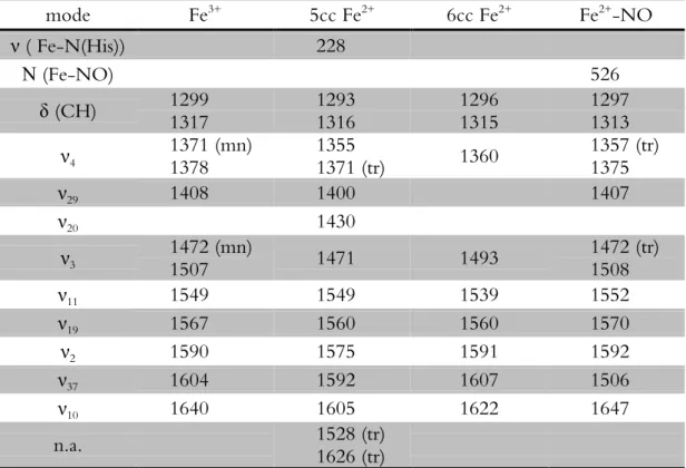
Table 2.2 Rate constants for the binding of NO to ferrous haemproteins ...34 Table 2.3 Vibrational modes of cytochrome c” in the ferric, ferrous and ferrous NO-bound state ...37
Table 2.4 Resonance Raman Frequencies for 5-coordinated nitrosyl adducts ...39 Table 2.5 Standard free energies of binding and of the red5cc-red6cc transition in
cytochrome c” ...41
Table 2.6 Standard free energies for the binding of NO, CO and CN- to different
haem proteins ...51
Table 3.1 Oligonucleotide primers used in cloning and site-directed mutagenesis of
cytochrome c” ...63
Table 3.2 Second order rate constants for the reduction of native cytochrome c” ....70
Table 3.3 Second order rate constants for the reduction of cytochrome c” mutants .74
Table 3.4 Effective protein charge ...77
Table 3.5 Rate constants for reduction of cytochrome c” at infinite ionic strengt ....77
Table 3.6 Reduction potentials of cytochrome c” at infinite ionic strength. Ratios of
the ET rate constants calculated from the kinetic results and the Marcus theory ...78
Table 4.1 Mole fractions of the oxidation stages of type I cytochrome c3 ...95 Table 4.2 Fitted matrices ( 12
0 2 1 0 LM
C
HAPTER
1
Introduction 3
C
ONTENTS
HAEM PROTEINS ... 3
CYTOCHROME C” ... 6
TETRAHAEM CYTOCHROMES C
3 ... 10
FURTHER SIGNIFICANCE OF MULTIHAEM PROTEINS ... 15
H
AEM PROTEINS
Haem proteins are one of the most versatile groups of proteins in nature. Found in all forms of life, they are involved in many different types of reactions such as electron transfer, oxygen transport and storage, oxygen reduction, nitric oxide production, sensing and transport, oxygenation, and reduction of peroxides. Most of this versatility arises from the presence of an iron-porphyrin prosthetic group – the haem. The nature of the haem, the presence or absence and the nature of the axial ligands to the iron atom, and the effect of the polypeptide chain of the protein on its surroundings all contribute to this diversity of properties. Of the different types of haem (Figure 1.1) the most common is haem b. It is present for example in
haemo-globin and myohaemo-globin, with a single axial iron ligand, where it serves as the binding site for oxygen. It is also present in catalase where it is part of the active site, assisting in the conversion of hydrogen peroxide into oxygen and water. Haem c, which
appears in most electron transfer proteins with two axial iron ligands, is very similar to haem b but is covalently attached to the polypeptide chain via thioether bonds. Other less common haems include, among others, haem a, present in cytochrome c oxidase,
character-4 Chapter 1
ized by the presence of saturated bonds in the porphyrin ring. In regard to the axial ligands, most haem proteins have at least one histidine coordinated to the haem. The sixth ligand is most commonly another histidine or a methionine, but asparagines (Leys et al. 2000), cysteines (Meier et al. 2001; Bamford et al. 2002), tyrosines (Fulop
et al. 1995; Arnoux et al. 2000) and others have also been observed. In some cases, the
sixth coordination site may also be empty or occupied by a water molecule. Not surprisingly, the nature of the aminoacid residues around the haem also contributes to the properties of the haem protein as different interactions can stabilize or destabilize reagents, products or intermediates.
Figure 1.1 (A) Different types of haem groups. (B) Porphyrin numbered according to
Introduction 5
Cytochromes c are a particular
group of haem proteins that are mainly involved in the transfer of electrons in respiratory pathways, both aerobic and anaerobic, covering a reduction potential range from -400 to +400 mV (Figure 1.2). They contain one or more covalently attached haems c and can be soluble or
membrane associated. The number of haems per protein can range from just one to more than twenty. The reason for the increase in the number of haems seems to be efficiency, since it not only maximises the likelihood of a successful electron
transfer, but also allows the transfer of more electrons at the same time. Moreover, the presence of more than one haem enables the occurrence of cooperativity, where the redox state of one centre influences the reduction potential of other centres, allowing the fine tuning of the reduction potential in order to optimize the interaction with other electron transfer partners in different physiological situations (Santos et al.
1984a). In this work two different kinds of cytochromes c were studied: the
single-haem cytochrome c” and a pair of tetrahaem cytochromes c3. The objective is to use
these cytochromes as models to further understand the mechanisms and functions of these proteins.
Rhodospirillaceaecytc2
Paracoccus denitrificanscytc550
Chlorobium limicolacytc555
Nitrosomonas europaeacytc551
Methylophilus methylotrophus cytc”
Desulfovibrionaceaecytc3
-400 -300 -200 -100 0 100 200 300 400
Em,7(mV)
Figure 1.2 Reduction potential range
6 Chapter 1
C
YTOCHROME
C
”
Cytochrome c” is a 14.2 kDa periplasmic protein isolated from the obligate
aerobe Methylophilus methylotrophus (Santos and Turner 1988). This organism is a
restrictive facultative methylotroph, meaning that it is capable of using single-carbon molecules like methanol or methylamines as both carbon and energy sources, but is also able to utilize a limited range of other organic compounds, such as glucose and fructose (Jenkins et al. 1987). It is considered an important organism for industrial
applications and has been used in the production of Single-Cell Protein since the 1980s (Windass et al. 1980). Other possible applications include the production of small metabolites and the isotope labelling of proteins or nucleic acids (Batey et al.
1996), with the main advantage of utilizing methanol, a relatively inexpensive substrate.
Cytochrome c” is a monomer with 124 aminoacid residues and a single haem
covalently bound via thioether bonds to cysteines 49 and 52. An extra pair of cysteines (96 and 104) forms a disulfide bond not far from the haem, which confers rigidity to the structure (Brennan et al. 2001) (Figure 1.3).
In the oxidised state the haem is coordinated by two histidinyl residues (H53 and H95) in a near-perpendicular orientation (Berry et al. 1990), but upon reduction
Introduction 7
upon reduction that has been attributed to the detached histidine; and one in which the pKa decreases from 6.4 to 5.4 that has been attributed to haem propionate 17. Therefore, a mechanism was proposed where, upon reduction at physiological pH, the detached histidine picks up a proton, resulting in no change in the charge around the haem. The movement of the detached histidine closer to the haem propionate, increasing the local charge and slightly lowering the pKa, can explain the
anti-electrostatic process observed in the propionate.
Figure 1.3 Structure of oxidised cytochrome c” (pdb: 1E8E (Brennan et al. 2001)). In red the
8 Chapter 1
Figure 1.4 UV-visible spectra of cytochrome c” oxidised (black), reduced 5-coordinated
(red) and reduced 6-coordinated (green) (Santos and Turner 1988; Brennan et al. 2001). The reduced 6-coordinated protein was obtained at pH 11.8 in the presence of 5 mM sodium dithionite.
Figure 1.5 pH dependence of the reduction potential of M. methylotrophus cytochrome c” at
25 °C. The dashed line is the best fit to a theoretical model that considers a single ionizing group. The solid line represents the best fit to a model that assumes two distinct and non-interacting ionizing groups influencing the reduction potential. Adapted from (Costa et al.
1992).
0 0.1 0.2 0.3 0.4
350 400 450 500 550 600
Wavelength (nm)
A
bs
or
ba
nc
e
0 0.02 0.04 0.06
460 510 560 610
Wavelength (nm)
A
bs
or
ba
nc
Introduction 9
It has been previously reported that cytochrome c” was able to bind cyanide in
both redox states, but only at high pH, and carbon monoxide at physiological pH, but only in the reduced form (Santos and Turner 1988). Nitric oxide binding had never been tested. Therefore, because of the growing importance of this small molecule and the similarities between cytochrome c” and cytochrome c’, which has been implicated
in NO metabolism in Paracoccus denitrificans (Moir 1999) and has been used as a model
to understand the mechanism of soluble guanylate cyclase, the human sensor for NO, we went on to study the binding of this ligand in detail as well as quantifying the binding of the other small molecules. Cytochrome c” proved to be highly selective for
NO when compared to other haem proteins. The binding of CO and CN- was rather
challenging because of the very low affinities of these ligands to cytochrome c”. This
limitation was overcome by performing the experiments at higher temperature, higher pH or in the presence of a high concentration of urea. However, a severe complication arises from the disulfide bond because of its tendency to become reduced when the haem iron was reduced in the conditions necessary for the binding, hence altering all of the properties of the cytochrome. The results of these studies are presented in chapter 2 of this thesis.
A previous study on the kinetics of reduction of cytochrome c” has shown that
the rate of reduction decreases with the pH (Coletta et al. 1997), as does the reduction potential (Figure 1.5). This result led us to investigate whether this decrease in the rate was due to changes in the electrostatic properties of the protein as was suggested or rather to the observed decrease in the driving force, which, according to the theory of Marcus, is one of the factors controlling the rate of electron transfer (Marcus and Sutin 1985). In fact, the theory of Marcus is the basis of the model used in the study of multihaem cytochromes (Catarino and Turner 2001). The changes in the driving force are used to obtain the kinetic parameters, and the method would fail if surface charge was a more important factor. Using cytochrome c” as a model it was
10 Chapter 1
reduction rates than the change in electrostatics, validating the approach used in multihaem cytochromes. Results are presented in chapter 3 of this thesis.
T
ETRAHAEM CYTOCHROMES
C
3Cytochromes c3 are a group of tetrahaem cytochromes present in
sulphate-reducing bacteria. These organisms are considered one of the oldest forms of bacterial life on earth (Barton and Fauque 2009). They are found in anaerobic environments, both aquatic and terrestrial and are located at the end of the microbial food chains, thus performing the important environmental role of further oxidizing the fermenta-tion products from other microorganisms (Muyzer and Stams 2008). They couple the oxidation of these small organic compounds, such as lactate or pyruvate, and molecu-lar hydrogen to the reduction of sulfate to sulfide (Hansen 1994). Sulfate-reducing bacteria are also industrially relevant for distinct reasons. On the one hand they are a source of major concern to industries like the petro-chemical because of the corrosion to the metal surfaces caused by sulfides (Hamilton 2003; Coetser and Cloete 2005). On the other hand they can also be applied beneficially in bioremediation, such as the removal of heavy metals from waste water (Lovley 2001).
The first cytochrome c3 was isolated in 1954 from Desulfovibrio vulgaris
(Ishimoto et al. 1954; Postgate 1954), but several others were later isolated from other
members of the Desulfovibrionaceae family. They are small (~15 kDa) and very stable
globular proteins. The polypeptide chains comprise 102-118 aminoacid residues and the four c-type haems have bis-histidinyl coordination. Despite low homology in aminoacid sequence (Magro et al. 1997), the general folding and the haem
architec-ture is strictly conserved (Matias et al. 2005) among them. Furthermore, the soluble cytochromes c3, called type I (TpIc3), possess a conserved patch of positive charges
Introduction 11
in the periplasm and are proposed to play a role in the hydrogen metabolism in sulphate-reducing bacteria (Matias et al. 2005) (Figure 1.6).
Figure 1.6 Proposed hydrogen metabolism in Desulvovibrio africanus. Hase: Hydrogenase;
12 Chapter 1
Molecular hydrogen is converted to electrons and protons by periplasmic hydrogenases. The resulting protons are released to the periplasm but the electrons are transferred to the transmembrane electron transfer complex and eventually are used in the reduction of the terminal electron acceptor, sulphate. This process generates a proton gradient across the membrane that is used to produce ATP. In the membrane, the electron acceptor is thought to be one of the multihaem cytochromes associated with the transmembrane complexes. Several different cytochromes have been identified so far, including a sixteen-haem cytochrome in D. vulgaris Hildenborough (Pollock et al. 1991), D. vulgaris Miyazaki (Ogata et al. 1993) and Desulfovibrio gigas
(Chen et al. 1994); a nine-haem cytochrome in Desulfovibrio desulfuricans ATCC 27774
(Liu et al. 1988; Matias et al. 1999), and a tetrahaem cytochrome c3, denoted type II
(TpIIc3), in Desulfovibrio africanus (Pieulle et al. 1996), D. vulgaris Hildenborough
(Valente et al. 2001) and D. gigas (Di Paolo et al. 2006). TpIIc3 are structurally similar
to the periplasmic ones but lack the lysine patch (Nørager et al. 1999). (Figures 1.7 and 1.8) Perhaps because of the absence of a lysine patch, TpIIc3 shows low reactivity
with hydrogenase, but the presence of TpIc3 increases the rate of electron transfer
between those two proteins (Pieulle et al. 1996). Hence, it is proposed that TpIc3 and
TpIIc3 are physiological partners, with TpIc3 receiving electrons from hydrogenase and
then delivering them to TpIIc3 associated to the transmembrane complex. However,
despite evidence of complex formation between TpIc3 and TpIIc3 (Pieulle et al. 2005),
Introduction 13
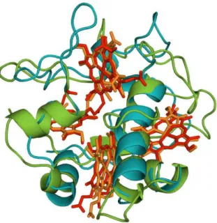
Figure 1.7 Superimposed 3-dimensional structures of TpIc3 (green, red haems, pdb: 2BQ4 (Pieulle et al. 2005)) and TpIIc3 (blue, orange haems, pdb: 3CAO (Nørager et al. 1999)).
Figure 1.8 Surface charge at pH 7 of TpIc3 (left, pdb: 2BQ4 (Pieulle et al. 2005)) and TpIIc3 (right, pdb: 3CAO (Nørager et al. 1999)). Positive charges are in blue and negative charges
14 Chapter 1
Due to their tightly packed haems, with distances of 11-18 Å between the iron atoms, the redox properties of each haem are affected by neighbouring ones (homotropic cooperativity). Moreover, the protonation state of some ionisable groups also interferes with the redox properties of the haem centres (heterotropic cooperativ-ity, also known as the redox-Bohr effect). The thermodynamic properties of several TpIc3 have been studied in detail (Turner and Catarino 2012). In all cases, cooperative
processes have been observed. At least one ionisable group is affected by the redox state of the protein with its pKa increasing with the reduction and decreasing with the
oxidation. This positive redox-Bohr effect (rB+) is easily explained by electrostatics
since it involves charges of opposite signs. Also easily explained by electrostatics is negative homotropic cooperativity, where the reduction of one haem makes the reduction of another haem less favourable, in this case because the charges have the same sign. However, most cytochromes c3 have one haem-haem interaction in which
the reduction of one haem facilitates the reduction of the other. These positive homotropic cooperativities must involve some conformational changes in the prox-imity of the haems, presumably a decrease in the exposure to the solvent, which stabilises the reduced state (Messias et al. 1998). This increased affinity for a second
electron following the uptake of the first one is the basis for the proposed concerted two-electron transfer step, i.e. the transfer of two electrons during the same
encoun-ter, that would efficiently channel the electrons from hydrogenase to the membrane acceptor.
In some TpIc3, the redox-Bohr group appears to be critical for a two-electron
transfer. In the fully oxidised state, the pKa of the ionisable centre is sufficiently low
that it is deprotonated at physiological pH. Upon receiving electrons from the periplasmic hydrogenase, the pKa increases, leading to the capture of one proton. This
in turn increases the reduction potential of the haems resulting in further reduction. The whole process is reversed at the membrane, with the pKa becoming lower as the
Introduction 15
ionisable centre is deprotonated. This results in energy transduction by converting weakly acidic protons into more acidic protons by dissipation of the reducing power of the coupled electrons. This mechanism has been called “proton thrusting” (Louro 2007).
Although the proteins possess the necessary thermodynamic properties, the two-electron transfer step has never been observed experimentally. Therefore, we sought to determine the nature of electron transfer between D. africanus TpIc3 and the
soluble D. africanus TpIIc3. We found that they are likely physiological partners but
the two-electron transfer step could not be observed, however it should not be ruled out. The results are presented in chapter 4.
F
URTHER
S
IGNIFICANCE OF MULTIHAEM
PROTEINS
The interest surrounding organisms that are able to perform extracellular reduction of insoluble metal oxides has been growing tremendously. These organisms couple the oxidation of electron donors to the reduction of extracellular electron acceptors. The acceptors can be large organic molecules (Lovley et al. 1996) or soluble
metal chelates (Coppi et al. 2007; Shelobolina et al. 2007), but those of greatest
interest are insoluble metal oxides, such as FeIII and MnIV oxides (Lovley
et al. 2004).
Coupling the oxidation of toxic organic compounds to the reduction of these metals could be an important method for bioremediation (Lovley et al. 1989; Lovley 1997).
Moreover, if the metal is on the surface of an electrode, it could be a way of generat-ing electricity (Lovley 2006a; Lovley 2006b). Organisms capable of extracellular metal reduction include the Gram-negative genera Geobacter and Shewanella (Shi et al. 2007), and the Gram-positive Thermincola potens (Carlson et al. 2012). Understanding how
16 Chapter 1
electron transport chains that transfer the electrons from the inner membrane to the extracellular medium.
C
HAPTER
2
H
IGHLY SELECTIVE LIGAND BINDING BY
M
ETHYLOPHILUS METHYLOTROPHUS
CYTOCHROME
C
”
Results published in:
Quintas, P. O., Catarino, T., Todorovic, S., and Turner, D. L. (2011) Highly selective ligand binding by Methylophilus methylotrophus cytochrome c", Biochemistry
50, 5624-5632.
Highly selective ligand binding by Methylophilus methylotrophus cytochrome c"
19
C
ONTENTS
ABSTRACT ... 20
INTRODUCTION ... 20
MATERIALS AND METHODS ... 23
Expression and purification of cytochrome c” ... 23
Binding of small molecules. ... 25
Effect of temperature ... 26
Effect of urea ... 27
Effect of DTT ... 28
Kinetic experiments ... 28
Resonance Raman Spectroscopy ... 29
RESULTS AND DISCUSSION ... 30
Binding of NO to cyt c” ... 30
Binding of CO and CN- to cyt c” ... 41
Effect of temperature in the reduced form ... 43
Effect of urea in the reduced form ... 45
20 Chapter 2
A
BSTRACT
Cytochrome c” (cyt c”) from Methylophilus methylotrophus is unusual insofar as
the haem has two axial histidine ligands in the oxidised form but one is detached when the protein is reduced. Despite having an axial site available for binding small ligands, we show here that only NO binds readily to the ferrous cyt c”. Binding of
CO, as well as CN-, on the other hand requires considerable structural reorganisation,
or reduction of the disulfide bridge close to the haem. Standard free energies for the binding of NO and CO reveal high selectivity of the ferrous cyt c” for NO, indicating
a possible physiological role. In this work we characterize in detail the kinetics of NO binding and the structural features of the Fe2+-NO adduct, by stopped-flow and
resonance Raman spectroscopy, respectively.
I
NTRODUCTION
Gaseous diatomic molecules are found in a wide variety of both unicellular and multicellular organisms. Their functions are usually related to the metabolism or signal transduction and vary depending on the organism. O2 is mostly recognized as
Highly selective ligand binding by Methylophilus methylotrophus cytochrome c"
21
molecule, much like NO, in processes of vasodilation and neurotransmission. It has also been involved in anti-inflammatory and anti-proliferative processes (Gullotta et al.
2012). Several haem proteins have been associated with the sensing, transport and synthesis of these molecules.
Binding of gaseous diatomic molecules to haem proteins is, therefore, of utmost importance for living cells. The haem group is indeed engineered to provide optimized binding of O2, NO and CO (i.e. XO), via effective back-donation of Fe(II)
dπx electrons to low-lying π* orbitals of these diatomic molecules (Soldatova et al.
2010). However, despite their almost identical size and shape, some haem proteins are able to discriminate these ligands. Selectivity is thought to be achieved by regulating the relative binding affinities to the ligands through electrostatic interactions with the nearby aminoacids and/or steric hindrance (Jain and Chan 2003). One example of this happens in haemoglobin and myoglobin (Springer et al. 1994; Olson and Phillips
1997). The haem is 5-coordinate, with a histidinyl residue as the proximal axial ligand. Despite the empty distal position, and the higher affinity of CO to the free haem, myoglobin is able to discriminate in favour of O2 because of the way the haem
pocket is organized. The presence of a histidine residue in the distal side of the haem is significant because it reduces the space available for ligand binding. CO usually binds perpendicularly to the haem while O2 binds at an angle, so, by hindering the
space on the distal side of the haem, it becomes harder for CO to bind. In fact, the binding of CO happens because of an opening of the haem pocket and not a tilting of the ligand. Moreover, the presence of this histidine residue helps stabilize the binding of the O2 molecule by electrostatic interactions, and thus increasing its affinity. Other
examples exist, such as the O2 sensor FixL (Gong et al. 1998; Gong et al. 2000). While
steric hindrance does not seem to play a role, the electrostatic interaction between O2
22 Chapter 2
the binding of one ligand triggers a response is the soluble guanylate cyclase (Zhao et al. 1999). In this case only NO activates the protein. The haem is coordinated by one
histidine residue and CO binds to the distal side. However, NO seems to bind to the proximal side, displacing the histidine, and this may be the trigger for its activation. A similar process happens in CooA (Aono et al. 1996; Reynolds et al. 2000), a CO
sensor, where the CO-bound form is 6-coordinated and the NO-bound form is 5-coordinated, but only the former activates the protein. The mechanisms that allow haem proteins to discriminate between diatomic ligands are therefore of considerable interest.
Here we have investigated the ligand selectivity by cytochrome c” (cyt c”), a
soluble monohaem protein isolated from the obligate aerobe Methylophilus methylotrophus that undergoes a redox-linked spin-state transition from low-spin (LS)
in the oxidised form to high-spin (HS) in the reduced form (Santos and Turner 1988) (see Figure 1.4). The axial ligands are two histidinyl residues with a near perpendicu-lar orientation in the oxidised form and a single proximal histidinyl residue in the reduced form (red5cc) (Berry et al. 1990). Previous work has also shown that
reduc-tion and alkylareduc-tion of a disulfide bridge located near the haem led to a 6-coordinated reduced form (red6cc), presumably with the previously detached histidine coordinated back to the haem iron (Brennan et al. 2001). So far, the physiological function of this cytochrome has not been determined, however, because of its ability to couple electron and proton transfer (Costa et al. 1992), it has been suggested that it could
serve as a model for more complex systems, such as cytochrome c oxidase (Xavier
2002).
In this work we addressed the ability of cyt c” to bind diatomic ligands.
Typi-cally, CO and O2 bind to ferrous haem, since their π* orbitals do not match the
contracted dπ orbitals in ferric proteins (Soldatova et al. 2010). NO is able to bind to
Highly selective ligand binding by Methylophilus methylotrophus cytochrome c"
23
binds CO only upon major conformational rearrangement, brought on by high temperature or pH, in the presence of high concentrations of urea, or upon reduction of the disulfide bond. Under these conditions, the protein undergoes a spin state transition that results in a red6cc species (Figure 1.4) which binds CO and CN- more
readily. Moreover, ferrous cyt c” does not bind O2, and the ferric protein does not
bind NO. Nevertheless, NO is able to bind to ferrous cyt c” at physiological pH and
temperature.
Binding of CO, CN- and NO, followed by UV-visible spectroscopy, was
performed in order to obtain binding affinities of those diatomic molecules for cyt c”.
Since CO and CN- do not bind at physiological pH and temperature, these
parame-ters, along with the concentration of urea, were varied so that the binding was achieved. The results were then extrapolated to physiological conditions for compari-son. The NO binding, as the only one that occurs at physiological conditions, was analysed further. Stopped-flow experiments were used to obtain the kinetic rate constants for the reaction and to understand its mechanism. In order to assess the structural implications of the binding of NO and evaluate the proposed mechanism, resonance Raman spectra of the NO adduct were also obtained. Detailed comparison of standard free energies for the binding of NO and CO revealed high selectivity of the ferrous cyt c” for NO, leading us to propose that it may be functionally relevant.
M
ATERIALS AND METHODS
E
XPRESSION AND PURIFICATION OF CYTOCHROME C”
The plasmid containing the cyt c” gene (pHS1) (Price et al. 2000) was
trans-formed into Escherichia coli strain BL21(DE3) harbouring plasmid pEC86 encoding
genes for cyt c maturation (Arslan et al. 1998). Plasmid pHS1 confers resistance to
24 Chapter 2
chloramphenicol. Cells were grown aerobically at 37 °C and 250 rpm in LB medium. To induce expression, IPTG was added to a concentration of 350 μM at a cell density of OD600 nm = 3. IPTG blocks the action of the lac repressor, allowing the
transcrip-tion of the genes under the control of the lac promoter, such as the one encoding for cyt c” in pHS1. After 3-4 hours cells were harvested by centrifugation (6500 ×g, 20
min, 10 °C). Periplasmic fractions were isolated by osmotic shock, by resuspending the cells in 160 mL of 20% sucrose in 30 mM Tris-HCl buffer (pH 7.5) containing 1 mM EDTA per litre of culture, followed by centrifugation (12500 ×g, 20 min, 4 °C). The pellet was then resuspended in 60 mL of cold 5 mM MgSO4, shaken for 10 min
in an ice bath and centrifuged in the same conditions.
Cyt c” was purified from the resulting supernatant by column chromatography
in three steps: (i) anion exchange on a Q-Sepharose column (GE Healthcare) equili-brated with 5 mM Tris-HCl pH 7.6 (cyt c” does not adsorb to this column and elutes
with the equilibrating buffer); (ii) cation exchange by applying the red fractions from (i) to an SP-Sepharose column (GE Healthcare) equilibrated with 5 mM Tris-HCl pH 7.6 and eluted with a salt gradient of 0-1M NaCl prepared in the same buffer; (iii) fractions containing cyt c” were concentrated by ultrafiltration (Amicon) and loaded
Highly selective ligand binding by Methylophilus methylotrophus cytochrome c"
25
Figure 2.1 SDS-PAGE gel of the purification of cytochrome c”. The lanes correspond to (from left to right): the molecular weight markers (lowest to highest: 14, 20, 30, 45, 66 and 97 kDa), the pool of fractions collected from the Q-Sepharose column, the SP-Sepharose column, and the Superdex-75 column (pure fractions).
B
INDING OF SMALL MOLECULES.
The binding of nitric oxide (NO) was followed by optical absorption spectros-copy in a Shimadzu UV-1203 spectrophotometer placed inside an anaerobic chamber (<2 ppm of O2). Because it may react with the NO releasing compound or NO itself,
care was taken to minimise the excess of sodium dithionite used to reduce cyt c”. The
addition of NO was performed by injecting a small volume of a diethylamine NONOate solution in 10 mM NaOH with a gas-tight syringe ([NONOate]final = 50
μM). The protein sample was prepared in 100 mM phosphate buffer (pH 7.5). At this pH, each NONOate molecule releases 1.5 molecules of NO (t½ = 16 min, at 25 °C,
pH 7.4). The amount of NO present in solution at each time was obtained by studying the decay of the NONOate peak under the same conditions (ε250nm = 6.5
mM-1 cm-1) (Keefer
26 Chapter 2
The binding of carbon monoxide (CO) and cyanide (CN-) was followed by
optical absorption spectroscopy on a Shimadzu UV-1603 spectrophotometer, using a quartz optical cell with a path-length of 10 mm, sealed with a silicone septum. Samples were prepared by injecting a few microliters of a concentrated protein solution into a degassed solution of the desired buffer. Reduction of the haem was achieved by adding excess sodium dithionite solution with a gas-tight syringe. In order to reduce the disulfide bond, 5 mM dithiothreitol (DTT) was added to some samples. The addition of CN- was performed by injecting a small volume of a
potassium cyanide solution in the same buffer as the sample ([CN-]
final = 10 mM). The
addition of CO was achieved by replacing argon in the headspace of the cuvette containing the protein sample with CO from a gas cylinder: this results in approxi-mately 1 mM of CO dissolved at 20 °C (Stephen et al. 1963).
In each case, the amount of bound and unbound protein was determined by treating each spectrum as a weighted sum of the spectra of the two pure forms (Figure 2.2).
E
FFECT OF TEMPERATUREThe temperature was controlled by a circulating water bath connected to the spectrophotometer. Samples were prepared in degassed 100 mM phosphate buffer (pH 7.5). After the addition of the ligand, the temperature was increased until full binding was achieved (approximately 60 °C for CO and 70 °C for CN-) and the
Highly selective ligand binding by Methylophilus methylotrophus cytochrome c"
27
Figure 2.2 Deconvolution of the UV-visible spectrum during CO binding experiments. The
experimental spectrum (grey) can be deconvoluted into a fraction of the pure CO-bound (blue) and reduced unbound forms (red), in this case 44% and 56%, respectively. Adjustment was performed by the least squares method. The residual is the difference between the spectrum resulting from the deconvolution (black) and the experimental spectrum (grey). The same strategy can be used to study any conversion between different forms.
E
FFECT OF UREASamples were prepared in degassed 100 mM phosphate buffer (pH 7) with various urea concentrations up to 6 M. Reversibility of the transition was studied by removing urea with a HiTrap desalting column (GE Healthcare). The effect of urea on the protein without any ligand was also studied using samples prepared in 100 mM phosphate buffer (pH 7.0) and 100 mM glycine/NaOH buffer at various pH values from 8.7 to 11.8.
-0.02 -0.01 0 0.01 0.02 R esi d u al 0 0.05 0.1 0.15 0.2 0.25 0.3
350 400 450 500 550 600
A bs or ba nc e Wavelength (nm) 0 0.005 0.01 0.015 0.02
480 510 540 570 600
28 Chapter 2
E
FFECT OFDTT
In addition to binding experiments in the presence of DTT, protein samples treated with 5 mM DTT were titrated with reduced methyl viologen to determine the state of the disulfide bond. Untreated protein was titrated as a control, with 6 M urea as a denaturant in each case. A stock solution of methyl viologen was reduced by zinc under an anaerobic atmosphere and its concentration was obtained from its UV-visible spectrum, using the value of absorbance at 605 nm and the extinction coeffi-cient of 13.7 mM-1
cm-1
(Watanabe and Honda 1982). 20 μL aliquots were added to the protein sample, followed by recording the UV-visible spectrum. The spectra recorded after each addition of methyl viologen were deconvoluted to obtain the contributions of the oxidised protein, the red5cc form, and the red6cc form, which are illustrated in Figure 1.4. Deconvolution was performed as shown in Figure 2.2, using three spectra instead of two.
K
INETIC EXPERIMENTSStopped-flow kinetic experiments were performed at a constant temperature of 25 °C using a SF-61 DX2 stopped-flow apparatus (Hi-Tech Scientific) placed inside an anaerobic chamber (<2 ppm of O2). Solutions of reduced protein and NO
Highly selective ligand binding by Methylophilus methylotrophus cytochrome c"
29
range). All reactions were performed under pseudo-first order conditions ([NO] >> [protein]) and traces were fitted with exponentials, or biexponentials when two processes were observed. Second order rate constants were obtained by measuring pseudo-first order rate constants as a function of [NO].
R
ESONANCER
AMANS
PECTROSCOPYAll RR measurements were performed with a confocal microscope coupled to a Raman spectrometer (Jobin Yvon U1000) equipped with 1200 l/mm grating and liquid-nitrogen-cooled back-illuminated CCD detector. Samples were placed in a quartz rotating cell and excited with the 413 nm line from a krypton ion laser (Coherent Innova 302). Oxidised (as purified) and sodium dithionite reduced cyt c”
samples (100-200 μM) were measured at room temperature in the presence or absence of NO with a laser power of 8 mW and accumulation times of 60 s. The samples of reduced cyt c” in the presence of NO were prepared in an anaerobic
chamber (<2 ppm of O2) by the addition of excess NONOate to the cell filled with
the protein solution in 10 mM Tris-HCl buffer, pH 7.6. UV-visible spectra were recorded after the Raman experiments to confirm the state of the sample.
30 Chapter 2
R
ESULTS AND DISCUSSION
B
INDING OFNO
TO CYT C”
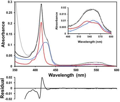
Binding of NO to ferrous cyt c” was followed via the characteristic blue shift of the Soret band (Figure 2.3 and Table 2.1), observed upon addition of small aliquots of NO releasing diethylamine NONOate to dithionite reduced protein. The UV-visible spectra were measured and in each case, the amount of NO-bound and free protein was determined by treating each spectrum as a weighted sum of the spectra of the two pure forms with electronic transitions at 398 and 426 nm.
At 20 °C and pH 7.5, NO binds to the reduced cyt c”, forming a 1:1 complex
with a dissociation constant of 0.37 μM, which corresponds to a ∆G° for the binding of -36.1 kJ/mol (Figure 2.4). The UV-visible spectra in the presence of excess NO show no evidence of further reaction such as nitrosylation of the cysteines. Binding is fully reversible since removal of NO results in the UV-visible spectra of the ligand-free native HS state of ferrous cyt c”.
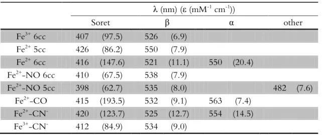
Table 2.1 UV-visible spectroscopic properties of cyt c” and its adducts.
λ (nm) (ε (mM-1 cm-1))
Soret β α other
Fe3+ 6cc 407 (97.5) 526 (6.9)
Fe2+ 5cc 426 (86.2) 550 (7.9)
Fe2+ 6cc 416 (147.6) 521 (11.1) 550 (20.4)
Fe2+
-NO 6cc 410 (67.5) 538 (7.9)
Fe2+-NO 5cc 398 (62.7) 535 (8.0) 482 (7.6)
Fe2+-CO 415 (193.5) 532 (9.1) 563 (7.4)
Fe2+-CN- 420 (123.7) 525 (12.7) 554 (14.5)
Highly selective ligand binding by Methylophilus methylotrophus cytochrome c"
31
Figure 2.3 (A) UV-visible spectra of oxidised cyt c” bound to cyanide (black) and reduced
cyt c” bound to cyanide (red) and carbon monoxide (green). (B) UV-visible spectra of reduced cyt c” in its native 5cc form (red) and reduced cyt c” bound to nitric oxide in the 6cc
form (black) and 5cc form (green). The shoulder at ~400 nm in the spectrum of the 6cc form arises from imperfect deconvolution.
Figure 2.4 Binding curve of NO to reduced cyt c” (100 mM phosphate buffer pH 7.5,
T = 20 °C, [c”] = 2.5 μM). The NO-bound fraction accounts for the concentration ratio of
NO-bound cyt c” to total cyt c”. The curve results from the fit to the experimental points of a
rearrangement of the equation Kd = [NO]free[unbound-cytc”]/[bound-cytc”]. The ∆G° value is obtained from ∆G° = RTln(1/Kd).
0 0.1 0.2 0.3 0.4 0.5
350 400 450 500 550 600
A bs or ba nc e Wavelength (nm) 0 0.01 0.02 0.03 0.04
460 510 560 610
A bs or ba nc e Wavelength (nm) 0 0.1 0.2 0.3 0.4
350 400 450 500 550 600
A bs or ba nc e Wavelength (nm) 0 0.01 0.02 0.03 0.04
460 510 560 610
A bs or ba nc e Wavelength (nm) A B 0 0.2 0.4 0.6 0.8 1
0 5 10 15 20
NO -bound f ra c ti on
32 Chapter 2
In the next step, stopped-flow experiments were carried out to study the kinetics of the reaction of reduced cyt c” with NO. An excess of ligand was used to ensure that the reaction occurred under pseudo-first-order conditions.
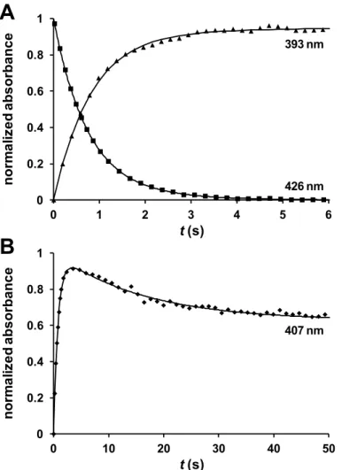
The reaction was followed by measuring the change in absorbance with time at 393, 407 and 426 nm, chosen by inspection of spectra obtained with the diode array, and corresponding to different mixtures of the NO-bound 5-coordinated (5cc), NO-bound 6-coordinated (6cc) and red5cc forms. The time-course traces show an increase in the absorbance at 393 nm and a decrease at 426 nm, while at 407 nm an increase is observed followed by a decrease (Figure 2.5). This suggests a mechanism in which NO binds to the reduced cyt c”, forming a 6cc NO-Fe-His intermediate, that is then converted to a 5cc NO-Fe form, with the detachment of the axial histidine. Each trace was fitted with a sum of two exponentials, as appropriate for consecutive reactions, to obtain first-order rate constants k1 and k2 for the NO binding and
histidine detachment, respectively. The rates show no dependence on the pH in the 6.5-7.8 range. Note that it is not necessary for the traces to represent pure forms in order to extract the rate constants; in fact the wavelengths used here have contribu-tions from more than one form. No change in the rates was observed when traces obtained at different wavelengths were used for the fitting.
The second-order rate constant for the binding of NO was determined from the slope of the straight line obtained by plotting the values of k1 at different NO
concentrations (Figure 2.6). The value obtained for kon (5.7 × 10³ M-1 s-1) is lower
than that determined for cytochrome c’ (cyt c’) and much lower than the ones for
myoglobin and soluble guanylate cyclase (sGC) (Table 2.2) (Moore and Gibson 1976; Makino et al. 1999; Andrew et al. 2002). The second process is clearly NO
inde-pendent, with a rate constant k2 of 0.06 ± 0.01 s
-1, suggesting that NO does not bind
to the proximal side of the haem, contrary to what was observed in the case of cyt c’
Highly selective ligand binding by Methylophilus methylotrophus cytochrome c"
33
Figure 2.5 Time-course traces at 393 and 426 nm (A) and 407 nm (B) for the reaction of 3
μM cyt c” with 300 μM NO (100 mM phosphate buffer pH 7.0, T = 25 °C). Solid lines represent the data fitted to the sum of two exponentials.
A
B
393 nm 426 nm 407 nm 0 0.2 0.4 0.6 0.8 10 1 2 3 4 5 6
nor m a li z e d a bs or ba nc e t(s) 0 0.2 0.4 0.6 0.8 1
0 10 20 30 40 50
34 Chapter 2
Figure 2.6 Dependence of k1 (red) and k2 (green) on the concentration of NO. Values of k1 and k2 were obtained by fitting time-course traces to a sum of two exponentials. Each point represents an average of separate fittings at different wavelengths, with the error bars repre-senting twice the standard deviation.
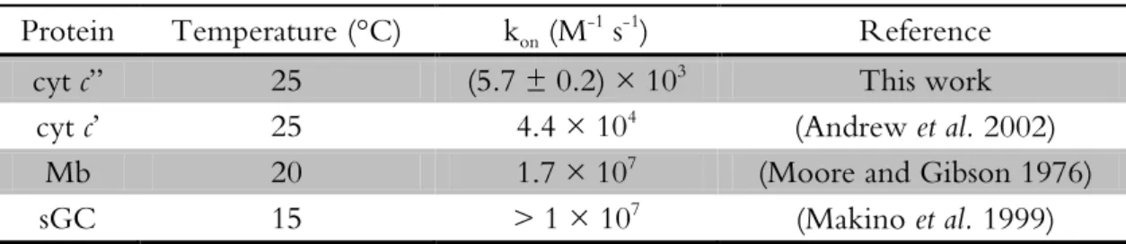
Table 2.2 Rate constants for the binding of NO to ferrous haemproteins.
Protein Temperature (°C) kon (M-1 s-1) Reference
cyt c” 25 (5.7 ± 0.2) × 103 This work
cyt c’ 25 4.4 × 10⁴ (Andrew et al. 2002)
Mb 20 1.7 × 10⁷ (Moore and Gibson 1976)
sGC 15 > 1 × 10⁷ (Makino
et al. 1999)
0 0.1 0.2 0.3 0.4 0.5
0 1 2 3 4 5 6
0 0.2 0.4 0.6 0.8 1
k
2(s
-1
)
k
1(s
-1