RESEARCH ARTICLE
The CD1/CD2 marker for specific detection of
Colletotrichum
lindemuthianum
is an iron transporter pseudogene
Pablo Gutiérrez1, Mauricio Salazar Yepes2, Juan F. Alzate Restrepo3, Katherin Vanegas Berrouet2 &
Mauricio Marín Montoya1
1Laboratorio de Microbiología Industrial y Laboratorio de Biología Celular y Molecular, Facultad de Ciencias, Universidad
Nacional de Colombia Sede Medellín, Colombia, A.A. 3840; 2Museo Micológico Universidad Nacional de Colombia, Sede
Medellín - MMUNM, Medellín, Colombia¸A.A. 3840; 3Centro Nacional de Secuenciación Genómica-CNSG, Facultad de
Medicina, Universidad de Antioquia, Medellín, Colombia, A.A. 1226
Author for correspondence:Mauricio Marín Montoya, e-mail: [email protected]
ABSTRACT
Colletotrichum lindemuthianum, the causal agent of anthracnose in common bean (Phaseolus vulgaris), is one of the most yield-limiting factors worldwide. Anthracnose affects the quality of pods by inducing black, sunken cankers and can also affect petioles, leaf veins and stems where it induces the typical anthracnose sunken lesions. A few years ago, a duplex PCR method that combines amplification of an ITS rDNA segment (CY1/CY2) together with an uncharacterized RAPD-derived amplicon (CD1/CD2) was developed for specific detection of C. lindemuthianum. This study shows that the CD1/CD2 marker corresponds to a portion of an iron permease (Ftr1) pseudogene in the vicinity of the gene encoding for a polyhydroxyproline-rich protein in Colletotrichum. Discrimination with Colletotrichum orbiculare is due to a 15 nt deletion in the CY1 annealing region. The potential of using this genomic region for phylogenetic analysis of the C. orbiculare species complex and detection of their related species is discussed.
Key words:Glomerella, Phaseolus vulgaris, anthracnose, PCR-based diagnosis.
INTRODUCTION
Colletotrichum lindemuthianum (Sacc. & Magnus)
Briosi & Cavara, the causal agent of anthracnose in common bean (Phaseolus vulgaris L.), is one of the most
yield-limiting factors for this crop in the world (Mahuku & Riascos, 2004; Schwartz et al., 2005). In the mountainous regions of South and Central America, severe anthracnose epidemics can lead to defoliation and reduce up to 95% of the crop yield when favorable weather conditions such as high humidity, moderate temperatures and high precipitation occur (Pastor-Corrales & Tu, 1989; Mahuku & Riascos, 2004; Schwartz et al., 2005). Bean anthracnose affects the quality of pods by inducing black, sunken cankers with a salmon-colored ooze centre containing millions of conidia (Melotto et al., 2000). Typical anthracnose symptoms such as the sunken lesions may also occur in petioles, leaf veins and stems (Melotto et al., 2000). In the case of infected bean seeds, they may have altered color but very often symptoms are absent, which constitute a problem when screening for disease-free material (Melotto et al., 2000; Schwartz et al. 2005). As this fungus is transmitted primarily by infected seeds and crop debris (Schwartz et al., 2005), the use of certified seeds is an important component in integrated disease management (Chen et al., 2013).
Traditionally, detection and identification of C. lindemuthianum has relied on its isolation using culture
media and the observation of fungal morphological structures that requires relatively long incubation time (10-14 days) and experience of the analyst (Chen et al., 2007; 2013). Certification of disease-free donor plants and seeds depends on the availability of accurate, sensitive and time-effective diagnostic tools such as conventional and real-time PCR (qPCR), which has been proposed to detect C. lindemuthianum quickly and reliably using species-specific
primers and or probes (Chen et al., 2007; 2013; Wang et al., 2008). DNA sequencing has also been used to differentiate closely related species within the main Colletotrichum
clades (Cai et al., 2009; Cannon et al., 2012; Damm et al., 2013). The partial annotated genomes of C. higginsianum
and C. graminicola, pathogens of maize (O’Connell et
al., 2012) and of C. orbiculare and C. gloeosporioides
isolated from strawberry (Gan et al., 2013) have identified pathogenicity-related genes.
Genomic regions most commonly used for the molecular characterization of fungi include ribosomal DNA (rDNA) and their internal transcribed spacers (ITS), mitochondrial DNA (mitDNA) (e.g. Cox1) and some protein
genes such as actin (ACT), beta-tubulin (TUB2), calmodulin
(CAL), chitin synthase I (CHS-1), manganese superoxide
dismutase (SOD2) and the translation elongation factor
1-α (EF1α) (Hyde et al., 2009; Cannon et al., 2012; Liu et al., 2013). In Colletotrichum, species definition based on
species-complexes, so a multilocus genotyping approach combining ITS with several other genes or their introns, such as the 900-bp intron of the glutamine synthetase gene and the 200-bp intron of the glyceraldehyde-3-phosphate dehydrogenase gene, is used (Liu et al., 2007; Cannon et al., 2012).
Several primers have been designed for the detection of C. lindemuthianum (Chen et al., 2007; 2013;
Liu et al., 2007; Wang et al., 2008); but their validity have been questioned and needs further confirmation because
C. lindemuthianum has high levels of genetic variation
(Apostolos et al., 2009; Pinto et al., 2012). In fact, C. lindemuthianum is a member of a clade of closely related
species that originally included C. orbiculare, C. trifolii,
and C. malvarum (Liu et al., 2007; Cannon et al., 2012).
Based on spore morphology, appressorium development, and rDNA sequence similarities, it was proposed that these species should be regarded as a single species (Sherriff et al., 1994). Recently, Damm et al. (2013), studied the phylogenetic relationships of these fungi using a multilocus genotyping approach based on ITS, glyceraldehyde-3-phosphate dehydrogenase (GAPDH), CHS-1, histone3
(HIS3), ACT, TUB2 and glutamine synthase (GS) sequences
of 42 strains of the C. orbiculare species complex.
Their results confirmed the existence of the four species previously known as belonging to this species complex (C. lindemuthianum, C. malvarum, C. orbiculare and C. trifolii)
and identified four new species: C. bidentis, C. sidae, C. spinosum and C. tebeestii.
Wang et al. (2008) developed a duplex PCR protocol for the detection of C. lindemuthianum. The first primer set,
CY1/CY2, targets differences in the ITS region common in C. lindemuthianum. The second set of primers, CD1/
CD2, was designed to amplify a DNA segment specific to
C. lindemuthianum identified through RAPD markers. This
primer amplifies a 638 bp segment and can distinguish C. lindemuthianum from C. orbiculare. Unfortunately, the
sequence of the reported target region was not published and no further characterization of this region was performed. In this study, by sequencing the CD1/CD2 amplicon and 454 pyrosequencing of one C. lindemuthianum isolate,
we demonstrate that the CD1/CD2 marker corresponds to a portion of an iron permease (Ftr1) pseudogene. The
potential of using this genomic region for phylogenetic analysis of the Orbiculare clade sensu Cannon et al. (2012)
and for the detection of their related species is discussed.
MATERIALS AND METHODS
Collection of isolates and DNA extraction
C. lindemuthianum isolates were obtained from leaves
and pods of bean plants grown in the province of Antioquia (Colombia), var. Cargamanto showing anthracnose symptoms. They were collected at the municipalities of San Vicente Ferrer (04°34′51″N 74°08′20″W, 2150 MASL; isolates A51, A53 and A93), Sonsón (05°42′44″N 75°18′50″W, 2475 MASL; isolate A77), Santa Rosa de Osos
(06°38′51″N 75°27′37″W, 2550 MASL; isolates A26, A37, A39, A76, and A90) and Urrao (006°18′55″N 76°08′03″W, 1800 MASL; isolates A47, A48 and A95). Isolate A83 from El Carmen de Viboral (06°05′06″N 75°20′19″W, 2150 MASL) previously identified as C. lindemuthianum was
used as positive control for PCR. Samples were disinfected using 1% sodium hypochlorite for 1 minute followed by a second wash using sterile distilled water. Isolates were cultured in Potato Dextrose Agar (PDA) supplemented with both penicillin and tetracycline (100 mg/L) at room temperature (20ºC-24ºC) for 5-7 days, transferred to a new PDA medium without antibiotics and identified morphologically using light microscopy.
DNA was extracted from mycelia using the CTAB method of Doyle & Doyle (1990) with modifications from Santa et al. (2012). Mycelia were obtained from liquid cultures in 2% malt extract (ME) after 16-20 days of growth at room temperature in the dark. Three to five hundred mg of mycelia macerated in liquid nitrogen were used for each extraction. The integrity of the DNA was verified by agarose gel electrophoresis (0.8%) and purity determined by absorbance readings at 260 nm and 280 nm using a Nanodrop 2000C (Thermo).
PCR amplifications
The identity of each isolate was further confirmed by PCR amplification and sequencing of the rDNA ITS region using universal primers ITS1 (5’ TCC GTA GGT GAA CCT GCG G 3’) e ITS4 (5’ TCC TCC GCT TAT TGA TAT GC 3’) (White et al., 1990). Duplex PCR was performed following the procedure and conditions reported by Wang et al. (2008) using C. lindemuthianum
specific primers CY1⁄CY2 (CY1: 5’-CTT TGT GAA CAT ACC TAA CC-3’; CY2: 5’-GGT TTT ACG GCA GGA GTG-3’) and CD1⁄CD2 (CD1: 5’-ACC TGG ACA CAT AAG TCA AAG-3’; CD2: 5’-CAA CAATGC CAG TAT CAG AG-3’). The size of the amplification products was confirmed using 1.8% agarose electrophoresis. Amplicons were purified using either the QIAquick PCR Purification or the QIAquick Gel Extraction kit (Qiagen). Sequencing of the PCR products was performed in both directions using the Big Dye Terminator kit (Applied Biosystems) in
an ABI Prism 3730XL sequencer (Applied Biosystems) at Macrogen (South Korea).
Pyrosequencing
(Medellín, Colombia). One complete PTP was sequenced for this study; reads below 50 bases were excluded from analysis.
Data analysis
Genome de novo assembly was carried out with the Newbler assembler v2.6 using the flags: -m -cpu 40 –urt. The contig containing the CD1⁄CD2 amplicon was identified using a local BLASTN search against the C. lindemuthianum
454 assembly data. Contigs containing Ftr1 homologs were
identified by a similar procedure but using a TBLASTN search and a set of Colletotrichum FTR1 proteins as query.
Gene annotation was done manually. Initially a BLASTX search against a non-redundant database at NCBI was performed. Intron positions were inferred by a multiple alignment of mRNA sequences encoding the closest Ftr1
homologs from Colletotrichum for each gene variant. GC
composition analyses were performed using a custom-made perl script that calculated the percent GC composition with a moving window of 100 bp. Sequence alignments were performed with MUSCLE (Edgar, 2004). Evolutionary analyses were conducted in MEGA5 (Tamura et al., 2011). Phylogenetic analysis relationship of sequences was inferred using the Neighbor-Joining method (Saitou & Nei, 1987) using Kimura’s two parameter model (Kimura, 1980) with 1000 bootstrap replicates (Felsenstein, 1985).
RESULTS
The CY1/CY2 and CD1/CD2 specific primers for
C. lindemuthianum were tested in 12 isolates identified at
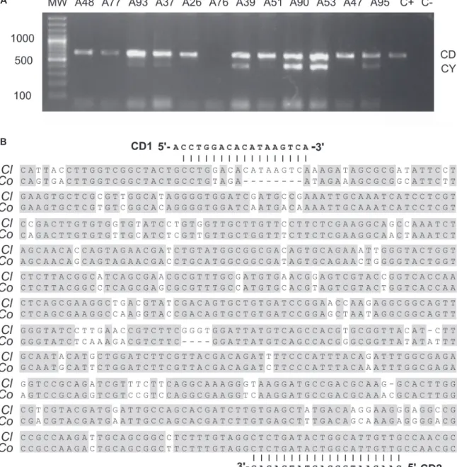
the basis of morphology and obtained at different bean-growing municipalities in the province of Antioquia, Colombia. As expected, two bands of 442 and 638 bp in size were observed after the duplex PCR in six isolates (Figure 1A). Amplification of the low molecular weight band, corresponding to the rDNA internal transcribed spacer, was not very consistent across the isolates, with repeated analyses revealing a failure to amplify in six samples, including the positive control (Figure 1A). This can probably be explained by polymorphisms at the primer binding sites in the Colombian C. lindemuthianum isolates
not observed in the original ITS data (Wang et al., 2008). The high molecular weight band was amplified in 11 isolates; the corresponding amplicon resulted in a stronger band compared to the ITS amplicon. For isolate A76, amplification failed for both primer sets, however, the duplex PCR was successful in the second trial (not shown). Sanger sequencing of the CD1/CD2 amplicon revealed complete conservation across isolates. With the expectation to find the CD1/CD2 target sequence in C. lindemuthianum, isolate
A83 was chosen for a high-throughput 454 pyrosequencing run which resulted in a total of 1,483,861 reads which assembled into 12212 unique sequences totaling 61,396,412 bp of the C. lindemuthianum genome. Using the CD1/CD2
amplicon sequence as query, an identical segment was
found in a 4,076 bp contig (C1027). The binding site of primer CD1 as located at positions 762-781 with one A/G mismatch at the 5’ position; primer CD2, on the other hand, is 100% complementary to the 1,379-1,398 region of C1027 (Figure 1B). According to this analysis, the exact size for the CD1/CD2 amplicon must be 636 bp in the Colombian
C. lindemuthianum isolates. A BLASTN search in the
NCBI database resulted in a single hit to a 1,134,473 bp unplaced genomic scaffold of C. orbiculare MAFF 240422
(gb: KB726112) (Gan et al., 2013).
In order to verify if the CD1/CD2 amplicon corresponds to a protein coding region a BLASTX search was performed. Surprisingly, the CD1/CD2 sequence shared significant similarity with the C-terminal end of high-affinity iron permeases, also known as FTR1. Even at very low stringency, BLASTX failed to detect significant similarity to the N-terminal protein of the FTR1 proteins. A similar result was obtained for the corresponding region in C. orbiculare (not shown). Within Colletotrichum, the
best scoring segment corresponds to residues 200-338 of a 366 a.a. FTR1 protein from C. graminicola (Figure 2A).
A 15-residue deletion mapping with the third intron of the
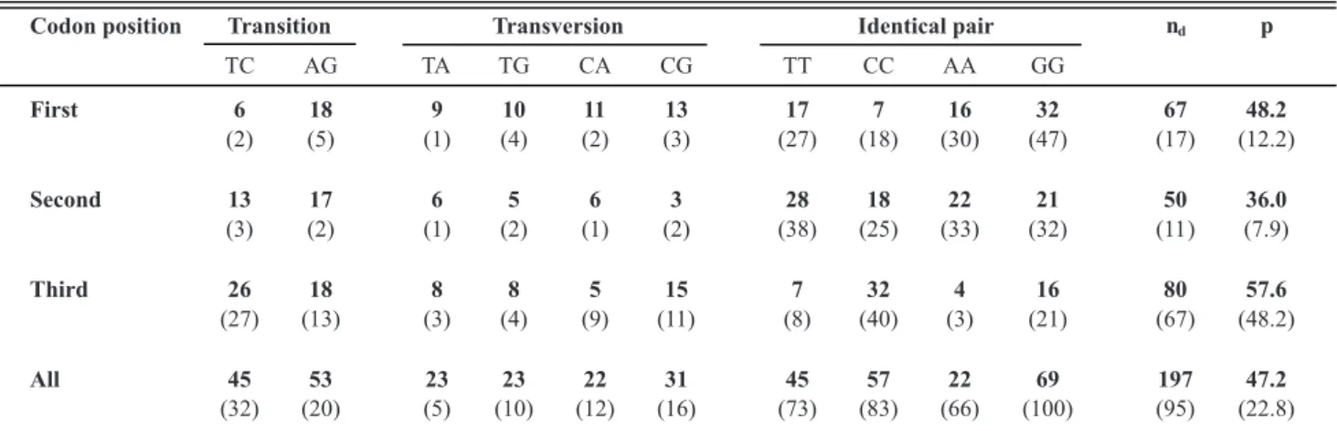
C. graminicola gene was found. Nucleotide changes at
each codon position in the C. lindemuthianum pseudogene
are shown in Table 1, comparing 417 nucleotide positions to their corresponding codon in C. gloeosporioides
(gb:KB020675) and C. graminicola (gb:380495603). The
former result, together with the presence of two stop codons in the translated aligned sequence, suggested the possibility that the CD1/CD2 region might be a Ftr1 pseudogene.
A search in the contig database from C. lindemuthianum
revealed the existence of at least two intact Ftr1 genes. The
first Ftr1 spans positions 15,346-16,736 of a 581,607 nt
contig (C0002) and has as closest homolog an Ftr1 gene
for C. orbiculare MAFF 240422 (gb:ENH77797). The
second Ftr1 gene was found at positions 7,652-8,929 of
contig 00366 (C00366: 24,549 bp) and is homologous to a different Ftr1 paralog from C. orbiculare MAFF 240422
(gb:ENH86230).
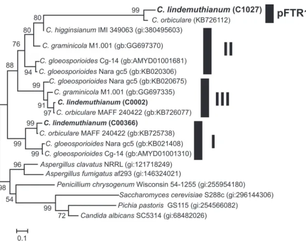
Phylogenetic analysis of available FTR1 proteins from Colletotrichum species revealed the existence of at
least three distinct clusters (Figure 2B). Clusters I and III include FTR1 encoded by contigs 0002 and 366 respectively; however no protein homologs of cluster II were found in our C. lindemuthianum 454 database. Interestingly, iron
permease EFQ33393 from C. graminicola, shown to be
similar to the CD1/CD2 amplicon (Figure 2B) clustered within group II suggesting that this amplicon could be a remnant of an ancient group II Ftr1 gene.
Genes encoding for group II FTR1 proteins in C. gloeosporioides Nara gc5, C. gloeosporioides cg-14, C. graminicola M1.001 and C. higginsianum are located
A
B
FIGURE 1 - Characterization of the CD1/CD2 amplicon in Colombian isolates of C. lindemuthianum. A. Duplex PCR
using primers CY1/CY2 and CD1/CD2 in 12 C. lindemuthianum isolates from Colombia; B. Partial sequence of contig
1027 from C. lindemuthianum L87 (Cl) containing the target of primers CD1/CD2. The corresponding segment in C. orbiculare is aligned below (Co).
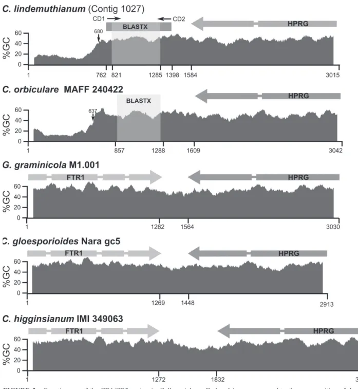
pseudogene in C. orbiculare. A side-by-side comparison
clearly indicates that the N-terminal portion comprising the first two exons of group II Ftr1 gene was lost in both C. lindemuthianum and C. orbiculare (Figure 3). This fact was
further supported by sequence composition analysis, which shows that this region has a higher AT content as compared to the corresponding segments in the Ftr1 pseudogen.
The average GC content of the Ftr1 genes, including their
introns, varies from 55.4-55.5% in close agreement with the average GC values observed for the C. lindemuthianum
(51.9%) and C. orbiculare (51.5%) pseudogenes. However,
local GC composition changes dramatically to a GC poor region (15.2-18.7%) in the segment where the N-terminal portion of the Ftr1 gene would be expected (Figure 3).
Phylogenetic analysis of the DNA segment comprising the CD1/CD2 region including all Ftr1 known genes from Colletotrichum revealed that the C. lindemuthianum and C. orbiculare pseudogenes have a close relation to group II Ftr1 (Figure 4). Predicted functional Ftr1 genes identified
A
B
FIGURE 2 - Sequence analysis of translated CD1/CD2 amplicon. A. Alignment of the C. graminicola iron permease
(EFQ33393) with the translated CD1/CD2 amplicon; B. Unrooted tree of iron permease protein sequences from Colletotrichum available in NCBI. C0002 and C00366 correspond to C. lindemuthianum proteins identified in this study.
Codon position Transition Transversion Identical pair nd p
TC AG TA TG CA CG TT CC AA GG
First 6 18 9 10 11 13 17 7 16 32 67 48.2
(2) (5) (1) (4) (2) (3) (27) (18) (30) (47) (17) (12.2)
Second 13 17 6 5 6 3 28 18 22 21 50 36.0
(3) (2) (1) (2) (1) (2) (38) (25) (33) (32) (11 ) (7.9)
Third 26 18 8 8 5 15 7 32 4 16 80 57.6
(27) (13) (3) (4) (9) (11) (8) (40) (3) (21) (67) (48.2)
All 45 53 23 23 22 31 45 57 22 69 197 47.2
(32) (20) (5) (10) (12) (16) (73) (83) (66) (100) (95) (22.8) TABLE 1 - Observed nucleotide changes at each codon position in the Colletotrichum lindemuthianum pseudogene. 417 nucleotide positions were analyzed and compared to their corresponding codon in C. gloeosporioides (gb:KB020675). The same analysis was performed on C. graminicola (gb:380495603, in parentheses).
nd= Total number of substitutions; p, percentage of changes
rate at the third codon position as changes in these positions normally result in silent changes at the protein level. Analysis of the mutation rates of the C. lindemuthianum
pseudogene revealed similar nucleotides changes at the first (48.2%) and second codon (36%) as in the third codon
position (57.6%). This clearly contrasts with mutational rates observed in the functional Ftr1 gene from C. graminicola
FIGURE 3 - Genetic map of the CD1/CD2 region in Colletotrichum.Ftr1 and hprg correspond to the gene position of the iron permeases and hydroxyproline rich glycoprotein; thin segments correspond to intron positions. The corresponding local GC content is plotted below and corresponds to the average of 100 residues surrounding the corresponding position. The compositional GC transition point in C. lindemuthianum and C. orbiculare is marked with an arrow; regions giving significant BLASTX results with FTR1 proteins are also indicated for these species. For C. lindemuthianum, the position of the CD1/CD2 is shown.
C. lindemuthianum pseudogene (47.2%) more than doubles
the changes observed in C. graminicola (22.8%).
DISCUSSION
The availability of pathogen-free seed is key for the control of bean anthracnose (Schwartz et al., 2005).
This requires highly sensitive and specific assays as those proposed by Wang et al. (2008) and Chen et al. (2013) based on duplex and qPCR, respectively. The use of molecular diagnosis, as a complement to traditional culture-dependent methods, in seed certification programs and quarantine vigilance of C. lindemuthianum would be very useful in the
disease (Pastor-Corrales & Tu, 1989; Mahuku & Riascos 2004). Unfortunately, our experience with the duplex PCR reported by Wang et al. (2008), using DNA from seeds, plant tissue (pods and leaves) and even C. lindemuthianum
mycelia indicates that the CY1/CY2 amplicon may not be detected using standard PCR conditions. These primers were designed from a restricted set of 24 ITS sequences and captured the variability of this region of C. lindemuthianum
and C. orbiculare while avoiding amplification from other Colletotrichum species and bean pathogens; however, it
is likely that for some strains these primers fail to capture sequence variants from other geographic regions such as Colombia. By contrast, the CD1/CD2 primer set proved to be more reliable as it worked well for fungal detection in both symptomatic and asymptomatic seeds and plantlets. These primers were obtained by the random amplification of polymorphic DNA technique and were shown to be highly specific to C. lindemuthianum (Wang et al., 2008);
unfortunately the authors did not report or characterized their target sequence. By sequencing the PCR product when using CD1/CD2 we showed that a pseudogene derived from the C-terminal end of a FTR1 high-affinity iron
permease was identified in both C. lindemuthianum and C. orbiculare. This hypothesis is supported by the presence of
stop codons, lack of exons 1 and 2, similar mutation rates at each position of the corresponding codons and mapping of this segment in the vicinity of the HPRG gene.
Sequence alignment clearly explained the specificity of the CD1/CD2 primer set; while primer CD2 is 100% complementary to its target region in both species, an 8 bp deletion in C. orbiculare disrupts binding of primer CD1
at the 3’ end. From this result it was also evident that a new forward primer, analogous to CD1, can be designed to specifically identify C. orbiculare, adding up a new marker
to identify this species to the sequence analysis of 900-bp intron of the glutamine synthetase gene used by Liu et al. (2007). Furthermore, each of the species in the Orbiculare clade (sensu Cannon et al., 2012) presented distinct host specificity: isolates of C. orbiculare, C. lindemuthianum,
and C. trifolii being pathogenic only on cucurbits, common
bean, and alfalfa, respectively, while de other species (C. malvarum, C. bidentis, C. sidae, C. spinosum and C. tebeestii) were found on different weeds (Damm et al.,
2013).
FTR1 proteins, together with iron transport multicopper oxidases (fet3), are part of a conserved
binary permease-oxidase complex involved in a high-affinity iron uptake in fungi (Philpott, 2006; Sutak et al., 2008). This system is essential for iron uptake in iron-limited environments and seems to be strongly related with pathogenesis in other fungal species such as Candida albicans and Aspergillus oryzae (Ramanan & Wang, 2000;
Knight et al., 2002; Sutak et al., 2008). However, this system does not appear to be necessary for all fungi as neither fet3
nor Ftr1 has been identified in complete genome analysis of Aspergillus nidulans (Philpott, 2006). With the exception of
C. lindemuthianum and C. orbiculare, three Ftr1 paralogs
where observed in all Colletotrichum species under study.
Little is known about the importance of these genes in the plant-pathogen interaction but it would not be surprising that they play a significant role in scavenging iron from their corresponding host during infection. In Rhizopus oryzae,
the high affinity iron permease gene (Ftr1) is required for
iron transport in iron-depleted environments and is required for full virulence in animal hosts (Ibrahim et al., 2010); a similar fact has been observed in the Ustilago maydis/maize
pathosystem (Eichorn et al., 2006). Multiple Ftr1 copies
may help Colletotrichum species to have a broader range of
possibilities to thrive in iron-poor environments. The lack of a functional Ftr1 copy could explain the narrow host range
of C. lindemuthianum and C. orbiculare in contrast with
wide host-range pathogens such as C. gloeosporioides.
Eukaryotic pseudogenes are classified as processed or nonprocessed. Processed pseudogenes are retrotransposed into the genome via an RNA intermediate and therefore lack introns, may possess relics of a poly(A) tail and are often flanked by target-site duplications (Zheng et al., 2007). Nonprocessed pseudogenes, on the other hand, arise from non-homologous recombination events. The evidence presented here suggests that the Ftr1 pseudogene of C. lindemuthianum is a nonprocessed pseudogene probably
resulting from a non–homologous recombination event near the exon 2-exon 3 junction of the Ftr1 gene. The
long branch joining the Ftr1 pseudogenes is expected as
pseudogenes normally show higher divergence rate than functional genes and are subject of neutral degeneration right after their formation (Subramanian & Kumar, 2003). Loss of this gene probably had no major consequences as the presence of two additional paralogs could compensate for this gene loss. The similarity between the Ftr1 pseudogene
in C. lindemuthianum and C. orbiculare strongly suggests
an evolutionary event that occurred previous to the separation of these closely related species (Liu et al., 2007; Cannon et al., 2012). The availability of the genomes of
C. lindemuthianum, currently under study by our group,
and C. orbiculare (Gan et al., 2013) will facilitate such
evolutionary studies as well as a deeper understanding of key aspects of the host-pathogen interaction of this species complex. All these information may help to test the hypothesis of separation of these taxa from Colletotrichum
at generic level due to its basal position in the phylogenectic studies developed for this genus (Cannon et al., 2012). Further work should explore the corresponding sequences in C. trifolii and C. malvarum to help clarify their close
evolutionary relationship within the Orbiculare clade. The discovery of CD1/CD2 C. lindemuthianum
specific marker by Wang et al. (2008) together with the molecular characterization presented in this study opens the possibility of designing new markers based on iron permease genes and/or pseudogenes for detecting different
Colletotrichum species and complement multilocus
phylogenetic analysis for this important group of plant pathogens.
ACKNOWLEDGEMENTS
This work was funded by Vicerrectoría de Investigaciones de la Universidad Nacional de Colombia, Grant: 20101009932. Convocatoria Nacional para el fortalecimiento de los Grupos de Investigación y Creación Artística de la Universidad Nacional de Colombia 2010-2012.
REFERENCES
Apostolos BJ, Koutita O, Klonari KT (2009) Molecular diversity and assessment of biological characteristics of Greek Colletotrichum lindemuthianum populations. Journal of Phytopathology 157:311-318.
Cai L, Hyde KD, Taylor PWJ, Weir BS, Waller JM, Abang MM, Zhang JZ, Yang YL, Phoulivong S, Liu ZY, Prihastuti H, Shivas RG, McKenzie EHC, Johnston PR (2009) A polyphasic approach for studying Colletotrichum. Fungal Diversity 39:183-204. Cannon PF, Damm U, Johnston PR, Weir BS (2012) Colletotrichum – current status and future directions. Studies in Mycology 73:181-213.
Chen YY, Conner RL, Boland GJ, Babcock C, Chang KF, Hwang SF, Balasubramanian PM (2007) A specific and sensitive method for the detection of Colletotrichum lindemuthianum in dry bean tissue. Plant Disease 91:1271-1276.
Chen YY, Conner RL, Gillard CL, McLaren DL, Boland GJ, Balasubramanian PM, Stasolla C, Zhou QX, Hwang SF, Chang KF, Babcock C (2013) A quantitative real-time PCR assay for detection of Colletotrichum lindemuthianum in navy bean seeds. Plant Pathology 62:900-907.
Damm U, Cannon PF, Liu F, Barreto R, Guatimosim E, Crous PW (2013) The Colletotrichum orbiculare species complex: important pathogens of field crops and weeds. Fungal Diversity 61:29-59. Doyle JJ, Doyle JL (1990) Isolation of plant DNA from fresh tissue. Focus 12:13-15.
Edgar RC (2004) MUSCLE: multiple sequence alignment with high accuracy and high throughput. Nucleic Acids Research 32:1792-1797.
Eichhorn H, Lessing F, Winterberg B, Schirawski J, Kämper J, Müller P, Kahmann R (2006) A ferroxidation/permeation iron
Plant Cell 18:3332-3345.
Felsenstein J (1985) Confidence limits on phylogenies: an approach using the bootstrap. Evolution 39:783-791.
Gan P, Ikeda K, Irieda H, Narusaka M, O’Connell RJ, NarusakaY TY, Kubo Y, Shirasu K (2013) Comparative genomic and transcriptomic analyses reveal the hemibiotrophic stage shift of Colletotrichum fungi. New Phytologist 197:1236-1249.
Hyde KD, Cai L, McKenzie EHC, Yang YL, Zhang JZ, Prihastuti H (2009) Colletotrichum: a catalogue of confusion. Fungal Diversity 39:1-17.
Ibrahim AS, Gebremariam T, Lin L, Luo G, Husseiny MI, Skory
CD, Fu Y, French SW, Edwards JE Jr, Spellberg B (2010) The
high affinity iron permease is a key virulence factor required for Rhizopus oryzae pathogenesis. Molecular Microbiology 77:587-604.
Kimura M (1980) A simple method for estimating evolutionary rate of base substitutions through comparative studies of nucleotide sequences. Journal of Molecular Evolution 16:111-120.
Knight SA, Lesuisse E, Stearman R, Klausner RD, Dancis A
(2002) Reductive iron uptake by Candida albicans: role of copper, iron and the TUP1 regulator. Microbiology 148:29-40.
Liu B, Walsiwa LA, Morelock TE, O’Neill NR, Carrell JC (2007) Comparison of Colletotrichum orbiculare and several allied Colletotrichum spp. for mtDNA RFLPs, intron RFLP and sequence variation, vegetative compatibility, and host specificity. Phytopathology 97:1305-1314.
Liu B, Cai L, Crous PW, Damm U (2013) Circumscription of the anthracnose pathogens Colletotrichum lindemuthianum and C. nigrum. Mycologia 105:844-860.
Mahuku SG, Riascos JJ (2004) Virulence and molecular diversity within Colletotrichum lindemuthianum isolates from Andean and Mesoamerican bean varieties and regions. European Journal of Plant Pathology 110:253-263.
Melotto M, Balardin RS, Kelly JD (2000) Host-pathogen interaction and variability of Colletotrichum lindemuthianum. In: Prusky D, Freeman S, Dickman MB (Eds.) Host specificity, pathology and host-pathogen interactions of Colletotrichum. St Paul, MN, USA. APS Press. pp. 346-361.
O’Connell RJ, Thon MR, Hacquard S, Amyotte SG, Kleemann J, Torres MF, Damm U, Buiate EA, Epstein L, Alkan N, Altmüller J, Alvarado-Balderrama L, Bauser CA, Becker C, Birren BW, Chen Z, Choi J, Crouch JA, Duvick JP, Farman MA, Gan P, Heiman D, Henrissat B, Howard RJ, Kabbage M (2012) Lifestyle transitions
in plant pathogenic Colletotrichum fungi deciphered by genome and transcriptome analyses. Nature Genetics 44:1060-1065. Pastor-Corrales MA, Tu JC (1989) Anthracnose. In: Schwartz H, Galvez G. (Eds.) Bean production problems in the tropics. Cali,
Colombia. Centro Internacional de Agricultura Tropical (CIAT). pp. 77-104.
Philpott CC (2006) Iron uptake in fungi: a system for every source. Biochimica et Biophysica Acta 1763:636-645.
Pinto JMA, Pereira R, Mota SF, Ishikawa FH, Souza EA (2012) Investigating phenotypic variability in Colletotrichum lindemuthianum populations. Phytopathology 102:490-497. Ramanan N, Wang Y (2000) A high-affinity iron permease essential for Candida albicans virulence. Science 288:1062-1064.
Saitou N, Nei M (1987) The neighbor-joining method: a new method for reconstructing phylogenetic trees. Molecular Biology and Evolution 4:406-425.
Santa CC, Marín MM, Díez MC (2012) Identificación del agente causal de la pudrición basal del tallo de vainilla en cultivos bajo cobertizos en Colombia. Revista Mexicana de Micologia 35:23-34.
Schwartz HF, Steadman JR, Hall R, Forster RL (2005) Compendium of Bean Diseases. 2nd Ed. St Paul, MN, USA. APS Press. Sherriff C, Whelan MJ, Arnold GM, Lafay J, Brygoo Y, Bailey JA (1994). Ribosomal DNA sequence analysis reveals new species groupings in the genus Colletotrichum. Experimental Mycology 18:121-138.
Subramanian S, Kumar S (2003) Neutral substitutions occur at a faster rate in exons than in noncoding DNA in primate genomes Genome Research 13:838-844.
Sutak R, Lesuisse E, Tachezy J, Richardson DR (2008) Crusade for iron: iron uptake in unicellular eukaryotes and its significance for virulence. Trends in Microbiology 16: 261-268.
Tamura K, Peterson D, Peterson N, Stecher G, Nei M, Kumar S (2011) MEGA5: Molecular Evolutionary Genetics Analysis using Maximum Likelihood, Evolutionary Distance, and Maximum Parsimony methods. Molecular Biology and Evolution 28:2731-2739.
Wang W, Tang JH, Wang YC (2008) Molecular detection of Colletotrichum lindemuthianum by duplex PCR. Journal of Phytopathology 156:431-437.
White TJ, Bruns T, Lee S, Taylor J (1990) Amplification and direct sequencing of fungal rRNA genes for phylogenetics. In: Innis AM, Gelfand, DH, Sninsky JJ, White TJ (Eds.) PCR Protocols: A guide to methods and applications. San Diego, CA, USA. Academic Press. pp. 315-322.
Zheng D, Frankish A, Baertsch R, Kapranov P, Reymond A, Choo SW, Lu Y, Denoeud F, Antonarakis SE, Snyder M, Ruan Y, Wei CL, Gingeras TR, Guigó R, Harrow J, Gerstein MB (2007) Pseudogenes in the ENCODE regions: consensus annotation, analysis of transcription, and evolution. Genome Research 17:839-851.