Medical applications of shape
memory alloys
1Departamento de Engenharia Mecânica e de Materiais, Instituto Militar de Engenharia, Rio de Janeiro, RJ, Brasil 2Departamento de Engenharia Mecânica, COPPE,
Universidade Federal do Rio de Janeiro, Rio de Janeiro, RJ, Brasil L.G. Machado1
and M.A. Savi2
Abstract
Shape memory alloys (SMA) are materials that have the ability to return to a former shape when subjected to an appropriate thermome-chanical procedure. Pseudoelastic and shape memory effects are some of the behaviors presented by these alloys. The unique properties concerning these alloys have encouraged many investigators to look for applications of SMA in different fields of human knowledge. The purpose of this review article is to present a brief discussion of the thermomechanical behavior of SMA and to describe their most prom-ising applications in the biomedical area. These include cardiovascu-lar and orthopedic uses, and surgical instruments.
Correspondence M.A. Savi
Departamento de Engenharia Mecânica
COPPE, UFRJ Caixa Postal 68.503 21945-970 Rio de Janeiro, RJ Brasil
E-mail: [email protected]
Research supported by CNPq.
Received July 3, 2002 Accepted December 4, 2002
Key words
•Shape memory alloys •Biomaterials
Introduction
Shape memory alloys (SMA) constitute a group of metallic materials with the ability to recover a previously defined length or a shape when subjected to an appropriate thermome-chanical load (1). When there is a limitation of shape recovery, these alloys promote high restitution forces. Because of these proper-ties, there is a great technological interest in the use of SMA for different applications.
Although a relatively wide variety of al-loys present the shape memory effect, only those that can recover from a large amount of strain or generate an expressive restitution force are of commercial interest. Particularly important among them are alloys based on Ni-Ti and on Cu, such as Zn-Al and Cu-Al-Ni (1). SMA based on Ni-Ti are the alloys most frequently used in commercial applica-tions because they combine good
mechani-cal properties with shape memory.
To-day, these applications are being developed in different fields of science and engineering. Basically, SMA present two well-defined crystallographic phases, i.e., austenite and martensite (3). Martensite is a phase that, in the absence of stress, is stable only at low temperatures; in addition, it can be induced by either stress or temperature. Martensite is easily deformed, reaching large strains (~8%) (1). Depending on the type of transformation experienced by these alloys, the crystal struc-ture of martensite can be either monoclinic or orthorhombic (4,5). When martensite is induced by temperature, it is called twinned martensite. The twinned martensite has 24 variants, i.e., 24 subtypes with different crys-tallographic orientations (6). On the other hand, when martensite is induced by stress, these 24 variants of twinned martensite be-come only one variant. As a consequence, there is a crystallographic orientation, aligned with the stress direction, which is called detwinned martensite. The austenite phase is stable only at high temperatures, having a single variant with a body-centered cubic crystal structure.
Martensitic transformation explains the shape recovery in SMA. This transformation occurs within a range of temperatures which varies according to the chemical content of each alloy (7). In general, four characteristic transformation temperatures can be defined:
MS and MF, which are the temperatures at
which the formation of martensite starts and ends, respectively, and AS and AF, which are
the temperatures at which the formation of austenite starts and ends, respectively.
Recent studies have shown that, depend-ing on specific conditions, some SMA can present another crystallographic phase known as R-phase. The R-phase transformation can appear before the martensitic transformation according to the following sequence: austenite → R-phase → martensite. The crystal
struc-ture of the R-phase is rhombohedric (4,5). Because of their remarkable properties, SMA can be used in a large number of
non-medical applications (8-10). SMA can solve problems in the aerospace industry, espe-cially those related to vibration control of slender structures and solar panels, and non-explosive release devices (11,12). Microma-nipulators and robotic actuators have been employed in order to mimic the smooth move-ment of human muscles (13,14). SMA are commonly used as external actuators or as SMA fibers embedded in a composite matrix so that they can alter the mechanical proper-ties of slender structures for the control of buckling and vibration (15).
Biomedical applications of SMA have been extremely successful because of the functional properties of these alloys, increas-ing both the possibility and the performance of minimally invasive surgeries (2,16,17). The biocompatibility of these alloys is one of the important points related to their bio-medical applications as orthopedic implants (18), cardiovascular devices (2), and surgi-cal instruments (16), as well as orthodontic devices and endodontic files (19-21).
This article presents a brief discussion of the thermomechanical behavior of SMA, and a description of their main applications in the biomedical field as cardiovascular and orthopedic devices and as surgical instru-ments.
Thermomechanical behavior
SMA present typical thermomechanical behaviors, like pseudoelasticity and shape memory effects (one-way and two-way). This section presents a short discussion of these behaviors, explaining the macroscopic phe-nomenological aspects related to each one (22).
Pseudoelasticity
Pseudoelasticity occurs whenever an SMA sample is at a temperature above AF
speci-men). Thus, one can consider an SMA sample subjected to a mechanical loading at a con-stant temperature above AF. The stress-strain
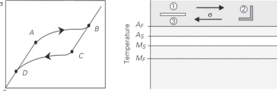
curve (σ-ε) in Figure 1, left side, illustrates the macroscopic behavior of SMA, showing the pseudoelastic phenomenon.
A mechanical loading causes an elastic response until a critical value is reached, point A, when the martensitic transformation
(austenite → martensite) arises, ending at point B. At this point, the crystal structure of the sample is totally composed of detwinned martensite. For higher stress values, SMA presents a linear response. During the un-loading process, the sample presents an elas-tic recovery (B→ C). From point C to D one
can note the reverse martensitic transforma-tion (martensite → austenite). From point D on, the sample presents an elastic discharge. When the loading-unloading process is fin-ished, SMA have no residual strain. How-ever, since the path of the forward martensi-tic transformation does not coincide with the reverse transformation path, there is a hys-teresis loop associated with energy dissipa-tion.
Another way to observe the pseudoelastic effect is indicated on the right side of Figure 1. First, let us consider an SMA at a temper-ature above AF, c. At this temperature, there
is only one phase, i.e., austenite. At a con-stant temperature, a mechanical loading is applied promoting the appearance of the detwinned martensite, d. During the un-loading process, reverse transformation takes place (detwinned martensite → austenite) and when load vanishes, e, the sample pre-sents no residual strain.
Shape memory effect
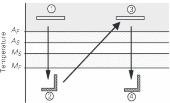
The second thermomechanical behavior that can be observed in SMA is the shape memory effect. Figure 2, left side, shows the stress-strain curve of an SMA sample at a low temperature (less than MF, the
tempera-ture below which only the martensitic phase
is stable) where the shape memory effect can be noted. When the sample is subjected to a mechanical loading, the stress reaches a criti-cal value, point A, when the transformation
of the twinned martensite into the detwinned martensite begins, ending at point B. When
the loading-unloading process is finished, the SMA sample presents a residual strain (point C). This residual strain can be
recov-ered by sample heating, which induces the reverse phase transformation. This is the shape memory effect, also known as one-way shape memory effect. This phenome-non can be understood from a motion of the hysteresis loop shown on the stress-strain curve in Figure 1. Since the temperature goes down, the hysteresis loop moves down as well.
The right side of Figure 2 presents an alternative way to observe the shape memory effect. At first, the SMA sample is at a temperature above AF, c. At this
tempera-ture, the sample has only the austenitic phase. When the temperature of the SMA sample
σ
ε
0
A
B
C
AF
AS
MS
MF
Temperature
1
3 2
4
Figure 2. Shape memory effect. For abbreviations, see legend to Figure 1. See text for explanation of process.
σ σ
ε
0
A B
C
D
AF
AS
MS
MF
Temperature
1
3
2
σ
Figure 1. Pseudoelasticity. AS, AF and MS and MF = temperature at which the formation of austenite and martensite starts and ends, respectively. σ-ε = stress-strain curve. See text
decreases and crosses the line related to MS,
the phase transformation begins to take place and the twinned martensite replaces the aus-tenite. This transformation is concluded when the sample temperature is below MF, d.
Under a constant temperature, a mechanical loading is applied (d→e), promoting the appearance of the detwinned martensite. When this load vanishes the sample presents a residual strain, e. The former shape of the sample can be recovered through a heating process (e→f) which causes the reverse martensitic transformation (detwinned mar-tensite → austenite).
Two-way shape memory effect
Another phenomenon concerning mar-tensitic transformation is the two-way shape memory effect. The primary characteristic of the two-way effect is associated with the presence of a specific phase in a specific setting. In this way, the sample has a shape in the austenitic state and another in the mar-tensitic state. The change of temperature produces a change in sample shape without any mechanical loading.
In order to obtain the two-way effect, it is necessary that the SMA sample be trained. Typically, there are two training procedures (23): shape memory effect cycling (cycles of shape memory effect) and the training through the appearance of the detwinned martensite, the stress-induced martensite training. Both induce considerable plastic strains.
Figure 3 shows a schematic presentation of the two-way effect. First of all, let us consider that a trained SMA sample is at a
temperature above AF, c. Sample cooling
promotes a phase change (austenite → mar-tensite), which leads to a change in shape, d. When the temperature is increased above
AF, the sample experiences another phase
transformation (d→e), recovering its ori-ginal shape, e. Another cooling returns the sample to its low temperature shape, f. It should be pointed out that, in contrast to the one-way shape memory effect, it is not nec-essary to apply mechanical loading in order to alter the sample’s shape at low tempera-ture.
Biocompatibility of shape memory alloys - Ni-Ti
Biocompatibility is the ability of a mate-rial to remain biologically innocuous during its functional period inside a living creature (24). This is a crucial factor for the use of SMA devices in the human body (25). A biocompatible material does not produce al-lergic reactions inside the host, and also does not release ions into the bloodstream. The period during which a biomaterial remains inside the human body is an important aspect to be considered concerning its use.
Generally, the biocompatibility of a ma-terial is strongly related to allergic reactions between the material surface and the inflam-matory response of the host. Several aspects can contribute to these reactions such as patient’s characteristics (health, age, immu-nological state, and so on), and material characteristics (rugosity and porosity of the surface and individual toxic effects of the elements present in the material) (25).
Several investigations have been con-ducted in order to establish the biocompati-bility of Ni-Ti-based alloys, and to exclude intrinsic hazards involved in their applica-tions (24,25). The analysis of aspects related to the biocompatibility of these alloys is performed by assessing each of their ele-ments, nickel and titanium, separately.
Nickel, although necessary to life, is a
AF
AS
MS
MF
Temperature
1 3
2 4
highly poisonous element (2). Studies have shown that persons having systematic con-tact with nickel present problems such as pneumonia, chronic sinusitis and rhinitis, nostril and lung cancer, as well as dermatitis caused by physical contact.
Unlike nickel, titanium and its compounds are highly biocompatible; moreover, due to their mechanical properties, they are usually employed in orthodontic and orthopedic im-plants (2). The oxidation reaction of tita-nium produces an innocuous layer of TiO2
which surrounds the sample. This layer is responsible for the high resistance to corro-sion of titanium alloys, and the fact that they are harmless to the human body.
Inquiries concerning the biocompatibili-ty of Ni-Ti alloys began shortly after their discovery in 1968. Corrosion analyses have shown that this alloy is easily changed to the passive condition in physiological solutions; moreover, its corrosion resistance is greater than that of stainless steel (24). In general, one can say that the properties of titanium confer good biocompatibility to Ni-Ti al-loys.
Applications of shape memory alloys
As mentioned earlier, the remarkable properties of SMA have promoted several investigations related to their applications in different fields of human knowledge. In this section we present a discussion of the bio-medical applications of SMA. Cardiovascu-lar applications are presented first, followed by orthopedic applications and the use of SMA in surgical instruments.
Cardiovascular applications
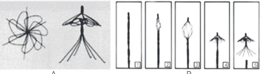
The first cardiovascular device devel-oped with shape memory was the Simon filter (25). The Simon filter (Figure 4) repre-sents a new generation of devices that are used for blood vessel interruption in order to prevent pulmonary embolism. Persons who
cannot take anticoagulant medicines are the major users of the Simon filter (26). The purpose of this device is to filter clots that travel inside the bloodstream. The Simon filter traps these clots that in time are dis-solved by the bloodstream (16). The inser-tion of the filter inside the human body is done by exploiting the shape memory effect. From its original shape in the martensitic state (Figure 4A) the filter is deformed and placed on a catheter tip. Saline solution flow-ing through the catheter is used to keep a low temperature, while the filter is placed inside the body. When the catheter releases the filter, the flow of the saline solution is stopped. As a result, the bloodstream pro-motes the heating of the filter that returns to its former shape. This procedure can be seen in Figure 4B (16).
The atrial septal occlusion device is em-ployed to seal the atrial hole (Figure 5) (20,26). The atrial hole is located between the two upper heart chambers upon the sur-face that splits the upper part of the heart into the right and left atria. The anomaly occur-ring when this hole is open can reduce life
1 2 3 4 5
Figure 4. Simon filter. A, Filter in the recovery form. B, Filter release. Taken from Ref. 26 (http://www.nmtmedical.com).
A B
Right atrium
Catheter
Vein
Septum Left atrium
Valve
Septum
PFO
Catheter
Tissue
1
3 B
A
2
CardioSEAL
expectancy. The traditional surgery that fixes this anomaly is extremely invasive and dan-gerous. The thorax of the patient is opened and the atrial hole is sewn. Because of the intrinsic risks of this surgery, several prob-lems might occur. The atrial septal occlusion device is an alternative to this surgery. This device is composed of SMA wires and a waterproof film of polyurethane (16). As is the case for the Simon filter, the surgery to place this device exploits the shape memory effect, being much less invasive than the traditional one. First, one half of the device
is inserted through a catheter by the vena cava up to the heart, in its closed form. Then, it is placed on the atrial hole and opened, recovering its original shape. Next, the sec-ond half of the device is placed by the same route as the first one, and then both halves are connected. This procedure seals the hole, avoiding blood flow from one atrium to the other. It is expected that the device will stay in the heart for an indefinite period of time since the heart tissue regenerates (26). Fig-ure 6A presents a scheme of the heart with the device in place.
Self-expanding stents, named after the dentist C.T. Stent, are another important car-diovascular application that is used to main-tain the inner diameter of a blood vessel. Actually, these devices are used in several situations in order to support any tubular passage such as the esophagus and bile duct (27), and blood vessels such as the coronary, iliac, carotid, aorta and femoral arteries (16). In this type of application, a cylindrical scaf-fold with shape memory (Figure 7) (28) is placed, for example, inside a blood vessel through a catheter. Initially, this scaffold is pre-compressed in its martensitic state. As the scaffold is heated, due to the body tem-perature, it tends to recover its original shape, expanding itself. This device can be used not only in the angioplasty procedure, in order to prevent another obstruction of a vessel, but also in the treatment of aneurysms for the support of a weakened vessel (16).
Orthopedic applications
SMA have a large number of orthopedic applications. The spinal vertebra spacer (Fig-ure 8) is one. The insertion of this spacer between two vertebrae assures the local rein-forcement of the spinal vertebrae, prevent-ing any traumatic motion durprevent-ing the healprevent-ing process. The use of a shape memory spacer permits the application of a constant load regardless of the position of the patient, who preserves some degree of motion (29). This Figure 6. Atrial septal occlusion device. A, Scheme of the heart with the device in place. B,
The first half of the device is placed in the left atrium. C, The second half of the device is placed in the right atrium. D, The catheter is withdrawn and the tissue begins its recovery. PFO = patent foramen ovale. Taken from Ref. 26 (http://www.nmtmedical.com).
Figure 7. Shape memory self-ex-panding stents. Taken from Ref. 28 (http://www.raychem.com).
device is used in the treatment of scoliosis (2). Figure 8 shows spinal vertebrae and a shape memory spacer. On the left side, the spacer is in the martensitic state, and on the right side, the spacer is in its original shape, recovered by the pseudoelastic phenomenon. Another application in the orthopedic area is related to the healing process of broken and fractured bones (30). Several types of shape memory orthopedic staplesare used to accelerate the healing process of bone frac-tures, exploiting the shape memory effect. The shape memory staple, in its opened shape, is placed at the site where one desires to rebuild the fractured bone. Through heating, this staple tends to close, compressing the separated part of bones. It should be pointed out that an external device performs this heating, and not the temperature of the body. The force generated by this process acceler-ates healing, reducing the time of recovery. Figure 9 presents an application of these staples during the healing process of a patient’s foot fracture.
With respect to the healing of fractured bones, one can also point out shape memory platesfor the recoveryof bones (31). These plates are primarily used in situations where a cast cannot be applied to the injured area, i.e., facial areas, nose, jaw and eye socket. They are placed on the fracture and fixed with screws, maintaining the original align-ment of the bone and allowing cellular re-generation. Because of the shape memory effect, when heated these plates tend to re-cover their former shape, exerting a constant force that tends to join parts separated by fractures, helping with the healing process (2). Figure 10 illustrates this device (31).
Orthopedic treatment also exploits the properties of SMA in the physiotherapy of semi-standstill muscles. Figure 11 shows gloves that are composed of shape memory wires on regions of the fingers (32). These wires reproduce the activity of hand muscles, promoting the original hand motion. The two-way shape memory effect is exploited in
Figure 11. Shape memory alloy glove. A, Low temperature position. B, High temperature position. Taken from Ref. 32 (http://www.amtbe.com).
A B
Ventral view
Atlas (C1) Axis (C2)
Left lateral view
Dorsal view
Atlas (C1) Axis (C2)
Thoracic vertebrae C7
T1
Sacrum (S1-5)
Coccyx
Sacrum (S1-5)
T12
L1
L5 L5
T12 T1 C7
L1 Atlas (C1) Axis (C2)
Sacrum (S1-5)
Coccyx Coccyx
T1 C7
T12 L1
L5
Cervical vertebrae
Lumbar vertebrae
Figure 8. Spinal vertebrae (A) and shape memory spacers (B) in the martensitic state (left) and in the original shape (right). Taken from Ref. 20 with permission.
A
A
B
Figure 10. Shape memory bone plates. A, Plates fixed upon a human jaw. B, Detail of the plate and the screw. Taken from Ref. 31 (http://database.cs.ualberta.ca/MEMS/sma_mems).
Four-hole, 2-mm miniplate Miniscrew, 2 mm
A B
Figure 9. A, Orthopedic staples. B, Staples placed in a human foot. C, X-ray of a human foot. Taken from Ref. 30 (http://www.labosite.com/anglais/pages/summary.html).
this situation. When the glove is heated, the length of the wires is shortened. On the other hand, when the glove is cooled, the wires return to their former shape, opening the hand. As a result, semi-standstill muscles are exercised.
Research for obtaining porous SMA is
currently underway. These alloys have a great potential application in orthopedic implants since their porosity enables the transport of body fluids from outside to inside the bone, which is in the healing process. This fact optimizes the treatment and also helps the fixation of the implant (33).
Applications to surgical instruments
In recent years, medicine and the medical industry have focused on the concept of less invasive surgical procedures (29). Follow-ing this tendency, shape memory surgical instruments have been created and are be-coming noticeable. Among the advantages of these tools, one can emphasize their flex-ibility as well as their possflex-ibility to recover their former shape when heated.
The SMA basketis used to remove kid-ney, bladder and bile duct stones (20). This basket is inserted into the human body in the same way as the Simon filter. Figure 12 presents a sequence of pictures related to the basket opening as it is heated.
The intra-aortic balloon pump (Figure 13) is used to unblock blood vessels during angioplasty. The device has an SMA tube whose diameter is reduced compared to poly-mer materials due to its pseudoelastic effect. Moreover, it also allows greater flexibility and torsion resistance when compared to the same tube made of stainless steel (16).
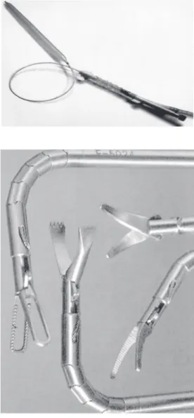
Laparoscopy is another procedure where SMA have been employed. Figure 14 shows some surgical tools where the actions of grippers, scissors, tongs and other mechan-isms are performed by SMA. These devices allow smooth movements tending to mimic the continuous movement of muscles. More-over, these devices facilitate access to intri-cate regions.
Final remarks
Applications of SMA to the biomedical field have been successful because of their Figure 12. Sequence of opening of the shape memory basket. Taken from Ref. 34 (http://
smet.tomsk.ru/eng/prod.htm).
Figure 13. Intra-aortic balloon pump. Taken from Ref. 16 with permission.
functional qualities, enhancing both the pos-sibility and the execution of less invasive surgeries. The biocompatibility of these al-loys is one of their most important features. Different applications exploit the shape memory effect (one-way or two-way) and the pseudoelasticity, so that they can be em-ployed in orthopedic and cardiovascular
ap-plications, as well as in the manufacture of new surgical tools. Therefore, one can say that smart materials, especially SMA, are becoming noticeable in the biomedical field. Probably, the adverse characteristic of bio-compatibility of nickel is one of the most critical point concerning the spreading use of Ni-Ti alloys.
References
1. Hodgson DE, Wu MH & Biermann RJ (1990). Shape Memory Alloys, Metals Handbook. Vol. 2. ASM International, Ohio, 897-902. 2. Mantovani D (2000). Shape memory alloys: Properties and
biomedi-cal applications. Journal of the Minerals, Metals and Materials Socie-ty, 52: 36-44.
3. Shape Memory Alloy Research Team (Smart) (2001). http:// smart.tamu.edu
4. Otsuka K & Ren X (1999). Recent developments on the research of shape memory alloys. Intermetallics, 7: 511-528.
5. Wu SK & Lin HC (2000). Recent development of TiNi-based shape memory alloys in Twain. Materials Chemistry and Physics, 64: 81-92.
6. Funakubo H (1987). Shape Memory Alloys. Gordon & Bleach, New York, NY, USA.
7. Shape Memory Applications, Inc. (2001). http://www.sma-inc.com 8. van Humbeeck J (1997). Shape memory materials: state of art and
requirements for future applications. Journal de Physique IV, 7: 3-12.
9. van Humbeeck J (1999). Non-medical applications of shape memory alloys. Materials Science and Engineering A, 273-275: 134-148. 10. Schetky LMcD (2000). The industrial applications of shape memory
alloys in North America. Materials Science Forum, 327-328: 9-16. 11. Denoyer KK, Erwin RS & Ninneman RR (2000). Advanced smart
structures flight experiments for precision spacecraft. Acta Astronautica, 47: 389-397.
12. Pacheco PMCL & Savi MA (2000). Modeling and simulation of a shape memory release device for aerospace applications. Revista de Engenharia e Ciências Aplicadas.
13. Webb G, Wilson L, Lagoudas DC & Rediniotis O (2000). Adaptive control of shape memory alloy actuators for underwater biomimetic applications. AIAA Journal, 38: 325-334.
14. Rogers CA (1995). Intelligent materials. Scientific American, Sep-tember: 122-127.
15. Birman V (1997). Theory and comparison of the effect of composite and shape memory alloy stiffeners on stability of composite shells and plates. International Journal of Mechanical Sciences, 39: 1139-1149.
16. Duerig TM, Pelton A & Stöckel D (1999). An overview of nitinol medical applications. Materials Science and Engineering A, 273-275: 149-160.
17. Pelton AR, Stöckel D & Duerig TW (2000). Medical uses of nitinol.
Materials Science Forum, 327-328: 63-70.
18. Chu Y, Dai K, Zhu M & Mi X (2000). Medical application of NiTi shape memory alloy in China. Materials Science Forum, 327-328: 55-62. 19. Airoldi G & Riva G (1996). Innovative materials: The NiTi alloys in
orthodontics. Bio-Medical Materials and Engineering, 6: 299-305. 20. Lagoudas DC, Rediniotis OK & Khan MM (1999). Applications of
shape memory alloys to bioengineering and biomedical technology.
Proceedings of the 4th International Workshop on Mathematical Methods in Scattering Theory and Biomedical Technology, Perdika, Greece, October 8-10, 1999.
21. Jordan L, Goubaa K, Masse M & Bouquet G (1991). Comparative study of mechanical properties of various Ni-Ti based shape memory alloys in view of dental and medical applications. Journal de Phy-sique IV, 1: 139-144.
22. Savi MA, Paiva A, Baêta-Neves AP & Pacheco PMCL (2002). Phe-nomenological modeling and numerical simulation of shape memory alloys: A thermo-plastic-phase transformation coupled model. Jour-nal of Intelligent Material Systems and Structures, 13: 261-273. 23. Zhang XD, Rogers CA & Liang C (1992). Modeling of the two-way
shape memory effect. Philosophical Magazine A, 65: 1199-1215. 24. Shabalovskaya SA (1995). Biological aspects of TiNi alloys surfaces.
Journal de Physique IV, 5: 1199-1204.
25. Ryhänen J (1999). Biocompatibility evolution of nickel-titanium shape memory alloy. Academic Dissertation, Faculty of Medicine, Univer-sity of Oulu, Oulu, Finland.
26. NMT Medical, Inc. (2001). http://www.nmtmedical.com 27. Medical Devicelink (2001). http://www.devicelink.com 28. Raychem (2001). http://www.raychem.com
29. Duerig TM, Pelton A & Stöckel D (1996). The use of superelasticity in medicine. Metall, 50: 569-574.
30. TECHNOSITE MEDICAL (2001). http://www.labosite.com/anglais/ pages/summary.html
31. SMA/MEMS Research Group (2001). http://database.cs.ualberta.ca/ MEMS/sma_mems
32. @medical technologies (2001). http://www.amtbe.com
33. Li B, Rong L, Li Y & Gjunter VE (2000). A recent development in producing porous NiTi shape memory alloys. Intermetallics, 8: 881-884.