Cryptosporidial Infection in Transgenic Undernourished
Mice
Orleaˆncio G. R. Azevedo1,3, David T. Bolick1, James K. Roche1, Relana F. Pinkerton1, Aldo A. M. Lima1,2, Michael P. Vitek4, Cirle A. Warren1, Reinaldo B. Oria´1,3*, Richard L. Guerrant1,2
1Division of Infectious Diseases and International Medicine, Center for Global Health, School of Medicine, University of Virginia, Charlottesville, Virginia, United States of America,2Laboratory of Infectious Diseases, Clinical Research Unit, Institute of the Brazilian Semi-Arid, School of Medicine, Federal University of Ceara, Fortaleza, Ceara´, Brazil,3Laboratory of the Biology of Tissue Healing, Ontogeny and Nutrition, Institute of the Brazilian Semi-arid, School of Medicine, Federal University of Ceara, Fortaleza, Ceara´, Brazil,4Duke University Medical Center, Department of Medicine, Durham, North Carolina, United States of America
Abstract
Apolipoliprotein E (apoE), a critical targeting protein in lipid homeostasis, has been found to have immunoinflammatory effects on murine models of infection and malnutrition. The effects of apoE in undernourished andCryptosporidium parvum -infected mice have not been investigated. In order to study the role of apoE in a model ofC. parvuminfection, we used the following C57BL6J mouse genetic strains: APOE-deficient, wild-type controls, and APOE targeted replacement (TR) mice expressing human APOE genes (E3/3; E4/4). Experimental mice were orally infected with 107-unexcysted-C. parvumoocysts between post-natal days 34–35 followed by malnutrition induced with a low-protein diet. Mice were euthanized seven days afterC. parvum-challenge to investigate ileal morphology, cytokines, and cationic arginine transporter (CAT-1), arginase 1, Toll-like receptor 9 (TLR9), and inducible nitric oxide synthase (iNOS) expression. In addition, we analyzed stool oocyst shedding by qRT-PCR and serum lipids. APOE4/4-TR mice had better weight gains after infection plus malnutrition compared with APOE3/3-TR and wild-type mice. APOE4/4-TR and APOE knockout mice had lower oocyst shedding, however the latter exhibited with villus blunting and higher ileal pro-inflammatory cytokines and iNOS transcripts. APOE4/4-TR mice had increased ileal CAT-1, arginase-1, and TLR9 transcripts relative to APOE knockout. Although with anti-parasitic effects, APOE deficiency exacerbates intestinal inflammatory responses and mucosal damage in undernourished andC. parvum -infected mice. In addition, the human APOE4 gene was found to be protective against the compounded insult of Cryptosporidiuminfection plus malnutrition, thus extending our previous findings of the protection against diarrhea in APOE4 children. Altogether our findings suggest that apoE plays a key role in the intestinal restitution and immunoinflammatory responses withCryptosporidiuminfection and malnutrition.
Citation:Azevedo OGR, Bolick DT, Roche JK, Pinkerton RF, Lima AAM, et al. (2014) Apolipoprotein E Plays a Key Role against Cryptosporidial Infection in Transgenic Undernourished Mice. PLoS ONE 9(2): e89562. doi:10.1371/journal.pone.0089562
Editor:Dipshikha Chakravortty, Indian Institute of Science, India
ReceivedSeptember 23, 2013;AcceptedJanuary 22, 2014;PublishedFebruary 28, 2014
Copyright:ß2014 Azevedo et al. This is an open-access article distributed under the terms of the Creative Commons Attribution License, which permits unrestricted use, distribution, and reproduction in any medium, provided the original author and source are credited.
Funding:This research was supported by NIH Research Grant#5R01HD053131 funded by the Eunice Kennedy Shriver National Institute of Child Health and Human Development (NICHD) and the NIH Office of Dietary Supplements (ODS). WNIH Cooperative Agreement U54 AI57168 for the Mid-Atlantic Regional Center for Excellence (MARCE) funded by the National Institute of Allergy and Infectious Diseases, and by Fogarty International Center grant TW006713-01. Orleancio de Azevedo was supported by the Center Global Infectious Diseases Research Training (GIDRT) program at NIH, grant 5 D43 TW006578-08/GIDRT. The funders had no role in study design, data collection and analysis, decision to publish, or preparation of the manuscript.
Competing Interests:The authors have declared that no competing interests exist. * E-mail: oria@ufc.br
Introduction
Cryptosporidiosis is a water-borne disease associated with the majority of parasitic protozoan outbreaks recently reported worldwide [1]. Immunocompromised hosts may fail to assemble efficient immune-inflammatory responses against Cryptosporidium infections (mainly Th-1-mediated cytokines), leading to intestinal barrier disruption and undernutrition [2].
Malnourished and immune-suppressed children are a particular risk group for Cryptosporidium infections, since they are more susceptible to acquiring the infection, more afflicted with a lasting morbidity, and at greater risk of disseminating the infection further, therefore amplifying its spread [3].
A reverberating vicious cycle of infection and malnutrition in young children may cause profound long-term deficits in physical
and cognitive development, even without overt diarrhea, which can be irreversible [4]. The predisposition and adverse outcomes from repeated or prolonged exposure to C. parvum and other enteric infections may additionally have a strong genetic compo-nent in terms of host-parasite and epigenetic interactions [5,6], which could affect the efficacy of nutritional interventions early in life in children at risk in endemic areas [7] and may have long-term consequences. Indeed, we have found that APOE4, the gene associated with increased susceptibility to Alzheimer’s disease, is actually protective against early childhood diarrhea and its associated cognitive impairment [8].
particles are internalized and metabolized [9]. The human APOE gene has 3 alleles at its locus on chromosome 19: APOE2, APOE3, the most frequent allele, and APOE4. APOE4 is often associated with late onset Alzheimer’s disease [10], poor recovery after neurological injury [11], and associated with increased oxidative stress [12].
ApoE-deficient mice have been shown to have impaired innate immune defenses in models of Listeria monocytogenes [13] and Klebsiella pneumonia [14] infections. In addition, in a model of maternal-offspring separation, undernourished APOE knockout mice failed to thrive after being re-fed, showing intestinal mucosal atrophy and poor IGF-1 response [15]. Therefore, we examined the effect of the apolipoprotein-E isoforms in a murine model ofC. parvum infection plus undernutrition, which we have recently validated [16]. The responses of human APOE targeted replace-ment and APOE knockout mice toC. parvumplus undernutrition were measured to determine whether we could confirm and extend our field studies showing the role of apolipoprotein E in the effects of childhood malnutrition and intestinal infection induced by C. parvum. This specific infection/undernutrition/APOE genotype mouse model has permitted us to examine whether pro-inflammatory states associated with APOE transgenic mice [12] would be beneficial against cryptosporidial infections.
Materials and Methods
Malnutrition protocol
Thirty-day-old C57BL/6J (APOE2/2, APOE+/+
, APOE3/3 and APOE4/4 targeted replacement male mice) were purchased from Taconic (Albany, New York). Mice were acclimated, body weight matched, and assigned to the experimental groups. Mice assigned to the undernourished groups received an isocaloric diet with 2% of protein (low-protein diet) (Harland Labs, Dublin, VA). Undernourished mice stayed under a low-protein diet for 7 days beforeC. parvumoocyst challenge and continued their diet until the end of the experiment. Nourished control groups received standard chow diet (20% of protein). Weights were monitored daily.
Experimental mice were sacrificed in CO2chambers seven days post-infection (mean age of 42 days old). Euthanasia was assured by cervical dislocation. After opening the abdominal cavity, approximately 1 cm-long ileum segment (proximal to the ileocecal valve) was removed and fixed in 4% paraformaldehyde. Another 2 cm-segment, proximal to the first segment, was removed and milked free of stool for quantitative real-time PCR for cryptospor-idial DNA analyses (tissues were frozen and stored in220uC until assay). A final 1-cm ileal segment was harvested and prepared for real time-PCR and cytokine assays, as described below (tissues were frozen immediately in liquid nitrogen and stored in280uC until further assays). The protocol described herein was approved and is in accordance with the Institutional Animal Care and Use Committee policies of the University of Virgı´nia.
Preparation and administration of inoculum
C. parvumunexcysted oocysts were obtained from experimentally infected calves (Iowa isolate; Waterborne, Inc., New Orleans, Louisiana). C. parvum oocysts were stored in phosphate-buffered saline (PBS) at 46C and used within 8 weeks of their receipt. The tube with oocysts was gently vortexed and incubated at room temperature for 10 min before use. Each infected mouse received 100ml of PBS plus freshly prepared unexcystedC. parvumoocysts in a recently vortexed solution (107 oocysts per mouse) by oral gavage directly into the stomach. Control mice received 100ml of PBS by oral gavage at the same time.
Stool collection and DNA extraction
Stools were collected in pre-weighed tubes daily after gentle abdominal stroking or milked free from the ileum after euthanasia for all groups and stored at2206C until DNA extraction. The DNA was extracted from the frozen stool samples using Qiagen QIAamp DNA Stool Kit (Qiagen, Inc., Germantown, Maryland) with some modifications. First, 400ml of ASL buffer was added to each sample, vortexed at 1,500 rpm overnight for complete homogenization. Samples were incubated at 82.56C for 5 min and then vortexed for 1 min at full speed. The supernatant was pipetted into a new tube containing 30ml of proteinase K to which 400ml of AL buffer was added and incubated at 70uC for 10 min with 400ml of absolute ethanol and mixed, according to manufacturer’s instructions. Finally, DNA was eluted in 200ml Elution Buffer and stored at220uC.
Morphological analyses
Ileal villus heights and crypt depths (n = 4 for each group) were measured using hematoxylin and eosin-stained slides on a light microscope (BH-2, Olympus, Tokyo, Japan), equipped with a high-resolution digital camera that was connected to a computer with an image capture program. Villus height was measured from the baseline to the villus apex. The crypt depth was measured from the baseline to the crypt bottom. All morphometric measurements were done blindly using NIH Image J 1.44 S (National Institutes of Health, Bethesda, MD) after proper calibration.
C. parvum stool detection by quantitative Polymerase Chain Reaction
Extracted DNA (5ml) was added to a master mix (20ml) to give a total reaction volume of 25ml per sample. The master mix was prepared by mixing 12.5ml of Bio-Rad iQ SYBR Green Supermix (Bio-Rad Laboratories, Hercules, California), 5.5ml of DEPC-treated nuclease free sterile water (Fisher Scientific, Pittsburgh, Pennsylvania), and 1.0ml (6.2 mM) each of both forward and reverse primers (Invitrogen, Carlsbad, California). The primers target the 18 s rRNA gene of the parasite as shown inTable 1 (GenBank no. AF164102). The reaction was performed in a Bio-Rad iCycler iQ multicolor PCR Detection System using iCycler software (version 3.0). Amplification consisted of 15 min at 95uC followed by 40 cycles of 15 sec at 95uC, 15 sec at 52uC, and 20 sec at 72uC, followed by 0.5-degree increments for 10 sec starting at 75uC and ending with 95uC for the Melt Curve. Fluorescence was measured during the annealing step of each cycle. Ct values of each run were compared to standards with known amounts ofC. parvumDNA and log transformed into number of organisms per mg of stool sample.
Inflammatory Cytokine Beads Assay (Luminex)
37uC for 15–20 min. The enzyme reaction was stopped with 2 N H2SO4 and absorbance was measured at 490 nm. Values were expressed as picograms/mililiter (pg/ml).
Serum lipid measurements
The samples were collected at the end of experiment (day 7 after infection). Blood was drawn in anesthetized animals through transcardiac puncture using a 1 ml-syringe. The blood was transferred to a PCR tube and coagulated at room temperature. Tubes were then centrifuged (3.000 rpm) for 10 minutes at 4uC. The serum was stored at 280uC until lipid analyses were performed according to the method of Roeschlau and colleagues [17].
CAT-1 and Arginase 1 qRT-PCR analyses (quantitative reverse transcriptase-polymerase chain reaction)
Total cellular RNA was obtained from each intestinal tissue using the RNeasy kits (Qiagen), and cDNA was synthesized from 1m, and cDNA wiScript (Biorad). For quantitative PCR analyses of cytokine mRNA abundance, cDNA was diluted 1:8; 4mL of this dilution were used for each PCR reaction. Reagents from the BioRad real-time PCR kit containing Sybr Green were used for quantitative PCR reactions. Primer sequences used in the experiment are shown in theTable 1.
The PCR conditions were: 95uC 10 min, 95uC 3 min, followed by 40 cycles of 95uC 30 s, 58uC 30 s, and 72uC 30 s, followed by a melt curve analysis. Data were analyzed and are presented based upon the relative expression method [16]. The formula used for calculation was: Relative expression = 22(SDCT- CDCT), where DCT is the difference in threshold cycle between the gene of interest (i.e., Arginase-1) and the housekeeping gene (b-actin)S=C. parvumchallenged mice andC= uninfected mice.
Statistical analysis
Analyses of weight loss/increase were expressed as percent change in baseline body weight. The analyses of stool shedding (number of parasites per mg of stool) were performed using Graph Pad Prism version 5.0 software (Media Cybernetics, CA). Statistical analyses were performed using ANOVA with Bonfer-roni post-hoc correction or unpaired Student T test when appropriate. A P value,0.05 was considered significant. Data are presented as mean 6 standard error, with the exception of serum lipid levels which are presented as mean 6 standard deviation.
Results
APOE 4/4 TR mice lost less weight when challenged by under nutrition and cryptosporidiosis
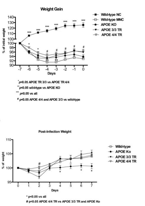
The inability to reach an expected growth rate has been proposed as a biological marker of under nutrition and/or enteric infections in animal models [15,18]. All experimental groups under the low protein diet showed significant short-term weight loss, as compared with the wild-type nourished controls, however APOE 4/4 targeted replacement mice had less weight loss with under nutrition compared with APOE 3/3 and wild-type undernourished mice. APOE knockout mice showed a slightly better weight adaptation than wild-type controls under the low-protein diet (Fig. 1A).
AfterCryptosporidium parvuminfection, undernourished APOE 4/ 4 TR mice had less weight decrements, at day 2 post-inoculum, when the parasite load was heavier (see below) when compared with undernourished APOE knockout and APOE 3/3 TR. APOE3/3 TR mice had the greatest weight loss on day 2. In addition, undernourished infected APOE knockout mice showed significant weight impairments after the 4th day of C. parvum challenge and showed poor growth catch-up after infection (Fig. 1B).
APOE 4/4 TR mice have significantly lower fecal C. parvum oocyst shedding
To better evaluate C. parvum infection in undernourished experimental mice, we assessed the quantity ofC. parvum oocyst shedding in stools using quantitative polymerase chain reaction. From day 3 after infection onward, the APOE 4/4 TR mice showed an accelerated pace of oocyst reduction. After one week post-infection, only one (1 out of 14, 7.4%) experimentally infected –APOE 4/4 TR mouse showed C. parvum shedding in stool (Fig. 2). In addition, the APOE knockout mice had slightly, but significantly lower oocyst shedding per milligram of stool (p,0.05) than APOE3/3 TR mice at days 1 and 3 post- infection, when the oocyst burden in stool was higher.
APOE 4/4 TR have improved intestinal villi when challenged by under nutrition and cryptosporidiosis
Undernourished uninfected mice showed ileal villus shortening and crypt derangement, with almost complete absence of mitoses. Cryptosporidial infections were associated with crypt hyperplasia, villus blunting, and inflammation in the lamina propria and submucosa as opposed to nourished controls (data not shown). Infected undernourished APOE knockout mice showed reduced villus height and more scattered villi as compared to wild-type and APOE4/4 TR. APOE 3/3 TR mice presented slightly reduced crypt depth as compared to wild-type mice (Fig. 3A, B and C). Furthermore, APOE4/4 TR showed better villus height as compared to APOE 3/3 TR and APOE knockout mice following infection and under nutrition (p,0.05) (Fig. 3A and B). In addition infected APOE 4/4 TR showed better villus/crypt ratio as opposed to all other challenged groups (Fig. 3D).
APOE 4/4 TR mice have lower serum triglyceride levels when challenged by under nutrition and
cryptosporidiosis
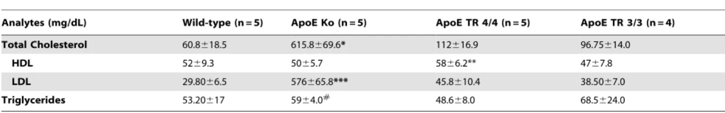
In order to find whether the host response to infections involves changes in lipid levels, we measured serum lipid fractions. FollowingC. parvuminfection in undernourished mice, we found significant increases (8 fold higher) in serum total cholesterol and
Table 1.Primers used in the study.
Primers Sequence (59-39)
Beta-actin AATTTCTGAATGGCCCAGGT TTTGTGTAAGGTAAGGTGTGC Arginase -1 TCTGCCAAAGACATCGTGTA
GGTAGCTGAAGGTCTCTTCC CAT – 1 CACTGCTGATCTGTGTACCT GTGGGGACATAAGATGCTCA RNA 18 sC. parvum CTGCGAATGGCTCATTATAACA
LDL-cholesterol in APOE knockout mice than in C57BL/6J controls.
The APOE 4/4 TR mice had a trend of increased serum HDL cholesterol levels when compared with the other groups. No significant differences were found between APOE 3/3 and APOE 4/4 TR mice. The APOE knockout mice had increased serum triglyceride levels in comparison to APOE 4/4 TR mice. In addition, albeit not significant, APOE 3/3 TR mice showed higher serum triglyceride levels (Table 2).
Undernourished APOE knockout mice have increased ileal pro-inflammatory cytokines with cryptosporidial infection
Ileal cytokine analyses revealed significantly higher IL-1b and IFN-clevels in APOE knockout mice followingC. parvuminfection and undernutrition as compared with respective wild-type controls. In general, there were heavier inflammatory cytokine responses associated with apoE deficiency. Furthermore, both APOE TR undernourished and infected mice showed lower ileal levels of IL-1b than APOE knockout and wild-type challenged mice.
Figure 1.A. Body weight gain (% initial weight) from experimental uninfected and undernourished groups under a low protein diet. APOE 4/4 targeted replacement (APOE 4/4 TR) mice (n = 17) showed 643 a better growth response in comparison with APOE 3/3 targeted replacement (APOE 3/3 TR) mice (n = 8).B.Body weight gain (% initial infection weight) from experimental mice challenged by a compounded malnutrition and Cryptosporidium parvuminsult. Undernourished mice were orally inoculated with 107- unexcysted oocysts diluted in 100ml of PBS. APOE deficient
IFN-c and TNF-a are key cytokines related to C. parvum eradication. They were both increased in wild-type undernour-ished infected mice in comparison with uninfected controls. On the other hand, IL-17 levels were lower in APOE knockout mice after infection and under-nutrition as compared with the APOE knockout uninfected controls and with undernourished infected wild-types. No significant differences were seen between APOE3/ 3 and APOE4/4 TR mice in all cytokines measured (Fig. 4).
APOE 4/4 TR mice showed increased CAT-1, arginase 1, and TLR-9 mRNA when challenged by under nutrition and cryptosporidiosis
In order to assess key mechanisms involved in host-parasite interactions and tissue repair, we addressed the cationic amino acid transporter (CAT-1), arginase-1, and Toll-like receptor 9. We found increased ileal expression of CAT-1 mRNA transcripts in the ileum from undernourished plus infected APOE 4/4 TR mice in comparison to wild-type and APOE-deficient mice on day 7 post-infection. In addition, APOE knockout mice had significantly reduced ileal CAT-1 mRNA expression as compared to wild-type mice (p,0.05). Similar changes were also seen with arginase 1, where the highest ileal levels were observed in APOE4/4 TR mice
(Fig. 5 A and B). In addition we found an increased ileal mRNA levels of TLR9 (Toll-like receptor 9) in APOE 4/4 TR group in comparison to APOE knockout mice (p,0.05). Increased ileal levels of inducible nitric oxide synthase (iNOS) mRNA were found in APOE knockout mice in comparison to all groups (p,0.05) (Fig. 5C and D).
Discussion
Children in developing areas are particularly susceptible to malnutrition associated with Cryptosporidium parvum infections, resulting in debilitating diarrhea as well as growth shortfalls [19]. Since nutritional states and the APOE genetic profile may directly affect the host response against infection [20,21] and therefore influence the risk of acquiring and spreading Cryptospo-ridia, we evaluated this infection in different undernourished C57BL6J genetically-engineered mice, including wild-type, APOE-deficient and APOE target-replacement mice.
In this study, we found that APOE-deficient mice had significantly lessC. parvum oocyst shedding measured by quanti-tative PCR in stools on the first and third days post-infection, when the infection peaks. In agreement with our findings, greater mucosal immune responses against orally-inoculated pathogens,
Figure 2. Fecal shedding of parasites in weaned undernourished C57BL/6 mice orally inoculated with 107-unexcysted Cryptosporidium parvumoocysts per mouse (given in100mL of PBS) on day 7 after the onset of the low protein diet.Results are shown in a log scale a mean6SEM.Cryptosporidium parvumstool oocyst shedding was determined by qRT PCR. Data were expressed as number of parasites per miligram of stool and percentage and number of infected mice with measurable oocyst shedding per day afterCryptosporidium parvum challenge. N above the bars means the number of mice still showing oocyst shedding.
via Th-1 responses, have been shown in APOE knockout mice [22]. In addition, our data suggest that these parasite-killing effects are independent of any increases in IL-17, which was actually found to be low in the ileal tissue of these animals. The Th-17-mediated pathway has been reported to coordinate the immune response against extracellular bacteria [23], suggesting that APOE deficient mice may be poorly adapted against the potential intestinal bacterial translocation due toC. parvum-induced intesti-nal barrier leakage.
Emulsion associated and free-apoE have been shown to bind to LPS (by its exposed hydrophilic domain involving arginine residues) and to redirect LPS from Kupffer cells to liver parenchymal cells, altering lipid metabolism and improving LPS clearance via bile from circulation and reducing LPS-induced lethality [24,25].
Although exhibiting more parasite elimination, undernourished APOE-deficient mice had greater inflammatory cytokine respons-es and mucosal atrophy in the ileal tissue one week-post inoculum, accompanied by greater weight deficits following 7 days of infection. This can be explained by a constitutive pro-inflamma-tory state that has been shown with ApoE deficiency in association with hypercholesterolemia [12]. Hypercholesterolemia found in the C. parvum-infected APOE knockout mice, even sustained following under nutrition, as shown in our study, may reflect impaired intestinal cholesterol delivery to the liver for metabolism. This effect may be associated with increased bile acid transport in the terminal ileum and diminished fecal bile acid loss, and higher serum and intestinal LDL-cholesterol levels [26].
The transient, slightly better gain weight seen in uninfected APOE knockout mice on the low-protein diet, as compared with wild-type controls, may reflect more available cholesterol, regardless of ApoE/lipoprotein delivery, used as an energy source for growth. Prentice and colleagues have shown that energy-enriched diets are critical for catch-up growth following acute enteric infections [27]. In our earlier studies, we found that neonatal undernourished APOE knockout mice failed to thrive after being re-fed [15]. These seemingly slightly contradictory findings may be explained by the low body-fat mass in the neonatal period that would render these mice with insufficient
cholesterol to support rapid growth or the high cholesterol phenotype in APOE knockout mice may occur later after weaning. Overproduction of TNF-a, IL-1b, and decreased IL-10 ileal levels seen in undernourished and infected ApoE knockout mice could be a consequence of the intestinal barrier disruption and increased luminal LPS-trafficking to the lamina propria. Infections and increased LPS intestinal transit (or cytokines activated by it), even in low amounts, could lead to a hypertriglyceridemic state altering liver lipid metabolism [28]. Endotoxin stimulates arginine transport via TNF-a signaling [29], thus impaired intestinal arginine uptake seen with ApoE deficiency (but improved in APOE4 targeted replacement mice) could contribute to poor bacterial removal and increased mucosal inflammation.
Lipoproteins and lipids present in the serum may contribute to the host innate immunity against pathogens [30]. Studies using APOE deficient mice confirmed the role of APOE in host susceptibility to endotoxemia and Klebsiella pneumoniae infection [14], while transgenic mice expressing human APOE3 and APOE4 genes revealed an isoform-specific effect of APOE on the proinflammatory response to lipopolysaccharide [31].
Toll-like receptors are key mediators of the innate immune system againstC. parvuminfections [32]. In our previous study, we have shown that undernourished C57BL6J mice had higher ileal mRNA transcripts for TLR2 and TLR4, but not TLR9, which was diminished byC. parvuminfection [16]. In the current study, we found that APOE4 mice had increased ileal TLR9 transcripts compared with APOE knockout mice. TLR9-immune mediated responses have been found important to control C. parvum infection in a neonatal mouse model [33].
APOE4 may also contribute to elimination of C. parvum infection with an inflammatory response that is more regulated compared to the uncontrolled cytokine production noted in ApoE deficient mice. ApoE also enhances microbial lipid antigen presentation (which can be found with increased bacterial intestinal translocation) to antigen-presenting cells via the low-density lipoprotein receptor (LDLR) which could culminate in natural killer T (NKT) cell activation and cytokine secretion [34,35], an effect that could attenuateC. parvuminfection.
Figure 3. A. Representative ileal histology from orallyCryptosporidium parvuminfected mice. Wild-type, APOE knock-out, and APOE targeted replacement mice (APOE 3/3 TR and APOE 4/4 TR) were fed with a low protein diet during 7 days then infected with 107-unexcystedCryptosporidium
parvumoocysts and euthanized seven days after infection. H&E6400. Scale bar 10mM.B. Ileal villus height;C. crypt depth, andD. villus-crypt ratio
from wild-type, APOE knock-out, and APOE targeted replacement mice (APOE 3/3 TR and APOE 4/4 TR). Morphometrics was done from hematoxylin and eosin stained-sections in at least four animals per group at low magnification. Data are presented as mean6SEM. Comparisons were performed by Students unpairedTtest. Villi and crypts were measured only when their full longitudinal axis was found.
doi:10.1371/journal.pone.0089562.g003
Table 2.Lipid profile of experimental undernourished mice followingCryposporidium parvuminfection (mice orally infected with 107unexcysted oocysts).
Analytes (mg/dL) Wild-type (n = 5) ApoE Ko (n = 5) ApoE TR 4/4 (n = 5) ApoE TR 3/3 (n = 4)
Total Cholesterol 60.8618.5 615.8669.6* 112616.9 96.75614.0
HDL 5269.3 5065.7 5866.2** 4767.8
LDL 29.8066.5 576665.8*** 45.8610.4 38.5067.0
Triglycerides 53.20617 5964.0# 48.6
68.0 68.5624.0
*p,0.001 ApoE Ko vs all;
** p = 0.05 ApoE TR 4/4 vs ApoE TR 3/3; *** p,0.001 ApoE Ko vs all;
#
Finally, APOE4 has been shown to up-regulate the L-arginine selective cationic protein transporter (CAT-1), part of the amino acid-polyamine-organocation (APC) superfamily, which has been associated with increased L-arginine uptake by neuroglia cells. Our data show that CAT-1 is up-regulated in the ileum of undernourishedC. parvuminfected-APOE4 targeted replacement mice compared to APOE3 controls. Increased uptake of L-arginine preferentially by enterocytes may enhance the intestinal barrier function, mucosal blood flow, and the immune system in addition to mucosal repair (cell proliferation and migration) following injury, by increasing polyamine synthesis and constitu-tive nitric oxide synthase (cNOS) activity [36]. Indeed, we have observed that arginine can enhance the killing of cryptosporidial parasites in our murine model, via both iNOS and arginase pathways [37]. In addition, protozoa could utilize arginine and therefore reducing the NO production by the host and weakening the innate immune defenses against infection [37].
Arginase-1 is an enzyme that catalyzes arginine’s conversion into ornithine, which may then enter into the polyamine pathway [38]. Taken with our previous findings and those Colton et al. see
below and reference [46], these findings with ApoE 4/4 targeted replacement transgenic mice suggest that increased uptake of arginine through CAT-1 activity and the arginine shift to the arginase pathway for improving mucosal restitution following C.parvum-induced mucosa injury in undernourished mice.
Recently, we have demonstrated that L-arginine supplementa-tion improved mucosal recovery followingC. parvuminfection plus undernutrition and that ileal arginase 1 was a marker of infection severity [37]. In contrast with that finding, APOE4 targeted replacement mice up-regulated arginase 1 which may indicate that mucosal recovery is still underway. More studies are warranted to evaluate the role of arginase 1 during C. parvum infection and mucosal recovery.
APOE4 targeted gene replacement and APOE knockout mice have been found with increased pro-inflammatory states [39], an effect that is potentially undesirable for neurodegenerative diseases, but beneficial during exposure to gut-infectious patho-gens early in life [40]. The beneficial effect of apoE4 in boosting host innate immunity against infectious agents is a potential explanation for the prevalence of the APOE4 in our genetic pool
Figure 4. Luminex assays from experimental mice for the following ileal pro-inflammatory cytokines: (A) Interleukin 1-b; (B) Interleukin-17; (C) Interferon-gamma; and (D) Tumor necrosis factor alpha. Experimental mice were challenged by a compounded malnutrition andCryptosporidium parvuminsult and samples were harvest on day 7 post-C. parvum inoculum. Wild-type, APOE knock-out, and APOE targeted replacement mice (APOE 3/3 TR and APOE 4/4 TR) were orally inoculated with 107-unexcysted oocysts diluted in 100ml of PBS. Groups have
at least 4 per groups and the results are shown as mean6SEM and are expressed in pg/ml. MNC = uninfected undernouorished control group. MNI = undernourished infected group.
[41], and recognized as a thrifty allele [42]. This gene is considered important to enhance energy usage and storage and to foster host defenses at times of food scarcity and when repeated bouts of infection outbreaks occur and energy usage for catch-up growth and fit immunity are critical requirements for immediate survival [43].
Interestingly, data from the Tsiname population in lowland Bolivia (an indigenous forager-farmer population, with high infectious morbidity and shortened life expectancy) when bearing APOE4 show lower C-reactive protein levels, suggesting low rates of environmental-related infections [44]. In our study, targeted transgenic mice expressing human apoE4 protein have an advantage in eliminating C. parvum oocysts after being exposed to a high load of parasitic inoculum supporting the concept of antagonistic pleiotropy for the APOE4 gene [45].
Altogether, our findings support that APOE4 is protective during C. parvum infections early in life, improving parasite elimination with enhanced intestinal mucosal recovery. These effects may be caused by boosting innate immunity but without the
overt inflammation seen with apoE deficiency and improving arginine uptake and arginase 1-driven mucosal restitution.
The APOE4 allele has been associated with increased nitric oxide production in platelet cells and in macrophages also is associated with increase expression of CAT-1 (cationic amino acid transporter), and with increased arginine uptake in brain microglia [46].
Our mouse model data support the beneficial effect of the APOE4 allele seen in cohort shantytown children afflicted with malnutrition and enteric infections who harbor this allele [20]. Although the orchestrated epithelial restitution and immune-inflammatory responses against malnutrition and enteric infections constitute a multi-factorial gene-to-gene interplay, the APOE gene appears to enable better intestinal adaptation against Cryptosporid-ium parvum.
One limitation of this study is the lack of the undernourished uninfected controls for comparisons of baseline levels for the APOE TR 4/4 and TR3/3 mice. In addition, we cannot rule out that the level of Cryptostoridium excystation after oral inoculation
Figure 5. Quantitative real-time PCR assays from experimental mice for the following ileal mRNA transcripts: (A) cationic amino acid transporter (CAT-1); (B) arginase 1; (C) Toll-like receptor 9 (TLR9); and (D) Inducible nitric oxide synthase (iNOS).Experimental mice were challenged by a compounded malnutrition andCryptosporidium parvuminsult and samples were harvested on day 7 post-C. parvum inoculum. Wild-type, APOE knockout, and APOE targeted replacement mice (APOE 3/3 TR and APOE 4/4 TR) were orally inoculated with 107
-unexcysted oocysts diluted in 100ml of PBS. Groups have at least 4 per groups and the results are shown as mean6SEM and expressed afterb-actin normalization.
may have varied in the different genetic mouse strains used, however, after 6 h of C. parvumchallenge in a model of human HCT-8 epithelial monolayers a considerable excystation of the oocysts is already seen [47].
In summary, our overall findings suggest that ApoE is one of the key players modulating the intestinal architecture and immune and inflammatory responses following malnutrition andC. parvum infection.
Acknowledgments
The authors would like to thank Gina Calabrese and Mary Stout for technical assistance and Glynis Kolling, Ph.D., for helpful comments to improve this manuscript.
Author Contributions
Conceived and designed the experiments: RG RO JR. Performed the experiments: OA DB. Analyzed the data: RP. Contributed reagents/ materials/analysis tools: RG RO JR. Wrote the paper: RG RO JR OA DB. Approved the final version of the manuscript: AL MV CW RO.
References
1. Karanis P, Kourenti C, Smith H (2007) Waterborne transmission of protozoan parasites: a worldwide review of outbreaks and lessons learnt. J Water Health 5: 1–38.
2. Marcos LA, Gotuzzo E (2013) Intestinal protozoan infections in the immunocompromised host. Curr Opin Infect Dis 26: 295–301. doi: 10.1097/ QCO.0b013e3283630be3.
3. Dillingham RA, Lima AA, Guerrant RL (2002) Cryptosporidiosis: epidemiology and impact. Microbes Infect 4: 1059–1066. doi: 10.1016/S1286-4579(02)01630-1.
4. Guerrant RL, Oria RB, Moore SR, Oria MO, Lima AA (2008) Malnutrition as an enteric infectious disease with long-term effects on child development. Nutr Rev 66: 487–505. doi: 10.1111/j.1753-4887.2008.00082.x.
5. Turan N, Ghalwash MF, Katari S, Coutifaris C, Obradovic Z., et al.(2012) DNA methylation differences at growth related genes correlate with birth weight: a molecular signature linked to developmental origins of adult disease? BMC Med Genomics 5: 10. doi: 10.1186/1755-8794-5-10.
6. Pinkerton RC, Oria RB, Kent JW, Jr., Kohli A, Abreu C., et al. (2011) Evidence for genetic susceptibility to developing early childhood diarrhea among shantytown children living in northeastern Brazil. Am J Trop Med Hyg 85: 893–896. doi: 10.4269/ajtmh.2011.11-0159.
7. Mitter SS, Oria RB, Kvalsund MP, Pamplona P, Joventino ES, et al.(2012) Apolipoprotein E4 influences growth and cognitive responses to micronutrient supplementation in shantytown children from northeast Brazil. Clinics (Sao Paulo) 67: 11–18. doi: 10.6061/clinics/2012(01)03.
8. Oria RB, Patrick PD, Blackman JA, Lima AA, Guerrant RL (2007) Role of apolipoprotein E4 in protecting children against early childhood diarrhea outcomes and implications for later development. Med Hypotheses 68: 1099– 1107. doi: 10.1016/j.mehy.2006.09.036.
9. Mahley RW, Rall SC, Jr. (2000) Apolipoprotein E: far more than a lipid transport protein. Annu Rev Genomics Hum Genet 1: 507–537. doi: 10.1146/ annurev.genom.1.1.507.
10. Strittmatter WJ, Bova HC (2002) Molecular biology of apolipoprotein E. Curr Opin Lipidol 13: 119–123.
11. Chapman J, Vinokurov S, Achiron A, Karussis DM, Mitosek-Szewczyk K, et al. (2001) APOE genotype is a major predictor of long-term progression of disability in MS. Neurology 56: 312–316.
12. Jofre-Monseny L, Minihane AM, Rimbach G (2008) Impact of apoE genotype on oxidative stress, inflammation and disease risk. Mol Nutr Food Res 52: 131– 145. doi: 10.1002/mnfr.200700322.
13. Roselaar SE, Daugherty A (1998) Apolipoprotein E-deficient mice have impaired innate immune responses to Listeria monocytogenes in vivo. J Lipid Res 39: 1740–1743.
14. De BN, Netea MG, Demacker PN, Kullberg BJ, van der Meer JW, et al. (2000) Apolipoprotein E-deficient mice have an impaired immune response to Klebsiella pneumoniae. Eur J Clin Invest 30: 818–822. eci715 [pii]. 15. Oria RB, Vieira CMG, Pinkerton RC, De Castro-Costa CM, Lopes MB, et al.
(2007) Apolipoprotein E knockout mice have accentuated malnutrition with mucosal disruption and blunted insulin-like growth factor responses to refeeding. Nutrition Research 26: 427–435. doi: 10.1016/j.nutres.2006.06.020. 16. Costa LB, JohnBull EA, Reeves JT, Sevilleja JE, Freire RS, et al. (2011)
Cryptosporidium-malnutrition interactions: mucosal disruption, cytokines, and TLR signaling in a weaned murine model. J Parasitol 97: 1113–1120. doi: 10.1645/GE-2848.1.
17. Roeschlau P, Bernt E, Gruber W (1974) Enzymatic determination of total cholesterol in serum. Z Klin Chem Klin Biochem 12: 226.
18. Coutinho BP, Oria RB, Vieira CM, Sevilleja JE, Warren CA, et al. (2008) Cryptosporidium infection causes undernutrition and, conversely, weanling undernutrition intensifies infection. J Parasitol 94: 1225–1232. doi: 10.1645/GE-1411.1.
19. Checkley W, Epstein LD, Gilman RH, Black RE, Cabrera L, et al. (1998) Effects of Cryptosporidium parvum infection in Peruvian children: growth faltering and subsequent catch-up growth. Am J Epidemiol 148: 497–506.
20. Oria RB, Patrick PD, Zhang H, Lorntz B, de Castro Costa CM, et al. (2005) APOE4 protects the cognitive development in children with heavy diarrhea burdens in Northeast Brazil. Pediatr Res 57: 310–316. doi: 10.1203/ 01.PDR.0000148719.82468.CA.
21. Vasunilashorn S, Finch CE, Crimmins EM, Vikman SA, Stieglitz J, et al. (2011) Inflammatory gene variants in the Tsimane, an indigenous Bolivian population with a high infectious load. Biodemography Soc Biol 57: 33–52.
22. De BN, Netea MG, Demacker PN, Verschueren I, Kullberg BJ, et al. (1999) Apolipoprotein E knock-out mice are highly susceptible to endotoxemia and Klebsiella pneumoniae infection. J Lipid Res 40: 680–685.
23. Serrano HA (2009) [Helper (TH1, TH2, TH17) and regulatory cells (Treg, TH3, NKT) in rheumatoid arthritis]. Reumatol Clin 5S1: 1–5. doi: 10.1016/ j.reuma.2008.11.012.
24. Rensen PC, Oosten M, Bilt E, Eck M, Kuiper J, et al. (1997) Human recombinant apolipoprotein E redirects lipopolysaccharide from Kupffer cells to liver parenchymal cells in rats In vivo. J Clin Invest 99: 2438–2445. doi: 10.1172/JCI119427.
25. Van OM, Rensen PC, Van Amersfoort ES, Van EM, Van Dam AM, et al. (2001) Apolipoprotein E protects against bacterial lipopolysaccharide-induced lethality. A new therapeutic approach to treat gram-negative sepsis. J Biol Chem 276: 8820–8824. doi: 10.1074/jbc.M009915200.
26. Bhat BG, Rapp SR, Beaudry JA, Napawan N, Butteiger DN, et al. (2003) Inhibition of ileal bile acid transport and reduced atherosclerosis in apoE2/2 mice by SC-435. J Lipid Res 44: 1614–1621. doi: 10.1194/jlr.M200469-JLR200.
27. Hoare S, Poppitt SD, Prentice AM, Weaver LT (1996) Dietary supplementation and rapid catch-up growth after acute diarrhoea in childhood. Br J Nutr 76: 479–490.
28. Feingold KR, Staprans I, Memon RA, Moser AH, Shigenaga JK, et al. (1992) Endotoxin rapidly induces changes in lipid metabolism that produce hypertriglyceridemia: low doses stimulate hepatic triglyceride production while high doses inhibit clearance. J Lipid Res 33: 1765–1776.
29. Pan M, Choudry HA, Epler MJ, Meng Q, Karinch A, et al. (2004) Arginine transport in catabolic disease states. J Nutr 134: 2826S–2829S.
30. Barcia AM, Harris HW (2005) Triglyceride-rich lipoproteins as agents of innate immunity. Clin Infect Dis 41 Suppl 7: S498–S503. 10.1086/432005 [doi]. 31. Zhang H, Wu LM, Wu J (2011) Cross-talk between apolipoprotein E and
cytokines. Mediators Inflamm 2011: 949072. doi: 10.1155/2011/949072. 32. McDonald V, Korbel DS, Barakat FM, Choudhry N, et al. (2013) Innate
immune responses against Cryptosporidium parvum infection. Parasite Im-munol 35: 55–64. doi: 10.1111/pim.12020.
33. Barrier M, Lacroix-Lamande S, Mancassola R, Auray G, Bernardet N, et al. (2006) Oral and intraperitoneal administration of phosphorothioate oligodeox-ynucleotides leads to control of Cryptosporidium parvum infection in neonatal mice. J Infect Dis 193: 1400–1407. doi: 10.1086/503748.
34. Chuang K, Elford EL, Tseng J, Leung B, Harris HW (2010) An expanding role for apolipoprotein E in sepsis and inflammation. Am J Surg 200: 391–397. doi: 10.1016/j.amjsurg.2009.10.017.
35. van den Elzen P, Garg S, Leon L, Brigl M, Leadbetter EA, et al. (2005) Apolipoprotein-mediated pathways of lipid antigen presentation. Nature 437: 906–910. doi: 10.1038/nature04001.
36. Li P, Yin YL, Li D, Kim SW, Wu G (2007) Amino acids and immune function. Br J Nutr 98: 237–252.
37. Castro IC, Oliveira BB, Slowikowski JJ, Coutinho BP, Siqueira FJ, et al. (2012) Arginine decreases Cryptosporidium parvum infection in undernourished suckling mice involving nitric oxide synthase and arginase. Nutrition 28: 678– 85. doi: 10.1016/j.nut.2011.09.011.
38. Wu G, Morris SM, Jr. (1998) Arginine metabolism: nitric oxide and beyond. Biochem J 336 (Pt 1): 1–17.
39. Vitek MP, Brown CM, Colton CA (2009) APOE genotype-specific differences in the innate immune response. Neurobiol Aging 30: 1350–1360. doi: 10.1016/ j.neurobiolaging.2007.11.014.
40. Finch CE, Morgan TE (2007) Systemic inflammation, infection, ApoE alleles, and Alzheimer disease: a position paper. Curr Alzheimer Res 4: 185–189. doi: 10.2174/156720507780362254.
41. Oria RB, Patrick PD, Oria MO, Lorntz B, Thompson MR, et al. (2010) ApoE polymorphisms and diarrheal outcomes in Brazilian shanty town children. Braz J Med Biol Res 43: 249–256. doi: 10.1590/S0100-879X2010007500003. 42. Corbo RM, Scacchi R (1999) Apolipoprotein E (APOE) allele distribution in the
43. Hoare S, Poppitt SD, Prentice AM, Weaver LT (1996) Dietary supplementation and rapid catch-up growth after acute diarrhoea in childhood. Br J Nutr 76: 479–490.
44. Vasunilashorn S, Crimmins EM, Kim JK, Winking J, Gurven M, et al. (2010) Blood lipids, infection, and inflammatory markers in the Tsimane of Bolivia. Am J Hum Biol 22: 731–740. doi: 10.1002/ajhb.21074.
45. Moreau PH, Bott JB, Zerbinatti C, Renger JJ, Kelche C, et al. (2013) ApoE4 confers better spatial memory than apoE3 in young adult hAPP-Yac/apoE-TR mice. Behav Brain Res 243: 1–5. doi: 10.1016/j.bbr.2012.12.043.