385
BILATERAL DEFICIT IN MULTIARTICULAR EXERCISE
FOR UPPER EXTREMITIES
ORIGINAL ARTICLE
Fernando Nazário-de-Rezende1,2
Eduardo G. Haddad1
Gilmar da Cunha Sousa3
Guilherme Goulart de Agostini3
João Elias Dias Nunes3
Moacir Marocolo Jr.1
1. UFTM – Master’s Program in Physical Education of the Federal University of Triângulo Mineiro – Uberaba – MG, Brazil.
2. FISIO2EX – Center for Research and Physical Evaluation in Human Performance of the Antônio Carlos President University – UNIPAC – Uberlândia, MG, Brazil.
3. CENESP – NIAFIS – FAEFI – UFU – Federal University of Uberlândia, MG, Brazil.
Mailing address:
FISIO2EX-UNIPAC - Universidade
Presidente Antônio Carlos
Rua Barão de Camargos, 695 - Centro Uberlândia – MG, Brasil
Cep: 38400-000
Email: [email protected]
LOCOMOTOR APPARATUS IN EXERCISE AND SPORTS
ABSTRACT
Muscular strength is an important component for physical activity and performance of daily living activities. A phenomenon usually observed is that the capacity of generating maximum strength is compromised when the homologous extremities bilaterally contract. This phenomenon is called bilateral deficit. Thus, the aim of this work was to compare the electrical activity of the deltoid muscle, medial portion, during unilateral and bilateral contractions in a converging articulated military press machine, with 90% of maximum voluntary load (MVL), in nine men aged between 20 and 30 years, stature of 174 ± 5cm and body mass of 78 ± 15 kg. The myoelectrical signals were obtained through placement of differential active surface electrodes by EMG System of Brazil, a reference electrode (ground) and a signal conditioner module (electromyograph), which provided numerical data in RMS (root mean square) for results analysis. Each signal collected picked only the concentric phase of the movement and it had duration of three seconds. The results evidenced that during bilateral and unilateral exercise with 90% of MVL, the electric activity of the non-dominant extremity was significantly higher than in the dominant one (p = 0.018). When the values obtained in the work of dominant extremity are summed with the work of non-dominant extremity in the bilateral exercise (2.231 ± 504µv) and compared with the values obtained in the unilateral work (2.663 ± 701µv), bilateral deficit was found (p = 0.018). According to our study, it was verified that the bilateral deficit phenomenon is present in the medium deltoid muscle in the converging multiarticular military press exercise in individuals familiarized with resistance exercises.
Keywords: multiarticular exercise, bilateral deficit, EMG.
INTRODUCTION
Muscular strength is an important component for physical activity and for the development of daily living activities. A generally observed phenomenon is that the capacity to generate maximum strength is compromised when the homologous extremities bilaterally contract. This phenomenon is named bilateral deficit, and it occurs when bilateral maximum voluntary strength is lower than the sum of the unilateral strength of the right and left extremities individually contract1,2.
According to Jakobi and Chilibeck1, bilateral deficit has received considerable attention in the literature. According to the authors men-tioned here, many dynamic studies have found bilateral deficit, con-sidering that isometric studies are more numerous and controversial. The literature related to resistance exercises refers to bilateral deficit as light neural decrease in recruiting of motor units during the development of bilateral tasks, when compared with the sum of unilateral tasks2. Such deficit has been studied through strength and electromyography, confirmed most of the times with the use of uniarticular exercises3-7 being controversial concerning EMG and strength in multiarticular exercises for lower and upper extremities3,8.
The strength acquisition and unilateral and bilateral contractions performance ratio was studied by Schantz et al.3 and Dieën et al.7, who stated that a unilateral muscular contraction results in
signifi-cant increase of neuromuscular activity, while a bilateral contraction results in decrease of this activity.
When load, power and maximum repetitions until exhaustion are analyzed, Simão et al.9,10 and Chaves et al.11 verified the existence of bilateral deficit related to maximal load, but the same episode did not occur with maximal muscular power9. It is supposed that uni and bilateral maximal contractions are characterized concerning their neuromuscular activation, by the recruiting of many muscular groups or in many frequencies through a parallel process of inter-muscular coordination. However, it is known that the intrainter-muscular coordination is a determining performance factor in sports in which maximal unilateral voluntary contractions are used12.
Studies related to bilateral deficit and uni and multiarticular exercises were conducted by Schantz et al.3, who verified decrease of 10% in maximal isometric voluntary strength during bilateral leg extension (multiarticular) and bilateral superiority of 4% in knee extension exercises (uniarticular) when compared with the unila-teral sum. These differences were not followed by the myoelectric signal, which obtained similar behavior during uni and bilateral contractions for the multiarticular exercise.
386
teral contractions. The electrodes used were surface, active, differential and single (Lynx Electronics Ltda., São Paulo, SP, Brazil), composed by two pure silver (Ag) rectangular parallel bars, each one 10mm long, 1mm wide and 10mm away from each other; 20mm wide x 41mm long and 5mm thick acrylic resin capsule; one meter long cable; 20 times gain; CMRR – Common Mode Rejection Ratio of 93dB and one plaque grounding electrode (Bio-logic Systems Corp. –SP Médica, Científica e Comercial Ltda., São Paulo, SP, Brazil), composed by a stain-less steel disc measuring 30mm of diameter and 1.5mm of thickness and cable of 1m attached, which was placed at the ulnar head of the volunteers with the purpose to eliminate external interference 16,17.
The electrodes were attached to the skin, positioned appro-ximately 4 ± 2cm away from the lateral border of the acromion, in a region where greater volume of muscle belly of the medial portion of the deltoid muscle was clear.
Electromyograph
The EMG collection of the studied muscles was obtained with a conditioning signal module (electromyograph), with simultaneous acquisition of up eight differential channels, channel entry impedance of 10GΩ in differential modules, 12 bits of resolution, low-pass filter of 20Hz to 5Hz and CMRR of 93db at 60Hz, entry band of –10a +10v and data acquisition system (Alc-EMG) which provided number data in RMS (root mean square) for results analyses. The electromyography was adjusted with 4,960 times gain, which guaranteed the necessary amplification for the analog-digital conversion process and sampling number of 6,000 and frequency per channel of 2,000Hz, resulting in total acquisition time of three seconds.
Goniometer
Knee and elbow joint angles were measured with the use of a CARCI plastic universal goniometer with 35 cm of length, used before the tests performance when the volunteer was already po-sitioned on the machine.
The knee joint was measured with the screw of the goniometer being placed on the lateral condyle of the femur, laterally aligned on the longitudinal axis of the thigh, from the major trochanter to the lateral condyle and on the axis between the head of the fibula until the lateral malleolus. The elbow joint had the goniometer aligned along the mean lateral line of the humerus, from the humeral head to the lateral epicondyle and from the mean lateral line of the radius to its styloid process 18.
The articular angles of the upper and lower extremities, in the be-ginning of the movement, have not been exactly delimited; however, the knee (106º ± 5º) and elbow joint positions (105º ± 5º) were similar to those adopted in their training routines.
Converging articulated military press
In order to determine load in one repetition maximum (1RM) and performance of bilateral exercise, a machine named Converging Articulated Military Press by MASTER, was used for the study. Such machine simulates the military press performed with dumbbells.
Movement performance
The volunteers sat in the machine with trunk and head rested on the rest and feet on the ground. After load selection with volun-teers already positioned, the electrodes were placed on the studied muscles in contralateral extremities are simultaneously activated4,7.
Herbert and Gandevia13 suggest that bilateral deficit in large muscles occurs due to problems in posture maintenance and con-sequently lower efficiency in strength transmission.
However, little is known about the bilateral deficit phenome-non and its correlation with different loads during performance of multiarticular exercises for the upper extremity in practitioners of resistance exercises. Thus, new tests related to bilateral deficit should be performed using multiarticular exercises for the upper extremity with the goal to better understand this phenomenon, since, according to Gardiner14 the mechanisms through which the bilateral deficit occurs are not known yet. Thus, our hypothesis is that bilateral deficit is evident by the highest number of muscles involved in the same task.
Keeping in mind the importance of a correct diagnosis of bila-teral deficit and resistance exercises, this study was developed with the aim to compare the electromyographic signals emitted by the right medial deltoid muscle (RMD) and left medial deltoid (LMD) during uni and bilateral contractions performed in multiarticular exercise with 90% of MVL.
METHOD
Sample
The medial deltoid muscle (MD) was uni and bilaterally analyzed through electromyography in nine men aged between 20 and 30 years, stature 174 ± 5cm and body mass 78 ± 15kg. Inclusion criteria of the study were: practice of resistance exercises for at least three months and without history of muscular or articular diseases which could interfere in the results.
General procedures
Before the electromyographic recording, the volunteers received information about the research, and were submitted to familiarization procedures. The volunteers received explanations and simulations about the most adequate posture for performance of the exercise, initial and final position of each movement, performance velocity and verbal command given by the electromyograph evaluator. Sub-sequently, they signed a consent form for participation in the study and publishing of the results according to resolution number 196/96 of the National Health Board. This study was approved by the ethics and research committee of the UFTM under protocol number 2,230. In order to establish a specific muscular preparation, the vo-lunteers performed three sets of 15 repetitions without overload.
Maximum voluntary workload test
All volunteers were submitted to a concentric bilateral maximal voluntary load test (MVL) one day before the collection, performed according to Nazário-de-Rezende et al.15. During the test, the load was added ranging from 1 and 2kg at every attempt. The load adopted for the experiment was of 90% of maximal to which all volunteers were submitted during the training sessions which pre-ceded the experiment date.
Electrodes
bila-387 RESULTS
The results obtained of the analysis of the mean of the RMS values of the electric activity of the nine volunteers and for each muscle evidenced that, during bilateral and unilateral exercise with 90% of MVL, the electric activity of the non- dominant ex-tremity was significantly predominant over the dominant one, as shown in figure 2 e tabela 1.
When the EMG values of the two limbs obtained in the bila-teral work are summed and compared with the values unilabila-terally obtained, also by the sum of the two limbs, significant differen-ces were found between the two analyzed variables (P = 0.018) (table 2 and figure 3).
In the comparison of the laterality between the bilateral do-minant deltoid versus unilateral dodo-minant deltoid, tendency to bilateral deficit with p = 0.054 was observed. In the comparison between the bilateral dominant deltoid with the unilateral non--dominant deltoid, the results were significantly higher for unilateral work with p = 0.015.
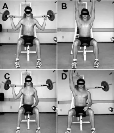
muscles. The movement started with the volunteer’s arms in semi abduction, forearms in flexion on the front plane, hands proned and head erect with eyes looking to the front (figure 1).
The movement occurred with arm abduction and forearm ex-tension simultaneously following the path allowed by the machine, being it the concentric phase of the exercise which had duration of three seconds.
Data collection was performed according to Hakkinen et al.5, and the bilateral tests preceded the unilateral ones. For each test (uni and bilateral) electromyographic means of three attempts were used to minimize accuracy of our collection and mean of the sta-tistical analysis was used.
Figure 1. Converging articulated military press exercise: beginning of bilateral movement (A) and end of bilateral movement (B); beginning of unilateral movement (C) and end of unilateral movement (D).
Figure 2. Mean and standard deviation of the RMS values of the electric activity of the bilateral dominant extremity (D BL) and bilateral non-dominant extremity (ND BL), unilateral dominant (D UL) and unilateral non-dominant (ND UL) with 90% of MVL for the nine volunteers. * (P < 0.05).
Recovery interval
The volunteers were told not to perform any training on the previous day of the EMG recording with the aim to avoid possible fatigue effects and alterations in the results. The volunteers, after finishing the movement, remained seated, kept upper extremities relaxed along the body during five minutes of rest between at-tempts, both for the EMG and MVL tests in order to avoid or mini-mize the fatigue effects and replace their energy6.
Statistical analysis
Initially, the Shapiro-Wilk’s W normality test was applied. After this test, normality of all sample groups was verified. Comparison of the two muscles (right medial deltoid added to left one, bilaterally and unilaterally) with load of 90% of MVL for the nine volunteers occurred with the application of the t test to the data under analy-sis, being the significance level established at p < 0.05 or 5%. The statistical program used was the Statistica 6.0, USA.
Contraction
Table 1. Values expressed in RMS (µν) of the electric activity of the deltoid muscle (medial portion) during unilateral and bilateral contractions performed with 90% of MVL. * (P < 0.05).
Muscular contraction (μν)
Volunteer Deltoid/bilateral Deltoid/unilateral
1 3.038 3.309
2 2.758 3.426
3 1.888 1.950
4 2.036 2.020
5 1.910 2.093
6 1.659 1.778
7 1.817 3.209
8 2.158 2.747
9 2.813 3.439
388
DISCUSSION
In order to improve the discussions related to bilateral deficit and verify possible unilateral and bilateral motor efficiency in the recruiting of muscle fibers, some issues should be clarified concer-ning the application of the bilateral deficit term. It was observed that this phenomenon may be found through EMG4, in strength3 and even in the EMG/strength ratio7 and studied concerning maximum repetitions19. Thus, for better understanding, we can define bilateral myoelectrical deficit as light or remarkable decrease in the EMG signal (UM recruiting) during the development of bilateral work when com-pared with the sum of unilateral work. Bilateral deficit on its turn can be defined as decrease in strength maximum quantity that a muscle or muscular group can generate during the development of bilateral work when compared with the sum of unilateral work. Endurance deficit of bilateral strength may be defined as the lowest capacity of strength maintenance during successive bilateral repetitions until exhaustion compared with the sum of unilateral repetitions.
The literature has not reached a consensus about the bilateral
Figure 3. Values expressed in RMS (µν) of electric activity of the deltoid muscle (medial portion) during unilateral and bilateral contractions performed with 90% of MVL. * (P < 0.05).
deficit phenomenon in multiarticular exercises for upper and lower limbs5,20,21. Our hypothesis was that a myoelectric deficit would not be evident for complex exercises such as converging articulated military press (i.e. forearm extension combined with arm abduc-tion). This hypothesis was based on the literature reviews which presented contradictory studies concerning the onset of myoelec-tric bilateral deficit in multiarticular exercises for upper and lower limbs such as in the leg press exercise (lower limbs). Schantz et al.3 found decrease of 10% in maximal voluntary isometric strength during bilateral leg extension (multiarticular) when compared with the unilateral with no difference in the electric signal of the vastus lateralis muscle, possibly for greater mechanic efficiency in the re-cruiting of muscle fibers during unilateral contractions compared with the bilateral ones.
Taniguchi20 found deficit of bilateral strength for leg press and bench press in men and women, as well as Janzen et al.21, ho verified bilateral deficit related to maximum load (kg) both for leg press and row exercise with no use of electromyographic analysis. Secher et al.8 found bilateral deficit in multiarticular exercises as leg press (lower limb), but not for the bench press (upper limb/multiarticular) in a group of younger volunteers.
Janzen et al.21 reported in their study that bilateral deficit appe-ars due to the neural inhibition during bilateral tasks compared with the unilateral contractions, being the nervous system more involved during contractions which involve multiarticular exercises; consequently, exercises which involve the movement of multiple joints may be more sensitive to bilateral deficit than exercises which involve movement in a single joint. Our results support this hy-pothesis and can be explained by the EMG analysis of an agonist muscle in multiarticular exercise, which favors higher level of inter-muscular coordination for both contractions in individuals trained in resistance exercises, facilitating hence motor performance for unilateral exercises. However, it is necessary that more sinergists are evaluated among themselves, since it is a multiarticular exercise which presents different strength moments in different joints in the entire range of motion.
Oda and Moritani4 studied the bilateral deficit during isometric strength and hand myoelectric activity. These authors concluded that bilateral deficit for strength and EMG is associated to the re-duction of movements related to cortical power, suggesting that this bilateral deficit is caused by an inter-hemisphere inhibition. Similar conclusions were taken by Dieën et al.7, who tested the hypothesis that inter-hemisphere inhibition may result in reduction of the neural drive in bilateral efforts when compared with unilateral efforts, both in small and large muscles, being the electromyogra-phic deficit similar to the strength deficit. It was concluded that the reduction of neural drive was the cause of the bilateral deficit, limiting performance in maximal contractions.
Although comparisons cannot be made, our findings disagree with the ones by Schantz et al.3 in which bilateral myoelectric deficit was not found either, possibly because in this study the authors assessed a single quadriceps muscle in isolation.
Reduction or elimination of bilateral deficit could be considered a neural adaptation to strength training, indicating added abili-ty to activate agonists in bilateral movements. Although bilateral activities reduce deficit, performance in unilateral exercises may
Contraction
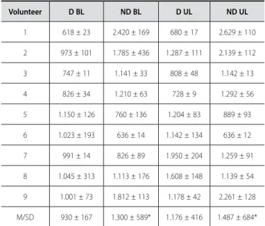
Table 2. Values expressed in RMS (µν) of the electric activity of the deltoid muscle (medial portion) during unilateral and bilateral contractions performed with 90% of MVL. * (P < 0.05).
Volunteer D BL ND BL D UL ND UL
1 618 ± 23 2.420 ± 169 680 ± 17 2.629 ± 110
2 973 ± 101 1.785 ± 436 1.287 ± 111 2.139 ± 112
3 747 ± 11 1.141 ± 33 808 ± 48 1.142 ± 13
4 826 ± 34 1.210 ± 63 728 ± 9 1.292 ± 56
5 1.150 ± 126 760 ± 136 1.204 ± 83 889 ± 93
6 1.023 ± 193 636 ± 14 1.142 ± 134 636 ± 12
7 991 ± 14 826 ± 89 1.950 ± 204 1.259 ± 91
8 1.045 ± 313 1.113 ± 176 1.608 ± 148 1.139 ± 54
9 1.001 ± 73 1.812 ± 113 1.178 ± 42 2.261 ± 128
389
constitute an important strategy with the aim to conserve strength, especially in relevant asymmetry situations7.
In pre-puberty children, the hypothesis of bilateral deficit for upper limb was tested by Germain et al.22 in the isometric flexion action of the dominant arm. It was demonstrated that no muscular activity decreased or alterations in recruiting were considered evi-dent in the electromyographic parameters. It was concluded that there was no strength or recruiting bilateral deficit. These data are not in agreement with the ones obtained by this investigation. Ho-wever, the lack of experience with strength exercises from the side of these children and the analysis of only one muscle may influen-ce the results mentioned above. It is important to categorize the movements studied to establish consistency; since bilateral deficit is an unstable phenomenon, its presence should be considered in the context of the studied movement, either uni or multiarticular for upper or lower extremity.
Behm et al.23 studied the increase of the bilateral muscular activation versus unilateral with multi and uniarticular contractions in trained and untrained volunteers in resistance exercises. The isometric activations of the quadriceps between single knee extension and squat exercises were tested. Significant differences have not been found between the maximum voluntary contraction of the dominant leg during uni and multiarticular exercises of leg extension. However, in untrained volunteers, the non-dominant leg during knee multiarticular knee extensions presented less strength than the dominant leg. According to these authors, bilateral deficit can be expressed due to the lower trust in the non-dominant limb, data which are in agreement with our study, since it became visible that the non-dominant side presented electric activity significantly higher than the dominant side for bilateral contractions. This episode was probably reached by greater neural drive for the non-dominant limb, by the existence of possible deficiencies in the intermuscular coordination levels during movement, causing greater recruiting of motor units. Opposite data were found by Simão et al.9,10 and
Chaves et al.11, in which unilateral differences were not observed in load and maximum muscular power6.
Some considerations must be mentioned concerning our fin-dings. The non-dominant side may have presented greater electric activity, since the lack of intermuscular coordination promotes lower synergist recruiting, requiring hence greater recruiting of motor units of the agonist.
Tassi et al.24 analyzed the bilateral behavior of one muscle of the thigh and, contrary to our findings, verified Strong potential of the dominant limb over the non-dominant. In those authors’ opinion, the dominant limb is more demanded in daily situations, providing considerable muscular development compared with the muscles of the non-dominant limbs. Thus, the daily muscular recruiting contri-butes to the anatomic and functional asymmetry.
It is possible that with the chronic effect of the strength training and the improvement of recruiting standard of the motor units, the differences between the muscular contractions of opposite si-des become less evident, perhaps due to the neural transfer effect mentioned by Moritani and De Vries25, Sale2, Simão6, Brentano and Pinto26. These data make us conclude that adaptation to chronic strength training eliminates bilateral deficit.
In practical terms, for prescription of neuromuscular training with use of multiarticular resistance exercises practice of unilateral exercise can be used as strategy, aiming higher level of intramus-cular coordination for trained subjects.
CONCLUSION
According to our study, it was verified that the bilateral deficit phenomenon is present for the medial deltoid muscle in the con-verging multiarticular military press exercise in individuals familiarized with resistance exercises.
All authors have declared there is not any potential conflict of interests concerning this article.
REFERENCES
1. Jakobi JM, Chilibeck Pd. Bilateral and unilateral contractions: possible differences in maximal voluntary force. Can J Appl Physiol 2001;26:12-33.
2. Sale DJ. Neural Adaptation to Resistance Training. Medicine & Science in Sports & Exercise 1988;20:135-45. 3. Schantz PG, Moritani T, Karlson E, Johansson E, Lundh A. Maximal voluntary force of bilateral and
unilateral leg extensoin. Acta Physiol Scand 1989;136:185-92.
4. Oda S, Moritani T. Movement-related cortical potentials during handgrip contractions with special reference to force and electromyogram bilateral deficit. Eur Jour Appl Physiol 1995;72:1-5. 5. Hakkinen K, Kalinen M, Linnamo V, Pastinen UN, Newton RU, Kraemer WJ. Neuromuscular adaptation
during bilateral versus unilateral strenght training in middle-aged and elderly men and women. Acta Physiol Scand 1996;158:77-88.
6. Simão R. Déficit bilateral – Comparação das cargas máximas no trabalho uni e bilateral. Revista Baiana de Educação Física 2001;2:15-21.
7. Dieën, JHV, Ogita F, Haan, F. Reduced Neural Drive in Bilateral Exertions: A Performance-Limiting Factor? Med Sci Sports Exerc 2003;35:111-8.
8. Secher NH, Rube N, Elers J. Strength of two- and one-leg extension in man. Acta Physiol Scand 1988;134:333-9.
9. Simão R, Monteiro WD, Araújo CGS. Potência Muscular Máxima na Flexão do Cotovelo Uni e Bilateral. Rev Bras Med Esporte 2001;7:157-62.
10. Simão R, Lemos A, Viveiros LE, Chaves CPG, Polito MD. Força muscular máxima na extensão da perna uni e bilateral. Revista Brasileira de Fisiologia do Exercício 2003;2:47-57.
11. Chaves CPG, Guerra CPC, Moura SRG, Nicoli AIV, Félix I, Simão R. Déficit bilateral nos movimentos de flexão e extensão de perna e flexão do cotovelo. Rev Bras Med Esporte 2004;10.
12. Kroll W. Central facilitation in bilateral versus unilateral isometric contractions. Am J Phys Med 1965;44:218-23.
13. Herbert RD, Gandevia SC. Muscle Activation in Unilateral and bilateral efforts assessed by motor nerve and cortical stimulation. J Appl Physiol 1996;80:1351-6.
14. Gardiner PF. Neuromuscular aspects of physical activity. Champaign: Human Kinetics, 2001. 15. Nazario-de-Rezende F, Sousa GC, et al. Electromyographic study of the rectus femoris and bíceps
femoris (long head) muscle during bilateral isotonic contraction in a 45o Leg Press Apparatus. Biosci. Journal 2006;22:95-104.
16. Portney, L. Eletromiografia e testes de velocidades de condução nervosa. In: O’Sullivan SB, Schmitz TJ. Fisioterapia: avaliação e tratamento. 2a. ed. São Paulo: Manole, 1993;183-223.
17. Mathiassen SE, Winkel J, Hagg GM. Normalization of Surface EMG amplitude from the upper trapezius muscle in ergonomic studies. J Eletromyogr Kinesiol 1995;4:197-226.
18. Gary L, Harrelson ED. Mensuração na reabilitação. In: Andrews JR, Harrelson GL, Wilk KE. Reabilitação Física das Lesões Desportivas. 2a. ed. Rio de Janeiro: Guanabara Koogan, 2000;42-60.
19. Monteiro WD, Simão R. Existe déficit bilateral na realização de 10 RM em exercícios de braços e pernas? Rev Bras Med Esporte 2006;12.
20. Taniguchi Y. Relationship between the modifications of bilateral defcit in upper and power limbs by resistance training in humans. Eur J Appl Physiol 1998;78:226-30.
21. Janzen CL, Philip D, Chilibeck K. Shawn Davison. The effect of unilateral and bilateral strength training on the bilateral deficit and lean tissue mass in post-menopausal women. Eur J Appl Physiol 2006;97:253-60. 22. Germain P, Germain Y, Taoutaou Z, Mimouni N, Halin R, Buttelli O. Déficit bilatèral chez l’ enfant prépubè-re non entraîné Bilateral Deficit for untrained pprépubè-repubertal childprépubè-ren. Science & Sports 2004;19:43-7. 23. Behm DG, Power KE, Drinkwater EJ. Muscle activation is enhanced with – and uniarticular bilateral
versus unilateral. Can J Appl Physiol 2003;28:38-52.
24. Tassi N, Filho JG, Gonçalves M, Vitti. M, Krool LB. Electromyographic bahavior of the Biceps Femuris mus-cle during knee extension and flexion performed on the leg press. Bras J Morphol Sci 1998;15:17-22. 25. Moritani MAT, De Vries HA. Neural Factors Versus Hypertrophy in the Time Course of Muscle Strength
Gain. Am J Phys Med 1979;58:115-30.